. 2009 May;37(8):2529-38.
doi: 10.1093/nar/gkp122. Epub 2009 Mar 5.

The Krüppel-like zinc finger protein Glis3 directly and indirectly activates insulin gene transcription

Affiliations

The Krüppel-like zinc finger protein Glis3 directly and indirectly activates insulin gene transcription

Yisheng Yang et al. Nucleic Acids Res. 2009 May.

Abstract

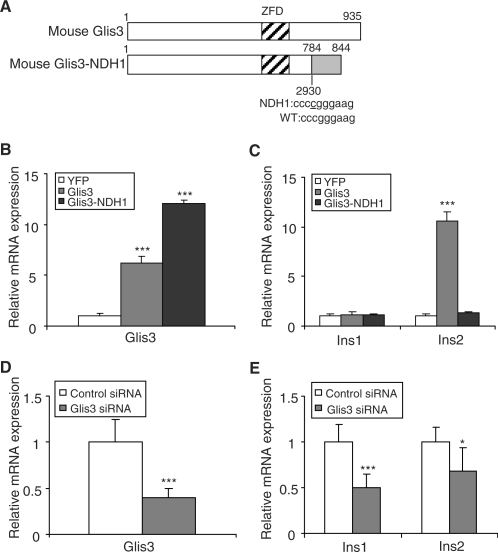
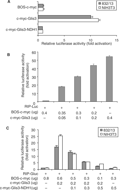
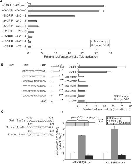
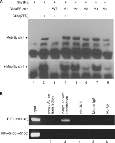
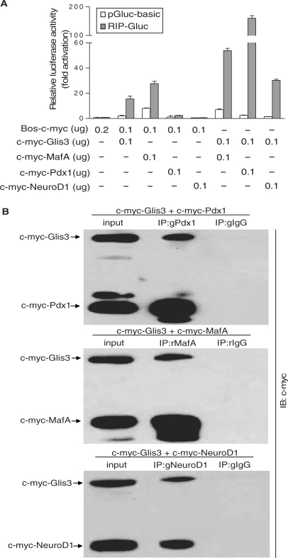
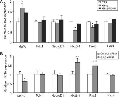
Glis3 is a member of the Krüppel-like family of transcription factors and is highly expressed in islet beta cells. Mutations in GLIS3 cause the syndrome of neonatal diabetes and congenital hypothyroidism (NDH). Our aim was to examine the role of Glis3 in beta cells, specifically with regard to regulation of insulin gene transcription. We demonstrate that insulin 2 (Ins2) mRNA expression in rat insulinoma 832/13 cells is markedly increased by wild-type Glis3 overexpression, but not by the NDH1 mutant. Furthermore, expression of both Ins1 and Ins2 mRNA is downregulated when Glis3 is knocked down by siRNA. Glis3 binds to the Ins2 promoter in the cell, detected by chromatin immunoprecipitation. Deletion analysis of Ins2 promoter identifies a sequence (5'-GTCCCCTGCTGTGAA-3') from -255 to -241 as the Glis3 response element and binding occur specifically via the Glis3 zinc finger region as revealed by mobility shift assays. Moreover, Glis3 physically and functionally interacts with Pdx1, MafA and NeuroD1 to modulate Ins2 promoter activity. Glis3 also may indirectly affect insulin promoter activity through upregulation of MafA and downregulation of Nkx6-1. This study uncovers a role of Glis3 for regulation of insulin gene expression and expands our understanding of its role in the beta cell.


Figures

Figure 1.
Figure 1.
Glis3 stimulates insulin gene transcription. (A) Schematic representation of the mouse Glis3 and Glis3-NDH1 constructs. (B, C) 832/13 cells were stably infected with mouse Glis3 and Glis3-NDH1 constructs or a YFP control retroviral construct. Mouse/rat Glis3 (B) and rat Ins1 and Ins2 (C) mRNA levels were quantified by qPCR relative to cyclophilin A. The Glis3 and Glis3-NDH1 values are shown relative to YFP-expressing cells and are expressed as the mean ± SD. (D, E) 832/13 cells were transiently transfected with siRNA against rat Glis3 or a control siRNA. Glis3 (D) and rat Ins1 and Ins2 (E) mRNA levels were quantified by qPCR relative to cyclophilin A. The relative Glis3, Ins1 and Ins2 expression levels in Glis3 siRNA transfected cells were compared to those in control siRNA-treated cells and presented as the mean ± SD. Each experiment was performed in triplicate and repeated three times. *P < 0.05; ***P < 0.001.
Figure 2.
Figure 2.
Transcriptional activity of Glis3 and Glis3-NDH1. (A) A β cell line (832/13) and a non-β cell line (NIH3T3) were co-transfected with RIP-Gluc, the internal control SRα-SEAP, and Glis3 or Glis3-NDH1 expression constructs as indicated. Gaussia luciferase assay was performed at 48 h after transfection. The fold change of RIP promoter activity caused by the c-myc-Glis3 constructs were compared to that of the BOS vector expressing the c-myc tag alone. Results are presented as mean ± SD. (B) Dose-dependent induction of RIP-mediated luciferase expression by Glis3. 832/13 cells were co-transfected with RIP-Luc and BOS-c-myc together with increasing amounts of c-myc-Glis3 as indicated. The amount of BOS-c-myc vector was adjusted to maintain the same amount of total DNA in each well. (C) Inhibition of Glis3-induced luciferase expression by the Glis3-NDH1 mutant. 832/13 or NIH3T3 cells were co-transfected with RIP-Gluc, c-myc-Glis3 and BOS-c-myc together with increasing amounts of c-myc-Glis3-NDH1 as indicated.
Figure 3.
Figure 3.
Identification of Glis3 DNA-binding sequence in RIP region. (A) The rat insulin 5′ flanking serial deletion constructs (0.2 µg) were co-transfected with BOS-c-myc (0.2 µg) or c-myc-Glis3 (0.2 µg). The activity of each construct is expressed relative to the background activity of BOS-c-myc (equal to 1.0). (B) The sequences and activities of the wild-type and mutation constructs (M1–M5) between −255 and −241 of RIP are shown. The mutated nucleotides are underlined. 832/13 cells were co-transfected with c-myc-Glis3 (0.2 µg) and the various RIP mutant constructs (0.2 µg) as indicated. Dual-luciferase assays were performed at 48 h after transfection. The activity of each construct was normalized to that induced by the co-transfection of BOS-c-myc and −280 RIP-Luc. (C) Alignment of the 15-bp sequences (Glis3RE) located between −255 and −241 of the rat Ins2 promoter compared to mouse and human. (D) Schematic view of five tandem copies of the Glis3RE fused upstream of the RIP TATA box to drive expression of luciferase [(Glis3RE)5-Luc]. 832/13 cells were co-transfected with the rat or human (Glis3RE)5-Luc with c-myc-Glis3 or c-myc-Glis3-NDH1 constructs. The activity of each construct was normalized to that obtained with BOS-c-myc. Each experiment was performed in triplicate and repeated three times. Minor variation between experiments in the basal expression with deletion of sequences between −280 and −240, with a decrease of 42.5% in (A) and 32% in (B).
Figure 4.
Figure 4.
Glis3 binds to the RIP Glis3 response element (Glis3RE). (A) Synthetic biotinylated RIP sequence from −265 to −230, containing Glis3RE, was used as a probe in EMSA with an in vitro translated Glis3 ZFdomain [Glis3 (ZFD)] peptide. The specificity of the protein–DNA complex (lane 2) was confirmed by competition with a 2-fold molar excess of unlabeled wild-type competitor (lanes 3) but not a 2-fold molar excess of the mutants M1 to M5 (lanes 4–8). The mobility shift bands with asterisk showed the results of similar experiment using 20-fold molar excess wild-type or five mutant competitors. The specific bands are indicated by arrows. (B) The c-myc-Glis3–DNA complex was immunoprecipitated by c-myc antibody from 832/13 cells which had been transfected with c-myc-Glis3. Immunoprecipitated DNA was purified and analyzed by PCR using primers specifically spanning the Glis3RE region (RIP −280 to +8) or region remote from Glis3RE (−5400 to −5150). Lane 1, total input DNA (1:100 dilution); lane 2, DNA immunoprecipitated with c-myc Ab without c-myc-Glis3 transfection; lane 3, DNA immunoprecipitated with c-myc Ab with c-myc-Glis3 transfection; lane 4, no DNA template; lane 5, immunoprecipitation with mouse IgG; lane 6, DNA precipitated in the absence of antibody.
Figure 5.
Figure 5.
Glis3 synergistically induces RIP-driven transcriptional activity with Pdx1, MafA and NeuroD1. (A) NIH3T3 cells were co-transfected with RIP-Gluc and c-myc-Glis3, c-myc-MafA, c-myc-Pdx1 and c-myc-NeuroD1 individually or in combination with c-myc-Glis3 as labeled. The relative luciferase activity is presented compared to that of Bos-c-myc with pGluc-basic (equal to 1.0). The background luciferase activity of pGluc-basic also is shown. (B) 832/13 cells were transfected with c-myc-Glis3 and c-myc-Pdx1, c-myc-MafA or c-myc-NeuroD1. Forty-eight hours after transfection, nuclear proteins were isolated and immunoprecipitated either with a rabbit anti-mouse MafA, goat anti-mouse Pdx1 or goat anti-mouse NeuroD1 antibody or appropriate control IgGs. The immunoprecipitates were analyzed with western blotting using anti-c-myc antibody. g: goat; r: rabbit.
Figure 6.
Figure 6.
Glis3 regulates islet-enriched transcription factors expression. (A) 832/13 cells were stably infected with Glis3, Glis3-NDH1 or the control YFP retroviral constructs, and MafA, Pdx1, NeuroD1, Nkx6-1, Pax6 and Pax4 mRNA levels were analyzed by qPCR relative to cyclophilin A. The expression values were normalized to those for YFP infected cells and presented as mean ± SD. (B) 832/13 cells were transiently transfected with siRNA against rat Glis3 or control siRNA. MafA, Pdx1, NeuroD1, Nkx6-1, Pax6 and Pax4 mRNA expression were analyzed by qPCR. The expression values were normalized to those for control siRNA treated cells and presented as mean ± SD. *P < 0.05; **P < 0.01; ***P < 0.001.

Similar articles

Cited by

References

    1. Kim YS, Lewandoski M, Perantoni AO, Kurebayashi S, Nakanishi G, Jetten AM. Identification of Glis1, a novel Gli-related, Kruppel-like zinc finger protein containing transactivation and repressor functions. J. Biol. Chem. 2002;277:30901–30913. - PubMed
    1. Zhang F, Nakanishi G, Kurebayashi S, Yoshino K, Perantoni A, Kim YS, Jetten AM. Characterization of Glis2, a novel gene encoding a Gli-related, Kruppel-like transcription factor with transactivation and repressor functions – roles in kidney development and neurogenesis. J. Biol. Chem. 2002;277:10139–10149. - PubMed
    1. Kim YS, Nakanishi G, Lewandoski M, Jetten AM. GLIS3, a novel member of the GLIS subfamily of Kruppel-like zinc finger proteins with repressor and activation functions. Nucleic Acids Res. 2003;31:5513–5525. - PMC - PubMed
    1. Attanasio M, Uhlenhaut NH, Sousa VH, O’Toole JF, Otto E, Anlag K, Klugmann C, Treier AC, Helou J, Sayer JA, et al. Loss of GLIS2 causes nephronophthisis in humans and mice by increased apoptosis and fibrosis. Nat. Genet. 2007;39:1018–1024. - PubMed
    1. Kim YS, Kang HS, Herbert R, Beak JY, Collins JB, Grissom SF, Jetten AM. Kruppel-like zinc finger protein Glis2 is essential for the maintenance of normal renal functions. Mol. Cell Biol. 2008;28:2358–2367. - PMC - PubMed

Publication types

MeSH terms