. 2009 Aug;50(8):3783-93.
doi: 10.1167/iovs.08-3051. Epub 2009 Mar 5.

Bacillus cereus-induced permeability of the blood-ocular barrier during experimental endophthalmitis

Affiliations

Bacillus cereus-induced permeability of the blood-ocular barrier during experimental endophthalmitis

Andrea L Moyer et al. Invest Ophthalmol Vis Sci. 2009 Aug.

Abstract

Purpose: The purpose of this study was to determine to what extent blood-retinal barrier (BRB) permeability occurred during experimental Bacillus cereus endophthalmitis and whether tight junction alterations were involved in permeability.

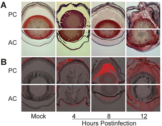
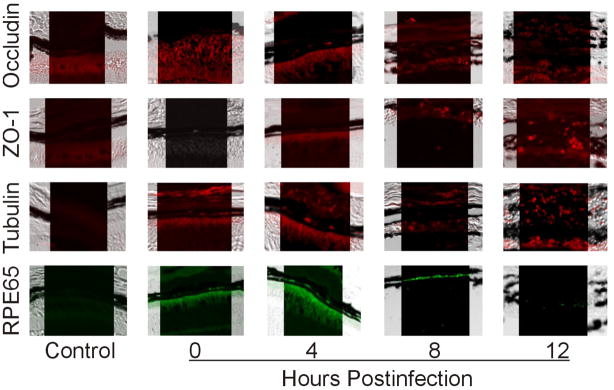
Methods: Mice were intravitreally injected with 100 colony-forming units of B. cereus, and eyes were analyzed at specific times after infection for permeability to fibrin and albumin, quantitation of intraocular plasma constituent leakage, production of inflammatory cytokines, and alterations in tight junction protein localization and expression at the level of the retinal pigment epithelium.

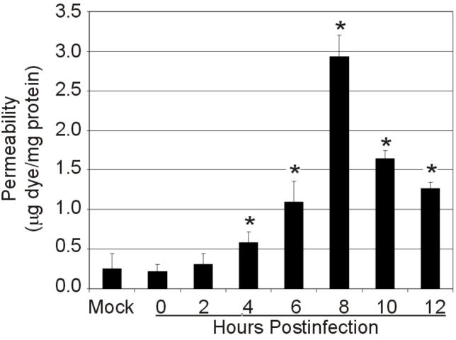
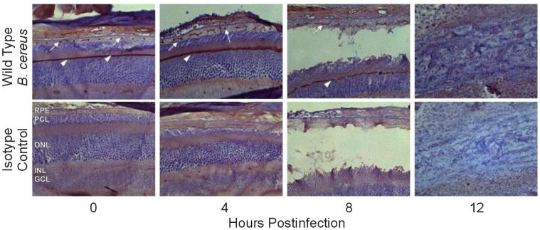
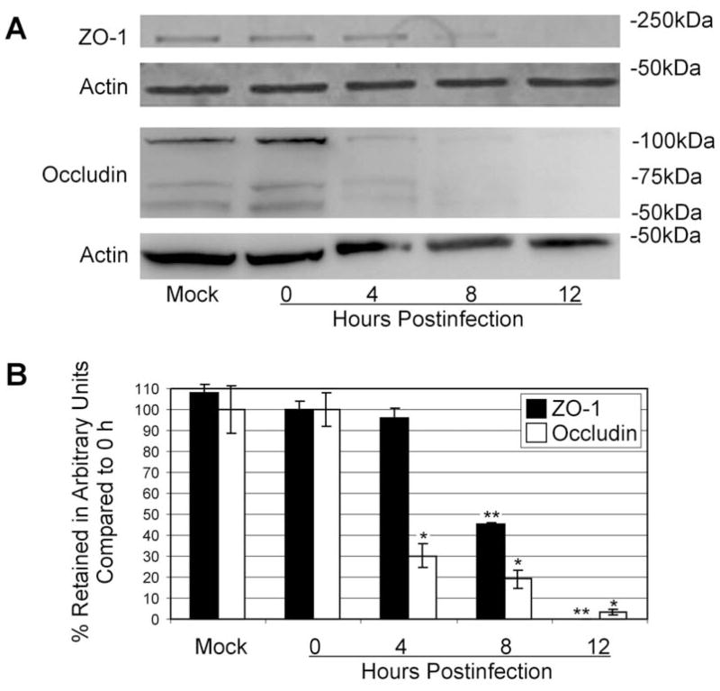
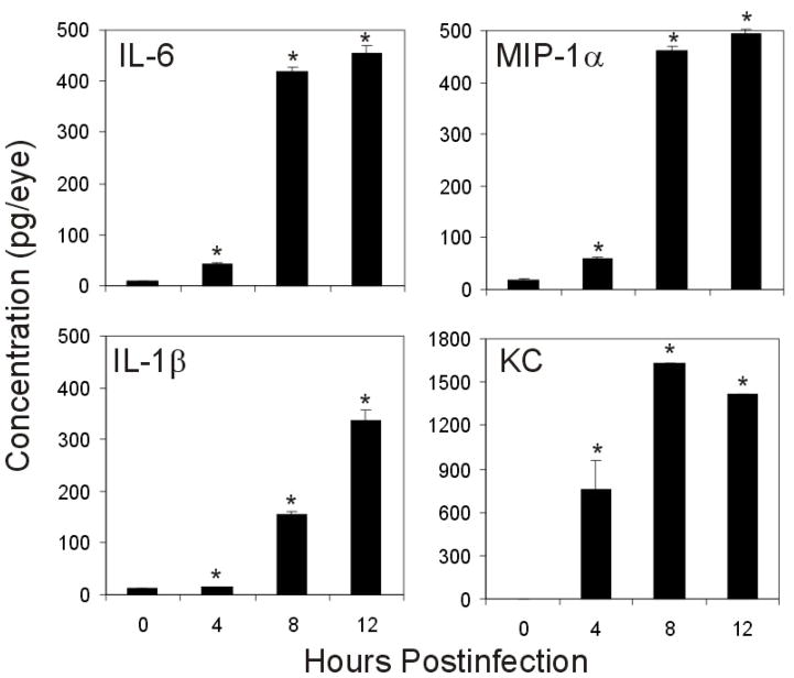
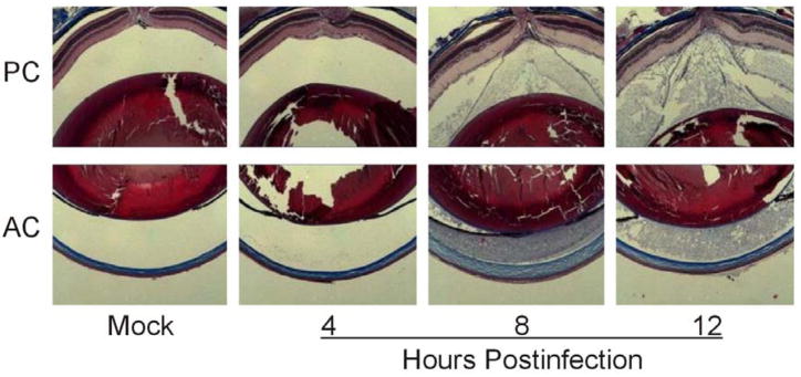
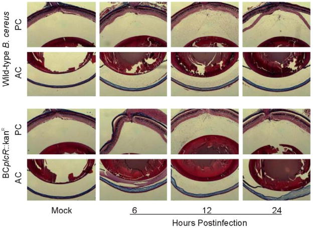
Results: B. cereus induced the leakage of albumin and fibrin into the aqueous and vitreous humor by 8 hours after infection. BRB permeability occurred as early as 4 hours and increased 13.30-fold compared with uninfected controls by 8 hours. Production of proinflammatory cytokines IL-6, MIP-1alpha, IL-1beta, and KC increased over the course of infection. In the retina, ZO-1 disruption began by 4 hours and was followed by decreasing occludin and ZO-1 expression at 4 and 8 hours, respectively. Tubulin condensation and RPE65 degradation occurred by 12 hours. A quorum-sensing mutant B. cereus strain caused BRB permeability comparable to that of wild-type B. cereus. Wild-type and mutant B. cereus sterile supernatants induced blood-ocular barrier permeability similarly to that of wild-type infection.

Conclusions: These results indicate that BRB permeability occurs during the early stages of experimental B. cereus endophthalmitis, beginning as early as 4 hours after infection. Disruption of tight junctions at the level of the retinal pigment epithelium may contribute to barrier breakdown. Quorum-sensing dependent factors may not significantly contribute to BRB permeability.


Figures

Figure 1
Figure 1. Fibrin and albumin infiltrate the posterior and anterior chamber during experimental B. cereus endophthalmitis
C57BL/6J mouse eyes were intravitreally injected with 100 CFU of B. cereus, injected with BHI (mock), or left untreated. Fibrin (A) and albumin (B) leakage into the anterior (AC) and posterior (PC) chambers was analyzed by trichrome staining and immunohistochemistry. Immunofluorescent images were overlaid onto brightfield images to enhance localization of albumin in the eye. Images represent three or more individual experiments. Images were acquired at 4× magnification.
Figure 2
Figure 2. BRB permeability to albumin during experimental B. cereus endophthalmitis
C57BL/6J mouse eyes were intravitreally injected with 100 CFU of B. cereus, injected with BHI (mock) or were not injected. Mice were injected into the tail vein with Evans blue dye 2 h prior to each time point. Albumin leakage into the retina was measured by dye quantification against a standard curve and normalized to total protein in each retina (data represents mean ± standard error of the mean of 8 or more eyes per group). Permeability increased significantly over mock-injected controls as early as 4 h postinfection (P=0.04).
Figure 3
Figure 3. Tight junction protein redistribution, tubulin condensation, and RPE65 degradation at the level of the RPE during experimental B. cereus endophthalmitis
Retinas of B. cereus-infected eyes were harvested and paraffin-embedded sections were used for histological analysis and immunohistochemistry. Intact retinas had little fluorescent signal in the RPE due to quenching by the pigment. However, at 12 h ZO-1 and occludin expression is intermittent and scattered. Additionally, tubulin condensed and RPE-65 expression was not detected by 12 h postinfection. Control samples were developed with secondary antibody alone. Images represent three or more individual experiments. Images were acquired at 100× magnification.
Figure 4
Figure 4. Lateral redistribution of ZO-1 in the retina during B. cereus endophthalmitis
Retinas of B. cereus infected eyes were harvested and paraffin-embedded sections were used for histological analysis and immunohistochemistry. Bleaching RPE pigment prior to immunohistochemistry resulted in a visible brown punctuate ZO-1 signal in the RPE (white arrows) as well as a continuous signal along the outer limiting membrane (white arrow heads). ZO-1 lateral redistribution was evident at 4 and 8 h postinfection (white arrows). By 12 h severe damage to retinal layers had occurred and ZO-1 signal was not detectable (top). Isotype controls confirm lack of nonspecific antibody (bottom). Images were acquired at 40× magnification.
Figure 5
Figure 5. Decreases in retinal occludin and ZO-1 expression during experimental B. cereus endophthalmitis
Expression of occludin and ZO-1 in retinas was analyzed by immunoblot and densitometry during B. cereus infection. Retinal occludin and ZO-1 expression was not detectable by 8 and 12 h postinfection, respectively (A). Densitometry indicated decreased ZO-1 and occludin expression over the infection course, with complete loss of detectable ZO-1 (**compared to 0 h ZO-1, P=0.00006) and >95% loss of occludin (*compared to 0 h occludin, P=0.0003) by 12 h (B).
Figure 6
Figure 6. Proinflammatory cytokines and chemokines related to barrier permeability are produced during experimental B. cereus endophthalmitis
C57BL/6J mouse eyes were intravitreally injected with 100 CFU of B. cereus, injected with BHI (mock), or were not injected, and production of proinflammatory cytokines and chemokines were analyzed by ELISA (data represents mean ± SEM of ≥ 8 eyes per group). Significant increases in cytokine production, above 0 h controls, occurred at 4, 8, and 12 h postinfection for KC (P=0.02, P<0.0001, P=0.03), IL-6 (P=0.001, P<0.0001, P<0.0001), IL-1β (P=0.03, P<0.0001, P=0.0001), and MIP-1α (P<0.0001, P<0.0001, P<0.0001).
Figure 7
Figure 7. plcR-deficient B. cereus induces barrier permeability similarly to wild-type B. cereus during experimental endophthalmitis
100 CFU of plcR-deficient (BCplcR::kanR) Bacillus was injected into C57BL/6J mouse eyes. Eyes were analyzed by histology and trichrome stained to detect fibrin in the aqueous and vitreous of the anterior (AC) and posterior chambers (PC). Fibrin leaked across the BOB, filling the aqueous and vitreous humor by 8 h postinfection. Images represent three or more individual experiments. Images were aquired at 5× magnification.
Figure 8
Figure 8. Sterile cell-free wild-type and plcR-deficient B. cereus supernatants are sufficient to induce BOB permeability
C57BL/6J mouse eyes were intravitreally injected with 0.5 μl sterile B. cereus supernatant prepared from either wild-type (BCWT) or plcR-deficient (BCplcR::kanR) cultures. Eyes were analyzed by histology. Fibrin (A) and albumin (B) leakage into the anterior (AC) and posterior (PC) chambers was analyzed by trichrome staining. Both wild-type and plcR-deficient Bacillus sterile supernatants caused BOB permeability to fibrin, with wild-type inducing fibrin leakage more rapidly than the mutant strain. Images represent three or more individual experiments. Images were aquired at 5× magnification.

Similar articles

Cited by

References

    1. Callegan MC, Engelbert M, Parke DW, II, Jett BD, Gilmore MS. Bacterial endophthalmitis: Epidemiology, therapeutics, and bacterium-host interactions. Clin Microbiol Rev. 2002;15:111–124. - PMC - PubMed
    1. Callegan MC, Kane ST, Cochran DC, Gilmore MS, Gominet M, Lereclus D. Relationship of plcR-regulated factors to Bacillus endophthalmitis virulence. Infect Immun. 2003;71:3116–3124. - PMC - PubMed
    1. Jepson MA, Schlecht HB, Collares-Buzato CB. Localization of dysfunctional tight junctions in Salmonella enterica serovar typhimurium-infected epithelial layers. Infect Immun. 2000;68:7202–7208. - PMC - PubMed
    1. Callegan MC, Booth MC, Jett BD, Gilmore MS. Pathogenesis of gram-positive bacterial endophthalmitis. Infect Immun. 1999;67:3348–3356. - PMC - PubMed
    1. David DB, Kirkby GR, Noble BA. Bacillus cereus endophthalmitis. Br J Ophthalmol. 1994;78:577–580. - PMC - PubMed

Publication types

MeSH terms