. 2009 Apr;174(4):1212-20.
doi: 10.2353/ajpath.2009.080458. Epub 2009 Mar 5.

Experimental autoimmune vasculitis: an animal model of anti-neutrophil cytoplasmic autoantibody-associated systemic vasculitis

Affiliations

Experimental autoimmune vasculitis: an animal model of anti-neutrophil cytoplasmic autoantibody-associated systemic vasculitis

Mark A Little et al. Am J Pathol. 2009 Apr.

Abstract

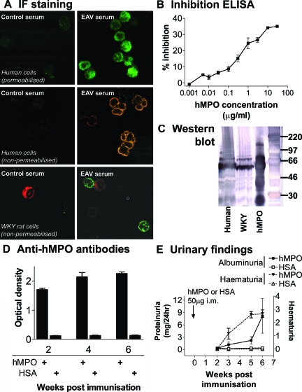
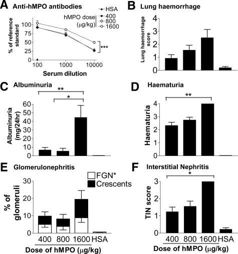
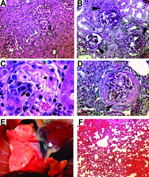
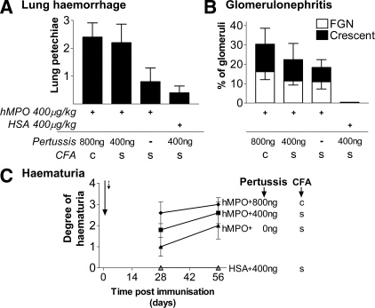
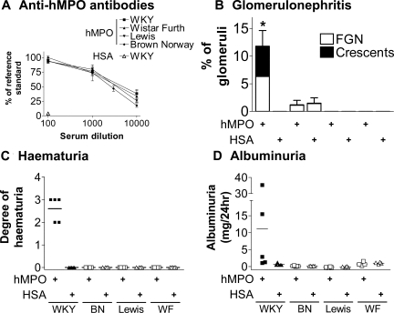
The morbidity burden associated with anti-neutrophil cytoplasmic autoantibody-associated vasculitis is increasing, and many novel biological therapies are now entering the drug development pipeline. There is thus an urgent need to develop a representative animal model to facilitate testing of these agents. We previously examined the effect of antineutrophil cytoplasmic autoantibody on leukocyte-endothelial interactions in WKY rats via immunization with human myeloperoxidase. We now seek to extend this model so that all animals reliably develop crescentic glomerulonephritis and lung hemorrhage. We also wish to investigate whether there is a genetic contribution to vasculitis development in this rat strain. Using escalating doses of human myeloperoxidase, we found that a dose of 1600 microg/kg induced pauci-immune crescentic glomerulonephritis and lung hemorrhage in all immunized animals. We also found that the addition of pertussis toxin and killed Mycobacterium tuberculosis to the adjuvant when immunizing with 400 microg/kg of myeloperoxidase resulted in crescentic glomerulonephritis and lung hemorrhage in all animals. However, when Lewis, Wistar Furth, or Brown Norway rats were immunized using a similar protocol, no animals developed hematuria or glomerulonephritis, despite having identical levels of anti-human myeloperoxidase antibodies. We conclude that, by adjusting the immunization regimen, all WKY rats immunized with myeloperoxidase develop experimental autoimmune vasculitis, thus facilitating future therapeutic studies. The resistance of Lewis rats to experimental autoimmune vasculitis provides a genetic basis for future studies of anti-myeloperoxidase antibody-associated vasculitis.


Figures

Figure 1
Figure 1
Antibody response and urinary changes in hMPO-immunized rats. A: Binding of EAV serum to human and WKY neutrophils. Permeabilized, paraformaldehyde-fixed human neutrophil suspensions (top) were incubated with serum (1:20) from rats immunized with HSA or hMPO and binding was detected with Alexa-488 anti-rat IgG. Middle: Binding patterns on human neutrophil cytospins fixed in 95% ethanol when incubated with serum (1:20) from rats immunized with saline and hMPO. Binding was detected with Alexa-568 anti-rat IgG. Bottom: Double-staining experiments on rat leukocyte cytospins using serum (1:20, detected with Alexa-488 anti-rat IgG, green) from rats immunized with HSA and hMPO (1600 μg/kg), and ED1 antibody against monocytes (detected with Alexa-633 anti-mouse IgG, red). All images were captured with a confocal microscope. Representative of three separate experiments. B: Specific inhibition of binding of serum from rats with EAV to hMPO immobilized on plastic by soluble hMPO. Serum (1:2000) was incubated with hMPO in the presence of increasing concentrations of soluble hMPO. The data points represent the mean ± range of two experiments. C: IgG from rats with EAV reacts with rat neutrophil lysates. Lysates of peripheral blood leukocytes were prepared from human (lane 1) and WKY (lane 2) blood and separated using sodium dodecyl sulfate-polyacrylamide gel electrophoresis under denaturing conditions. Purified hMPO (as used to induce EAV) was also run as a positive control (lane 3). The blot was probed with Ig prepared from animals immunized with hMPO (EAV IgG 1 mg/ml). Binding was detected using alkaline phosphatase conjugate. D: WKY rats immunized with hMPO develop high titers of anti-hMPO antibodies. Anti-hMPO antibodies in selected serum samples (1:100) collected at various time points after immunization with 400 μg/kg of hMPO or HSA were measured using ELISA. The bars represent the mean ± SEM optical density (n = 5 in each group). E: WKY rats immunized with hMPO develop hematuria and albuminuria throughout time. Hematuria was assessed semiquantitatively with a urinary dipstick. Urinary albumin excretion rate was measured with ELISA at various time points. The bars represent the mean ± SEM optical density (n = 5 in each group).
Figure 2
Figure 2
Vasculitis phenotype 6 weeks after immunization with a dose range from 400 to 1600 μg/kg of hMPO or HSA in WKY rats. A: Anti-hMPO antibodies in serially diluted serum were assessed using ELISA. B: Lung hemorrhage was scored macroscopically at the time of sacrifice. Bars represent the mean ± SEM. C: Urinary albumin excretion rate was measured with ELISA. Bars represent the mean ± SEM. D: Hematuria was assessed semiquantitatively with a urinary dipstick. Bars represent the mean ± SEM. E: Glomerular changes (focal proliferative glomerulonephritis and crescent formation) were assessed blindly using H&E and periodic acid-Schiff-stained sections. Bars represent the mean ± SEM. F: Severity of TIN was assessed blindly using H&E-stained sections. Bars represent the mean ± SEM. For all data, n = 4 to 12 separate animals for each dose of hMPO. Statistically significant differences between groups are demonstrated with asterisks, *P < 0.05, **P < 0.01, ***P < 0.05.
Figure 3
Figure 3
Histological changes 6 weeks after immunization with a dose range from 400 to 1600 μg/kg of hMPO or HSA in WKY rats. A: Normal appearing glomeruli, tubules, and interstitium of rats immunized with 400 μg/kg of HSA. Periodic acid-Schiff. B: Crescentic glomerulonephritis (arrowheads) in a rat immunized with 400 μg/kg of hMPO, periodic acid-Schiff. C: Fibrinoid necrosis (arrow) in a rat immunized with 800 μg/kg of hMPO, H&E. D: Circumferential crescent formation (arrowhead) in a rat immunized with 1600 μg/kg of hMPO, periodic acid-Schiff. E: Macroscopic appearance of lung petechiae in a rat immunized with 1600 μg/kg of hMPO. F: Alveolar hemorrhage in a rat immunized with 800 μg/kg of hMPO, H&E. Original magnifications: ×10 (A); ×20 (B, F); ×100 (C); ×40 (D).
Figure 4
Figure 4
Renal and pulmonary effects of pertussis toxin and customized CFA in EAV. WKY rats were immunized with 400 μg/kg of hMPO or HSA in either standard (s) or customized (c) CFA. In addition, rats received 400 to 800 ng of pertussis toxin or vehicle intraperitoneally on days 0 (arrow) and 2 (dotted arrow). A: Lung hemorrhage was scored blindly at the time of sacrifice on day 56. Bars represent the mean ± SEM. B: Glomerular changes 6 weeks after immunization were assessed blindly using H&E- and periodic acid-Schiff-stained sections. One hundred percent of animals immunized with 400 μg/kg of hMPO in customized CFA with 800 ng of pertussis toxin developed crescentic GN. Bars represent the mean ± SEM. C: Rats were immunized as in A and hematuria was assessed at days 28 and 56. Each point represents the mean ± SEM. For all data n = 5 in each group.
Figure 5
Figure 5
Phenotype 6 weeks after immunization with 400 μg/kg of hMPO or HSA in WKY, Brown Norway (BN), Lewis, and Wistar Furth (WF) rats. A: Anti-hMPO antibodies in serially diluted serum were measured using ELISA 6 weeks after immunization with HSA or hMPO. Each point indicates the mean ± SEM for each rat strain. B: Glomerular changes 6 weeks after immunization were assessed blindly using H&E- and periodic acid-Schiff-stained sections. Bars represent the mean ± SEM, *P < 0.05. C: Hematuria was assessed semiquantitatively with a urinary dipstick. Each data point represents an individual animal and the bars indicate the mean. D: Urinary albumin excretion rate was assessed with ELISA. Each data point represents an individual animal and the bars indicate the median. n = 4 to 5 rats in each group.

Similar articles

Cited by

References

    1. Davies DJ, Moran JE, Niall JF, Ryan GB. Segmental necrotising glomerulonephritis with antineutrophil antibody: possible arbovirus aetiology? Br Med J (Clin Res Ed) 1982;285:606. - PMC - PubMed
    1. van der Woude FJ, Rasmussen N, Lobatto S, Wiik A, Permin H, van Es LA, van der Giessen M, van der Hem GK, The TH. Autoantibodies against neutrophils and monocytes: tool for diagnosis and marker of disease activity in Wegener’s granulomatosis. Lancet. 1985;1:425–429. - PubMed
    1. Niles JL, McCluskey RT, Ahmad MF, Arnaout MA. Wegener’s granulomatosis autoantigen is a novel neutrophil serine proteinase. Blood. 1989;74:1888–1893. - PubMed
    1. Falk RJ, Jennette JC. Anti-neutrophil cytoplasmic autoantibodies with specificity for myeloperoxidase in patients with systemic vasculitis and idiopathic necrotizing and crescentic glomerulonephritis. N Engl J Med. 1988;318:1651–1657. - PubMed
    1. Jennette JC, Hoidal JR, Falk RJ. Specificity of anti-neutrophil cytoplasmic autoantibodies for proteinase 3. Blood. 1990;75:2263–2264. - PubMed

Publication types

MeSH terms