. 2009 Apr;174(4):1564-71.
doi: 10.2353/ajpath.2009.080452. Epub 2009 Mar 5.

Membrane type-1 matrix metalloproteinase potentiates basic fibroblast growth factor-induced corneal neovascularization

Affiliations

Membrane type-1 matrix metalloproteinase potentiates basic fibroblast growth factor-induced corneal neovascularization

Tatsuya Onguchi et al. Am J Pathol. 2009 Apr.

Erratum in

  • Am J Pathol. 2013 Jul;183(1):326

Abstract

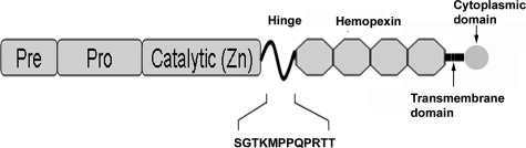
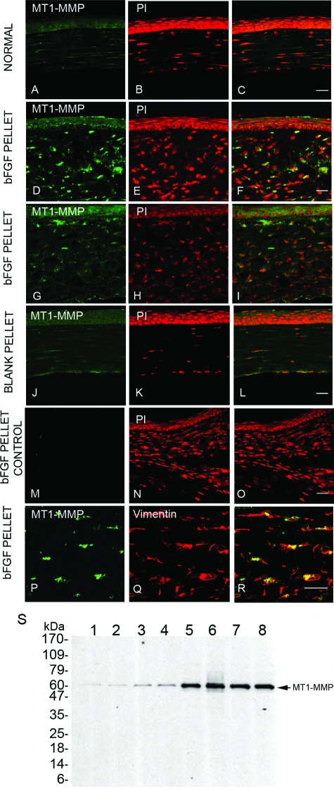
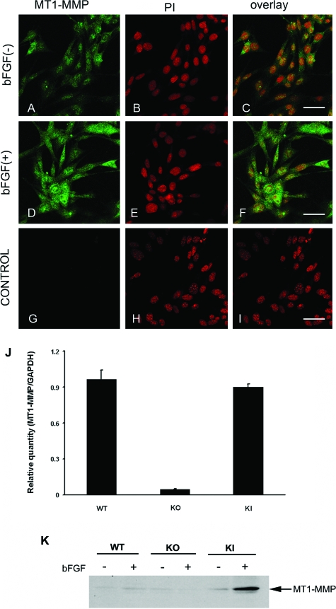
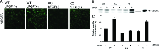
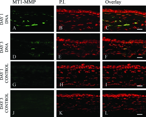
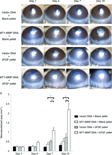
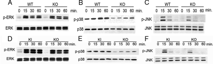
Corneal neovascularization is one of the leading causes of blindness. The aim of this study was to evaluate the pro-angiogenic role of corneal fibroblast-derived membrane type-1 matrix metalloproteinase (MT1-MMP) on basic fibroblast growth factor (bFGF)-induced corneal neovascularization in vivo and in vitro. Immunohistochemical studies demonstrated that MT1-MMP was expressed in keratocytes and immortalized corneal fibroblast cell lines. Vascular endothelial growth factor protein levels were increased after bFGF-stimulation of wild-type fibroblast cells compared with MT1-MMP knockout fibroblast cells. Corneal vascularization was significantly increased after a combination of bFGF pellet implantation and naked MT1-MMP DNA injection in wild-type mouse corneas compared with either bFGF pellet implantation or naked MT1-MMP DNA-injected corneas. Western blotting analysis of the phosphorylation levels of the key signaling molecules (p38, JNK, and ERK) demonstrated that phosphorylation levels of both p38 and JNK were diminished after bFGF stimulation of MT1-MMP knockout cells compared with wild-type and MT1-MMP knockin cells. These results suggest that MT1-MMP potentiates bFGF-induced corneal neovascularization, likely by modulating the bFGF signal transduction pathway.


Figures

Figure 1
Figure 1
Schematic representation of MT1-MMP and the location of the amino acid sequence (SGTKMPPQPRTT) that was used to generate the rabbit anti-MT1-MMP antibody.
Figure 2
Figure 2
MT1-MMP immunolocalization in normal and bFGF-induced mouse corneas. Normal uninjured mouse eyes were enucleated, frozen in OCT, and corneal sections were stained with anti-MT1-MMP antibody (A) and PI (B); overlay image (C). Seven days after the blank (controls) or 120-ng bFGF pellet implantation, mouse eyes were enucleated and the sections were stained with anti-MT1-MMP antibody (D, G, J and P), PI (E, H, and K), or anti-vimentin antibody (Q); overlay images (F, I, L, O, and R). Antibody pre-incubated with cognate peptide was used to stain the bFGF pellet-inserted cornea as a control (M, N, and O). S: Lysates from individual bFGF-stimulated mouse corneas,,, and untreated controls,,, were immunoblotted with anti-MT1-MMP antibody. Scale bars = 20 μm.
Figure 3
Figure 3
MT1-MMP immunolocalization in corneal fibroblast cell lines. Unstimulated wild-type (WT) corneal fibroblast cell lines were immunostained with anti-MT1-MMP antibody (A) and PI (B). Wild-type corneal fibroblast cells stimulated with bFGF (20 ng/ml) were immunostained with anti-MT1-MMP antibody (D) and PI (E); overlay images (C, F, and I). Antibody preincubated with cognate peptide was used as a control (G–I). J: Real-time PCR analysis of MT1-MMP expression in wild-type, MT1-MMP KO, and MT1-MMP KI keratocytes was performed. K: Western blot analysis was performed using MT1-MMP antibody in wild-type, MT1-MMP KO, and MT1-MMP KI keratocytes treated with and without bFGF. Scale bars = 50 μm.
Figure 4
Figure 4
VEGF-A expression in corneal fibroblasts. A: Wild-type (WT) and KO corneal fibroblasts were starved for 18 hours and treated with 20 ng/ml of bFGF for 48 hours. They were then fixed, permeabilized, and stained with antibody to VEGF-A. Corneal fibroblasts were treated with bFGF in the same way, and the cell lysates were subjected to Western blot analysis with antibodies to VEGF-A (B; VEGF-A control is shown) as well as real-time PCR analysis (C). Scale bar = 20 μm.
Figure 5
Figure 5
MT1-MMP protein expression after naked MT1-MMP or vector DNA injection. Either MT1-MMP-encoding DNA (AF) or empty vector DNA (GL) was injected into mouse corneas, and the eyes were enucleated at days 1 and 3 and the corneal sections were stained with anti-MT1-MMP antibody (A, D, G, and J) and PI (B, E, H, and K); overlay images (C, F, I, and L). Data are representative of three independent experiments. Scale bars = 20 μm.
Figure 6
Figure 6
Enhanced bFGF-induced corneal NV after a combination of bFGF pellet implantation and naked MT1-MMP DNA plasmid injection. The blank pellets were implanted immediately after MT1-MMP DNA (E–H) or vector control DNA (A–D) was injected into corneal stroma. Likewise, the bFGF pellets were implanted immediately after MT1-MMP DNA (M–P) or vector control DNA (I–L) was injected into corneal stroma. Photographs were taken on days 1, 4, 7, and 10 after surgery. Q: Graphic representation of at least five independent experiments (mean ± SEM, *P < 0.05).
Figure 7
Figure 7
Activation of MAP kinases by bFGF stimulation in wild-type (WT) and MT1-MMP KO cell lines. MT1-MMP KO and wild-type cell lines were serum-starved for 18 hours and treated with bFGF for 15, 30, or 60 minutes. The cell lysate was subjected to sodium dodecyl sulfate-polyacrylamide gel electrophoresis with subsequent Western blot analysis with antibodies against phospho-ERK1/2, ERK1/2 (A), phospho-p38, p38 (B), or phospho-JNK and JNK (C). Comparison of the phosphorylation of MAP kinases on bFGF stimulation in MT1-MMP KO and KI cell lines. Cell lines were serum-starved for 18 hours and stimulated with bFGF for 15, 30, and 60 minutes. Cell lysates were subjected to sodium dodecyl sulfate-polyacrylamide gel electrophoresis with subsequent Western blot analysis with antibodies against phospho-ERK1/2, ERK1/2 (D), phospho-p38, p38 (E), or phospho-JNK and JNK (F).

Similar articles

Cited by

References

    1. Tallquist MD, Soriano P, Klinghoffer RA. Growth factor signaling pathways in vascular development. Oncogene. 1999;18:7917–7932. - PubMed
    1. Yancopoulos GD, Davis S, Gale NW, Rudge JS, Wiegand SJ, Holash J. Vascular-specific growth factors and blood vessel formation. Nature. 2000;407:242–248. - PubMed
    1. Gale NW, Yancopoulos GD. Growth factors acting via endothelial cell-specific receptor tyrosine kinases: VEGFs, angiopoietins, and ephrins in vascular development. Genes Dev. 1999;13:1055–1066. - PubMed
    1. Kwon YS, Kim JC. Inhibition of corneal neovascularization by rapamycin. Exp Mol Med. 2006;38:173–179. - PubMed
    1. Kenyon BM, Voest EE, Chen CC, Flynn E, Folkman J, D'Amato RJ. A model of angiogenesis in the mouse cornea. Invest Ophthalmol Vis Sci. 1996;37:1625–1632. - PubMed

Publication types

MeSH terms

Substances