. 2009 Apr;174(4):1280-90.
doi: 10.2353/ajpath.2009.080409. Epub 2009 Mar 5.

Ets transcription factors control epithelial maturation and transit and crypt-villus morphogenesis in the mammalian intestine

Affiliations

Ets transcription factors control epithelial maturation and transit and crypt-villus morphogenesis in the mammalian intestine

Paul Jedlicka et al. Am J Pathol. 2009 Apr.

Abstract

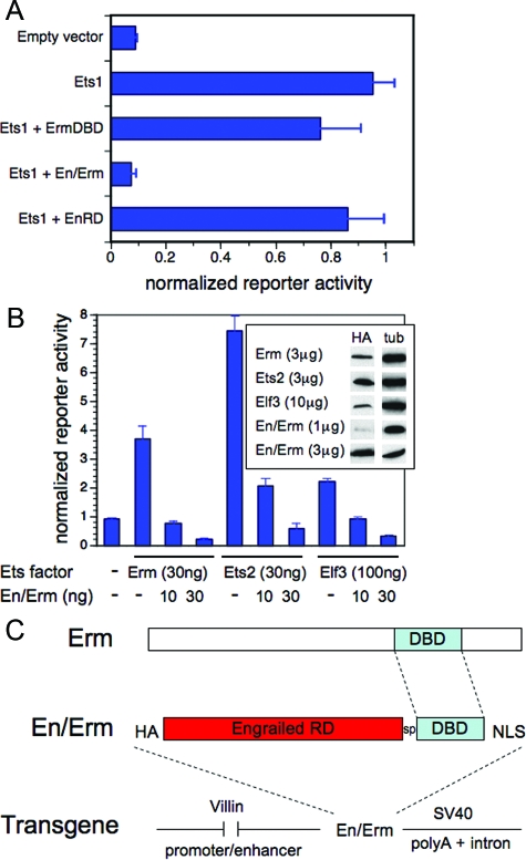
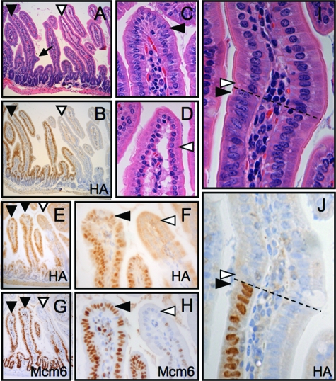
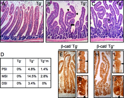
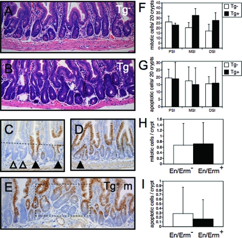
Members of the Ets transcription factor family are widely expressed in both the developing and mature mammalian intestine, but their biological functions remain primarily uncharacterized. We used a dominant repressor transgene approach to probe the function of epithelial Ets factors in the homeostasis of the crypt-villus unit, the functional unit of the small intestine. We show that targeted expression in small intestinal epithelium of a fusion protein composed of the Engrailed repressor domain and the Erm DNA-binding domain (En/Erm) results in marked disruption of normal crypt-villus homeostasis, including a cell-autonomous disturbance of epithelial maturation, increased epithelial transit, severe villus dysmorphogenesis, and crypt dysmorphogenesis. The epithelial maturation disturbance is independent of the regulation of TGFbetaRII levels, in contrast to Ets-mediated epithelial differentiation during development; rather, regulation of Cdx2 expression may play a role. The villus dysmorphogenesis is independent of alterations in the crypt-villus boundary and inappropriate beta-catenin activation, and thus appears to represent a new mechanism controlling villus architectural organization. An Analysis of animals mosaic for En/Erm expression suggests that crypt nonautonomous mechanisms underlie the crypt dysmorphogenesis phenotype. Our studies thus uncover novel Ets-regulated pathways of intestinal homeostasis in vivo. Interestingly, the overall En/Erm phenotype of disturbed crypt-villus homeostasis is consistent with recently identified Ets function(s) in the restriction of intestinal epithelial tumorigenesis.


Figures

Figure 1
Figure 1
Transcriptional blocking activity of the En/Erm dominant repressor in vitro. HeLa cells were transfected with the reporter plasmid 8x(EBS)-TK-luciferase and the indicated plasmid DNA expression constructs. A: Transfected DNA amounts were: Ets1, 50 ng; ErmDBD, 250 ng; En/Erm, 150 ng; EnRD, 50 ng (chosen to normalize for differences in construct expression levels). B: Transfected DNA amounts were as shown. Reporter activity, determined by quantitative luminometry, was normalized to the activity of the co-transfected Renilla-luciferase construct; results are expressed as mean and SD of triplicate transfections. All constructs were expressed from the plasmid pSG5, and all except Ets1 have an N-terminal HA epitope tag. Protein expression (B, inset) was determined by immunoblotting with antibody against HA, and tubulin (tub) as loading control. C: Modular organization of the villin-En/Erm dominant repressor transgene. The transgene consists of the 12.4-kbp villin promoter-enhancer fragment, a HA epitope tag, the Drosophila Engrailed repressor domain (RD), a 7-amino acid (GGGSGGG) spacer (sp), the Erm DNA-binding domain (DBD), a 7-amino acid (PKKKRKV) SV40 large T-antigen nuclear localization sequence (NLS), and a SV40 intron and polyA tail.
Figure 2
Figure 2
Transgene expression and impaired enterocyte maturation in En/Erm-expressing small intestine. HA immunostaining of small intestine from control (A) and transgenic (B) animals shows the expression of En/Erm protein specifically in epithelial nuclei of transgenic animals. En/Erm expression was strong in the villus epithelium (B, open arrowhead), and was also observed in the superficial aspects of crypts (B, filled arrowhead), but not the deep aspects of crypts (B, arrow); no immunostaining was observed in control animals (A). H&E-stained sections of small intestine from control (C; detailed views of villus in E and F) and transgenic (D; detailed view of villus in G and H) animals; note morphological resemblance of transgenic enterocytes at the villus tip to enterocytes at the villus base (filled arrowheads, enterocytes at villus base; open arrowheads, enterocytes at villus tip).
Figure 3
Figure 3
Characterization of enterocyte maturation disturbance in En/Erm transgenic animals. Histochemical (PAS) and immunohistochemical (iAP, iFabp, Mcm6, and Ki-67) staining of small intestine from control (Tg: A, C, E--G, K--M, and Q) and transgenic (Tg+: B, D, H--J, N--P, and R) animals. Arrowheads in A and B: PAS-positive glycocalyx on enterocytes; arrowheads in C and D: iAP expression in superficial aspect of enterocytes (insets: detailed views of villus tips); arrowheads in E--J: iFABP expression in enterocyte cytoplasm (F and I: detailed views of villus tips in E and H, respectively; G and J: detailed views of villus bases in E and H, respectively); arrowheads in K--P: Mcm6 expression in enterocyte nuclei at villus base (filled arrowheads) and villus tip (open arrowheads; L and O: detailed views of villus tips in K and N, respectively; M and P: detailed views of villus bases in K and N, respectively; filled arrowheads in Q and R: upper limit of residual Ki-67 immunopositivity in villus enterocytes; open arrowheads in Q and R: solitary ectopic Ki-67 immunopositivity in transgenic superficial villus enterocytes (inset: detailed view).
Figure 4
Figure 4
BrdU immunostaining of small intestine from control (Tg: A, C, and E) and transgenic (Tg+: B, D, and F) animals pulsed in vivo with BrdU and analyzed 2 hours (A and B) or 24 hours (C--F) later (filled arrowheads: crypt epithelial cells; open arrowheads: villus epithelial cells; arrows: upper limit of epithelial cell transit after BrdU incorporation in the crypt; PSI: proximal small intestine; DSI: distal small intestine).
Figure 5
Figure 5
Characterization of enterocyte maturation disturbance in En/Erm mosaic animals. H&E-stained (A) and HA-immunostained (B) small intestinal focus mosaic for En/Erm expression (filled arrowhead: En/Erm expressing villus; open arrowhead: nonexpressing villus; arrow: incipient villus branch). Detailed views of the tips of the En/Erm expressing (C) and nonexpressing (D) from A (arrowheads: enterocytes). HA (E and F) and Mcm6 (G and H) immunostaining of a small intestinal focus mosaic for En/Erm expression (arrowhead designations are as in A--D). As above, En/Erm expression appears as immunopositivity in epithelial nuclei (filled arrowhead; contrast with absence of nuclear immunopositivity, shown by open arrowheads, in adjacent villus). Detailed view of H&E-stained (I) and HA-immunostained (J) individual villus mosaic for En/Erm expression. Note immature morphology of En/Erm-expressing enterocyte (filled arrowhead), but maturation appropriate for position along villus axis (dashed line) of adjacent nonexpressing enterocyte (open arrowhead).
Figure 6
Figure 6
Analysis of potential mediators of the disturbed epithelial maturation phenotype. TGFβRII (A and B)- and Cdx2 (C and D)-immunostained small intestine from control (Tg) and transgenic (Tg+) animals. Note similar level of expression of TGFβRII in control and transgenic animals (arrowheads in A and B), and inappropriate persistence of Cdx2 expression throughout villi of transgenic animals compared with controls (arrowheads in C and D). TGFβRII is cytoplasmic, whereas Cdx2 is nuclear. Insets: Magnified views of villus epithelium.
Figure 7
Figure 7
Villus architectural dysmorphogenesis in En/Erm-expressing small intestine. A–C: H&E-stained sections of small intestine from control (Tg) and transgenic (Tg+) animals (arrow: villus branching; arrowheads: villus turns; asterisk: villus bridging). D: Quantitation of villus branching in control (Tg, n = 8) and transgenic (Tg+, n = 3) animals, and transgene-expressing foci in mosaic animals (Tg+m, n = 2), in proximal (PSI), mid (MSI), and distal (DSI) small intestine. β-Catenin immunostaining in control (E–G) and transgenic (H–J) animals. Note nuclear staining (filled arrowheads, G and J) limited to epithelial cells at bases of crypts, and membranous (filled arrowheads, F and I) but no nuclear (open arrowheads, F and I) staining in villi, in both transgenic and control animals.
Figure 8
Figure 8
Crypt alterations in En/Erm-expressing small intestine. A and B: H&E-stained small intestine from control (Tg) and transgenic (Tg+) animals. Note crypt disorder, including increased variation in crypt position and size in transgenic animals (B) relative to controls (A). C–E: HA immunostaining of small intestine in mosaic transgenic animals (open arrowheads: non-expressing crypts; black arrowheads: En/Erm-expressing crypts; dashed line in E: crypt-villus boundary; dashed box in G: crypt dysmorphogenesis in a large En/Erm-expressing mosaic focus. Crypt mitotic (F) and apoptotic (G) activity in proximal (PSI), mid (MSI), and distal (DSI) small intestine of control (Tg, n = 7 and 8, respectively) and transgenic (Tg+, n = 3 for each) animals. Average mitotic (H) and apoptotic (I) cells per En/Erm nonexpressing (En/Erm, white bars) and expressing (En/Erm+, black bars) crypts in mosaic animals (49 total expressing and nonexpressing crypts along the entire small intestine were scored for each; values are expressed as average and SD). Mitotic and apoptotic cells were scored by their characteristic morphology on H&E-stained sections, in well-oriented, fully-visualized crypts; none of the comparisons between experimental and control groups yielded statistically significant differences (P < 0.05).

References

    1. Oikawa T, Yamada T. Molecular biology of the Ets family of transcription factors. Gene. 2003;303:11–34. - PubMed
    1. Sharrocks AD. The ET: S-domain transcription factor family. Nat Rev Mol Cell Biol. 2001;2:827–837. - PubMed
    1. Gutierrez-Hartmann A, Duval DL, Bradford AP. ETS transcription factors in endocrine systems. Trends Endocrinol Metab. 2007;18:150–158. - PubMed
    1. Graves BJ, Petersen JM. Specificity within the ets family of transcription factors. Adv Cancer Res. 1998;75:1–55. - PubMed
    1. Sementchenko VI, Watson DK. Ets target genes: past, present and future. Oncogene. 2000;19:6533–6548. - PubMed

Publication types

Substances