. 2009 Apr;174(4):1504-14.
doi: 10.2353/ajpath.2009.080592. Epub 2009 Mar 5.

RNA interference-mediated inhibition of erythropoietin receptor expression suppresses tumor growth and invasiveness in A2780 human ovarian carcinoma cells

Affiliations

RNA interference-mediated inhibition of erythropoietin receptor expression suppresses tumor growth and invasiveness in A2780 human ovarian carcinoma cells

Gyorgy Paragh et al. Am J Pathol. 2009 Apr.

Abstract

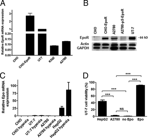
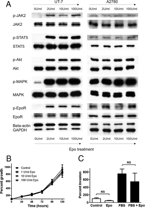
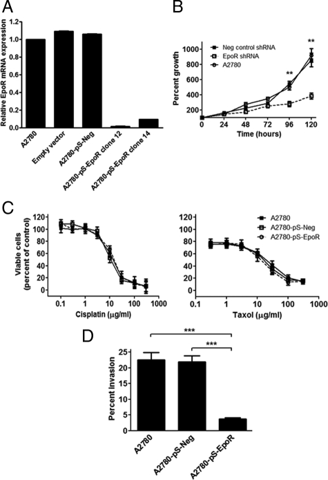
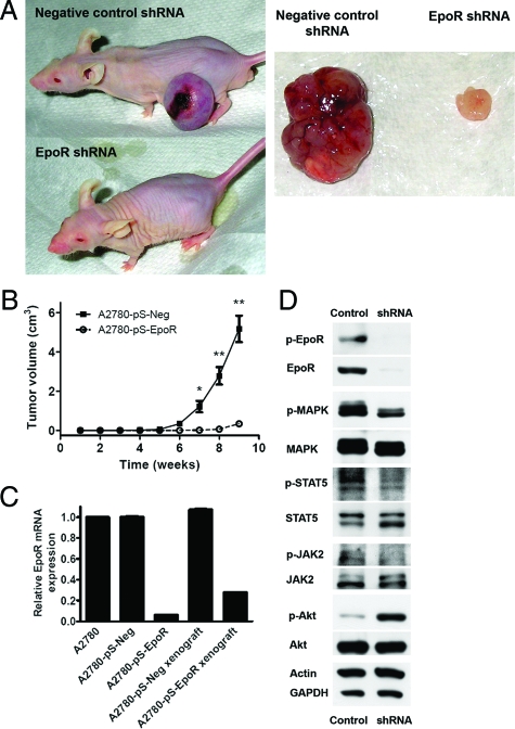
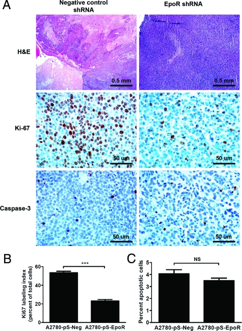
Although recombinant human erythropoietin (rHuEpo) has revolutionized the treatment of anemia, recent clinical trials suggested that rHuEpo use may be associated with decreased survival in cancer patients. Although the expression of erythropoietin (Epo) receptor (EpoR) has been demonstrated in various human cancers, the effect of exogenous Epo on the growth and therapy resistance of EpoR-bearing tumor cells is unclear at present. In the current study, we examined the hypothesis that EpoR may contribute to tumor growth independent of Epo in A2780 human ovarian carcinoma cells. A2780 human ovarian carcinoma cells showed high levels of EpoR expression, but lacked expression of Epo mRNA and biologically active Epo protein under both normoxic and hypoxic conditions. Exogenous Epo did not stimulate EpoR-mediated signaling, proliferation, invasiveness, or resistance to cytotoxic drugs in A2780 cells. In contrast, specific inhibition of EpoR expression using a short hairpin RNA (shRNA) expression plasmid resulted in markedly reduced proliferation and invasiveness in vitro. In addition, inhibition of EpoR expression led to abrogated in vivo ovarian cancer cell growth in a tumor xenograft system and resulted in decreased EpoR signaling. Our findings suggest that EpoR may be constitutively active in some cancer cells in the absence of Epo and provide the first evidence for a potential role of an Epo-independent, EpoR-mediated pathway in the growth of some human cancers.


Figures

Figure 1
Figure 1
A: A2780 cells express high levels of erythropoietin receptor (EpoR) mRNA. Relative EpoR expression levels are presented relative to the EpoR mRNA level in UT-7 erythroleukemia cells. Quantitative RT-PCR assays were run in triplicate; error bars indicate the SEM. Three independent experiments yielded similar results. B: A2780 ovarian carcinoma cells express EpoR protein. Protein extracts were prepared from A2780 cells, A2780 cells stably transfected with an EpoR-specific shRNA-expressing vector (A2780-pS-EpoR), and UT-7 cells and subjected to Western blotting. As negative and positive controls, CHO cells and CHO cells stably transfected with human full-length EpoR (CHO-EpoR) were used, respectively. A specific immunoreactive band was seen at ∼66 kDa in the positive control CHO-EpoR, A2780, and UT-7 cells, whereas no corresponding band was seen in the negative control CHO cells. Note that A2780-pS-EpoR cells show a marked decrease in the intensity of the specific band at ∼66 kDa. The blots were stripped and reprobed with β-actin and GAPDH as loading controls. For the image of the full blot, please see Supplemental Figure S1 at http://ajp.amjpathol.org. C: Erythropoietin (Epo) mRNA is not expressed in A2780 cells. Real-time quantitative RT-PCR results are presented relative to the EpoR mRNA level in UT-7 cells under normoxic conditions. No significant Epo mRNA expression was seen in A2780 cells, similar to known Epo nonexpressing UT-7 and the negative control CHO cells under either normoxic conditions or after exposure to hypoxia (2% O2) for 6 hours. In contrast, HepG2 cells, which are known to express Epo, showed two orders of magnitude higher Epo mRNA expression levels, which was further elevated by exposure to hypoxia. Quantitative RT-PCR assays were run in triplicate; error bars indicate SEM. Three independent experiments yielded similar results. D: A2780 cells fail to express biologically active Epo and cannot promote the survival of Epo-dependent UT-7 cells. Epo-dependent UT-7 cells were grown in a transwell co-culture system in the presence of HepG2 or A2780 cells. As a positive and negative control, UT-7 cells were also grown in the presence and absence of Epo (0.4 U/ml), respectively. Cell viability was tested after 8 days of culture using the Trypan Blue dye exclusion assay. Four wells were used for each experimental condition, error bars represent SEM. Results were analyzed by pair-wise comparison using the Mann-Whitney test (NS, not significant; P > 0.05) (*P < 0.05; **P < 0.01; ***P < 0.001).
Figure 2
Figure 2
A: Exogenous erythropoietin (Epo) treatment fails to stimulate downstream Epo receptor (EpoR) signaling in A2780 cells. Exogenous Epo (2 to 100 U/ml) treatment of UT7 erythroleukemia and A2780 ovarian carcinoma cells was performed for 5 minutes after overnight Epo and serum starvation. Strong induction of the phosphorylation of EpoR signaling pathway mediators was found in UT7 cells, whereas exogenous Epo did not stimulate phosphorylation of the same proteins in A2780 cells. Of note, a high baseline level of phosphorylation of EpoR signaling components was seen in A2780 cells even in the absence of exogenous Epo. Data are representative of three independent experiments yielding similar results. B: Exogenous Epo has no effect on the proliferation of A2780 cells. The effect of exogenous Epo (0 to 100 U/ml) on the proliferation of A2780 ovarian carcinoma cells was studied using a modified MTT assay. Eight wells were used for each time point; error bars represent the SEM (P > 0.05, one-way analysis of variance). Three independent experiments yielded similar results. C: Exogenous Epo has no effect on the invasiveness of A2780 cells. The effect of exogenous Epo on the in vitro invasiveness of A2780 ovarian carcinoma cells was examined using a Matrigel invasion assay. Cells were plated in the invasion chambers in culture medium containing 0.5% FBS, whereas the lower compartments contained medium with 0.5% FBS with or without 10 U/ml Epo. As a positive control, 10% FBS (designated as FBS on the figure) was used in the lower compartments (with or without 10 U/ml Epo). Assays were performed using six inserts per treatment group. Results are presented as mean percent invasion from one representative experiment; error bars represent SEM. Three independent experiments yielded similar results.
Figure 3
Figure 3
A: Erythropoietin receptor (EpoR) was specifically down-regulated in A2780 ovarian carcinoma cells. Greater than 90% reduction in EpoR mRNA expression was found in A2780 cells stably transfected with EpoR-specific shRNA expression plasmids in clones 12 and 14 compared with parent cells and cells transfected with negative scrambled shRNA-expressing vectors (A2780-pS-Neg). Quantitative real-time RT-PCR assays for EpoR were performed in triplicates and expression results were normalized to the geometric mean of BACT, SDHA, and HPRT1 expression. Normalization to GAPDH expression data gave similar results. Error bars indicate the SEM. Three independent experiments yielded similar results. B: Down-regulation of EpoR in A2780 cells results in decreased in vitro cell proliferation. A modified MTT assay was used to determine the proliferation of A2780 cell clones stably transfected with EpoR-specific shRNA expression plasmids (A2780-pS-EpoR). Inhibition of EpoR expression in A2780-pS-EpoR cells resulted in a significantly decreased proliferation rate compared with parent A2780 or A2780 cells transfected with a negative control scrambled shRNA expression plasmid (A2780-pS-Neg). Data are presented as mean percent growth compared with time 0; error bars represent SEM (**P < 0.01, Student’s t-test). C: Down-regulation of EpoR expression does not alter the sensitivity of A2780 cells to the cytotoxic drugs cisplatin and taxol. A modified MTT assay was used to assess the sensitivity of A2780 cells stably transfected with EpoR-specific shRNA expression plasmids (A2780-pS-EpoR) to cis-platin and taxol. Inhibition of EpoR expression in A2780-pS-EpoR cells did not result in altered sensitivity of the cells to the cytotoxic drugs compared with parent A2780 or A2780 cells transfected with a negative control scrambled shRNA expression plasmid (A2780-pS-Neg). Data are presented as the mean percentage of viable cells compared with untreated cells; error bars represent SEM. Three independent experiments yielded similar results. D: Down-regulation of EpoR resulted in decreased invasiveness of A2780 ovarian carcinoma cells in vitro. In vitro invasiveness of A2780 cells stably transfected with EpoR-specific shRNA expression plasmids (A2780-pS-EpoR) was assessed using the Matrigel invasion assay with 10% FBS containing medium as a chemoattractant. Inhibition of EpoR expression in A2780-pS-EpoR cells resulted in significantly decreased invasiveness compared with parent A2780 or A2780 cells transfected with a negative control scrambled shRNA expression plasmid (A2780-pS-Neg). Results are presented as the mean percentage of cells invading through Matrigel-coated compared with control membranes; error bars represent SEM. Three independent experiments yielded similar results. ***P < 0.001.
Figure 4
Figure 4
A: Down-regulation of EpoR expression in A2780 ovarian carcinoma cells using vector-based RNA inhibition resulted in markedly diminished tumor growth in vivo in a xenograft system. The figure shows representative images of NCr nude mice 9 weeks after implantation of A2780 cells transfected with a negative control scrambled shRNA-expressing plasmid (A2780-pS-Neg cells, negative control shRNA) and A2780 cells transfected with EpoR-specific shRNA-expressing plasmids (A2780-pS-EpoR cells, EpoR shRNA) (n = 5 for each group). The right part of the figure shows representative images of the A2780-pS-Neg and A2780-pS-EpoR xenograft tumors excised from the animals 9 weeks after implantation. B: Down-regulation of EpoR expression resulted in decreased growth of xenograft tumors in vivo. A2780 cells stably transfected with EpoR-specific shRNA-expressing plasmids (A2780-pS-EpoR) showed significantly diminished tumor growth in a mouse xenograft model compared with cells transfected with a negative control scrambled shRNA-expressing plasmid (A2780-pS-Neg). Cells were implanted in the flanks of NCr nude mice and tumor size was measured weekly by calipers. Results are presented as mean tumor volume (n = 5); error bars represent the SEM (*P < 0.05, **P < 0.01; Student’s t-test). C: The expression of EpoR mRNA was down-regulated in xenograft tumors of A2780 cells transfected with EpoR-specific shRNA-expressing plasmids (A2780-pS-EpoR). Quantitative real-time RT-PCR assays for EpoR mRNA expression were performed in triplicate. Expression results were normalized to the geometric mean of BACT, SDHA, and HPRT1 expression levels and expressed relative to EpoR mRNA levels in parent A2780 cells. Error bars represent SEM. Three independent measurements yielded similar results. D: Down-regulation of EpoR expression in A2780 ovarian carcinoma cells resulted in decreased phosphorylation of EpoR-signaling component proteins in xenograft tumors. A2780-pS-EpoR xenografts showed markedly decreased expression of EpoR and p-EpoR proteins by Western blotting verifying the stability of gene down-regulation in vivo. The phosphorylation of MAPK, STAT5, and JAK2 was decreased in A2780-pS-EpoR tumors compared with A2780-pS-Neg controls. Interestingly, an elevation in the phosphorylation status of Akt was found in the tumors showing EpoR down-regulation. Detection of GAPDH and β-actin levels served as loading controls.
Figure 5
Figure 5
A: H&E-stained slides of the xenografts showed large areas of central necrosis in tumors of A2780 cells transfected with negative control scrambled shRNA expression plasmids (A2780-pS-Neg) consistent with hypoxia, whereas the small tumors formed by A2780 cells transfected with EpoR-specific shRNA-expressing plasmids (A2780-pS-EpoR) showed only minute foci of necrosis and individual cell death. Immunohistochemical assays for Ki-67 and cleaved capsase-3 showed markedly reduced tumor cell proliferation, but no significant change in apoptotic activity in xenografts of A2780-pS-EpoR cells, respectively. B: The proliferation rate as assessed by the Ki-67 index was markedly lower in A2780-pS-EpoR xenografts expressing EpoR-specific shRNA compared with negative control A2780-pS-Neg tumors. Data are presented as mean percent Ki-67-positive tumor cells determined in 10 random areas of the tumors away from necrotic tumor regions; error bars represent the SEM (***P < 0.0001, Mann-Whitney test). C: The rate of apoptotic activity, as determined by immunohistochemistry for cleaved caspase-3, was not significantly different in xenografts of A2780-pS-EpoR cells compared with negative control A2780-pS-Neg tumors. Data are presented as mean percent cleaved caspase-3-positive tumor cells determined in 10 random areas of the tumors away from necrotic tumor regions; error bars represent SEM (NS: P > 0.05, Mann-Whitney test). Original magnifications: ×25 (top); ×200 (middle and bottom).

Similar articles

Cited by

References

    1. Jelkmann W. Molecular biology of erythropoietin. Intern Med. 2004;43:649–659. - PubMed
    1. Ebert BL, Bunn HF. Regulation of the erythropoietin gene. Blood. 1999;94:1864–1877. - PubMed
    1. Witthuhn BA, Quelle FW, Silvennoinen O, Yi T, Tang B, Miura O, Ihle JN. JAK2 associates with the erythropoietin receptor and is tyrosine phosphorylated and activated following stimulation with erythropoietin. Cell. 1993;74:227–236. - PubMed
    1. Bittorf T, Buchse T, Sasse T, Jaster R, Brock J. Activation of the transcription factor NF-kappaB by the erythropoietin receptor: structural requirements and biological significance. Cell Signal. 2001;13:673–681. - PubMed
    1. Miura Y, Miura O, Ihle JN, Aoki N. Activation of the mitogen-activated protein kinase pathway by the erythropoietin receptor. J Biol Chem. 1994;269:29962–29969. - PubMed

Publication types

MeSH terms