. 2009 Mar 24;106(12):4584-90.
doi: 10.1073/pnas.0900774106. Epub 2009 Mar 5.

Identification of the phosphorylation sites on the E3 ubiquitin ligase Pellino that are critical for activation by IRAK1 and IRAK4

Affiliations

Identification of the phosphorylation sites on the E3 ubiquitin ligase Pellino that are critical for activation by IRAK1 and IRAK4

Hilary Smith et al. Proc Natl Acad Sci U S A. .

Abstract

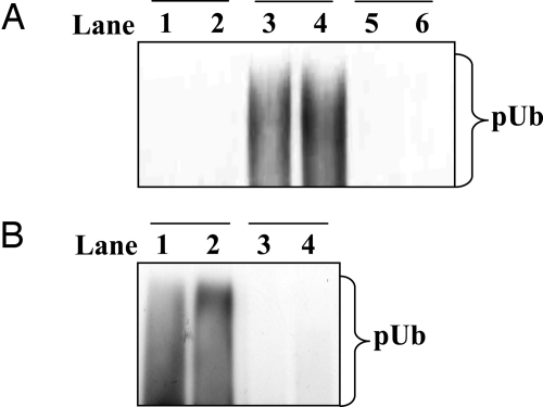
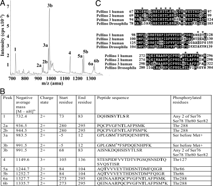
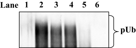
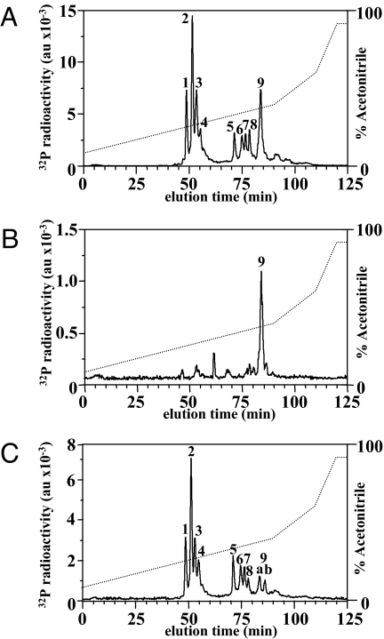
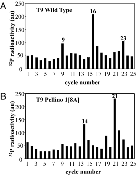
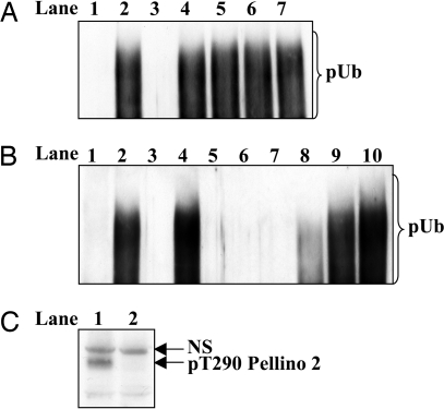
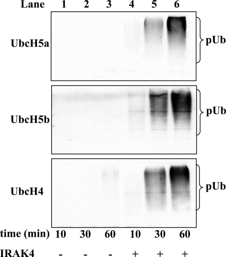
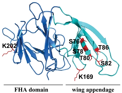
The E3 ubiquitin ligase Pellino can be activated by phosphorylation in vitro, catalyzed by IL-1 receptor-associated kinase 1 (IRAK1) or IRAK4. Here, we show that phosphorylation enhances the E3 ligase activity of Pellino 1 similarly with any of several E2-conjugating enzymes (Ubc13-Uev1a, UbcH4, or UbcH5a/5b) and identify 7 amino acid residues in Pellino 1 whose phosphorylation is critical for activation. Five of these sites are clustered between residues 76 and 86 (Ser-76, Ser-78, Thr-80, Ser-82, and Thr-86) and decorate a region of antiparallel beta-sheet, termed the "wing," which is an appendage of the forkhead-associated domain that is thought to interact with IRAK1. The other 2 sites are located at Thr-288 and Ser-293, just N-terminal to the RING-like domain that carries the E3 ligase activity. Unusually, the full activation of Pellino 1 can be achieved by phosphorylating any one of several different sites (Ser-76, Thr-86, Thr-288, or Ser-293) or a combination of other sites (Ser-78, Thr-80, and Ser-82). These observations imply that dephosphorylation of multiple sites is required to inactivate Pellino 1, which could be a device for prolonging Pellino's E3 ubiquitin ligase activity in vivo.


Conflict of interest statement

The authors declare no conflict of interest.

Figures

Fig. 1.
Fig. 1.
The IRAK-catalyzed activation of Pellino 1 requires the phosphorylation of Pellino 1. (A) The E3 ubiquitin ligase activities of three different Pellino complexes were compared by using Ubc13–Uev1a as the E2-conjugating enzyme. Lanes 1 and 2 show the activity of a phosphorylated IRAK1–unphosphorylated Pellino 1 complex, lanes 3 and 4 show a phosphorylated IRAK1–phosphorylated Pellino 1 complex, and lanes 5 and 6 show unphosphorylated Pellino 1 in the absence of IRAK1. The preparation of each complex and their assay are detailed in Experimental Procedures. pUb, polyubiquitin. (B) GST–Pellino 1 was phosphorylated with IRAK4 (lanes 1 and 2) or left unphosphorylated by incubation without IRAK4 (lanes 3 and 4). GST–Pellino 1 was then freed from IRAK4 and assayed for E3 ligase activity as described in Experimental Procedures.
Fig. 2.
Fig. 2.
Identification of phosphorylation sites in Pellino 1 by LC/MS. (A) Pellino 1 was phosphorylated by IRAK1 in vitro and subjected to SDS/PAGE, and the band corresponding to phosphorylated Pellino 1 was excised, digested with trypsin, and analyzed by LC/MS with precursor of 79 scanning (27). (B) Identification of the major peaks in A. Phosphorylated residues are shown in bold, and methionine sulfoxide is indicated by M*. The first 5 residues of phosphopeptides 3a and 3b, precede the initiating methionine (M+) of Pellino 1 for reasons explained in Results. (C) Comparison of the amino acid sequences surrounding the identified phosphorylation sites in human and Drosophila Pellinos. The major sites of IRAK1/4-catalyzed phosphorylation identified in this study are denoted by *. The white letters on a black background indicate identities between the sequences and black letters on a gray background indicate conservative replacements.
Fig. 3.
Fig. 3.
Activation of wild-type and mutant forms of Pellino 1 by IRAK4 in vitro. Pellino 1 was phosphorylated with (lanes 2–6) or without (lane 1) IRAK4, and its E3 ligase activity was measured with Ubc13-Uevla for 3 min as described in Experimental Procedures. Lanes 1 and 2, wild-type Pellino 1; lane 3, Pellino 1[7A]; lane 4, Pellino 1[8A]; lane 5, Pellino 1[10A]; lane 6, Pellino 1[10A] in which Ala-125 and Ala-127 were back-mutated to Ser and Thr, respectively. In Pellino 1[7A] residues 76, 78, 80, 82, 86, 127, and 288 all were mutated to Ala. Pellino 1[8A] contained the same mutations as Pellino 1[7A] except for the further mutation of Ser-70 to Ala. Pellino 1[10A] contained the same mutations as Pellino 1[8A] except for the further mutation of Ser-125 and Ser-293 to Ala (see Fig. 2C for the amino acid sequences of these regions).
Fig. 4.
Fig. 4.
Separation of tryptic phosphopeptides from Pellino 1. 32P-labeled Pellino 1 obtained by incubation with Mg-[γ32P]ATP and either IRAK4 (A and B) or IRAK1 (C) was subjected to SDS/PAGE, the gel was stained with Coomassie blue, and Pellino 1 was excised, digested with trypsin, and subjected to RP-HPLC on a Vydac C18 column equilibrated in 0.1% (vol/vol) trifluoroacetic acid. The column was developed with an acetonitrile gradient in 0.1% (vol/vol) trifluoroacetic acid. 32P radioactivity in arbitrary units (au) is shown by the full line, and the acetonitrile gradient is indicated by the diagonal broken lines. (A and C) Wild-type Pellino 1. (B) Pellino 1[8A].
Fig. 5.
Fig. 5.
Solid-phase sequencing of peptide T9 from Fig. 4. Peak T9 from wild-type Pellino 1 (Fig. 4A) and Pellino 1[8A] (Fig. 4B) were subjected to solid-phase sequencing (36) to identify the cycles of Edman degradation at which 32P radioactivity (filled bars) was released from the phosphopeptides present in these fractions.
Fig. 6.
Fig. 6.
Activation of different mutants of Pellino 1. Wild-type and mutated forms of Pellino 1 were phosphorylated with (lanes 2–7) or without (lane 1) IRAK4, and their E3 ligase activity was measured for 3 min as described in Experimental Procedures with Ubc13-Uevla. Pellino 1[10A] is a form of Pellino 1 in which the 10 phosphorylation sites at residues 70, 76, 78, 80, 82, 86, 125, 127, 288, and 293 are all mutated to Ala. (A) Lanes 1 and 2, wild-type Pellino 1; lane 3, Pellino 1[10A] in which Ala-125 and Ala-127 had been back-mutated to Ser and Thr, respectively; lane 4, same as lane 3, except for the additional back mutation of Ala-288 to Thr; lane 5, same as lane 3 except for the additional back mutation of Ala-293 to Ser; lane 6, same as lane 3, except that both Ala-288 and Ala-293 were back-mutated to Thr and Ser, respectively; lane 7, Pellino 1 in which Thr-288 and Ser-293 (but no other residue) was mutated to Ala. (B) Lanes 1 and 2, wild-type Pellino 1; lane 3, Pellino 1[10A] in which Ala-125 and Ala-127 were back-mutated to Ser and Thr, respectively; lanes 4, 5, 6, 7, and 8 show the activities of Pellino 1[10A] in which Ala-76 alone, Ala-78 alone, Ala-80 alone, Ala-82 alone, or Ala-86 alone, respectively were back-mutated to the Ser or Thr present in wild-type Pellino 1; lane 9, Pellino 1[10A] in which Ala-78, Ala-80, and Ala-82 were backmutated to Ser, Thr, and Ser, respectively; lane 10, Pellino 1[10A] in which Ala-78, Ala-80, Ala-82, and Ala-86 were back-mutated to Ser, Thr, Ser, and Thr, respectively. (C) DNA vectors encoding HA-Pellino 2 and either wild-type flag–IRAK1 (lane 1) or the catalytically-inactive flag–IRAK1[K239A] mutant (lane 2) were cotransfected into IRAK1-null IL-1R cells. Twenty-four hours later the cells were lysed and 25 μg of extract protein was subjected to SDS/PAGE and immunoblotted with a phospho-specific antibody that recognizes Thr-288 of Pellino 1 and the equivalent residue in Pellino 2 (Thr-290). NS, nonspecific band recognized by the antibody.
Fig. 7.
Fig. 7.
Effect of phosphorylation of Pellino 1 on its E3 ligase activity in the presence of different E2-conjugating enzymes. Pellino 1 was phosphorylated by incubation for 30 min at 30 °C without (lanes 1–3) or with (lanes 4–6) IRAK4, and its E3 ligase activity then measured for the times indicated by using UbcH5a (Top), UbcH5b (Middle), or UbcH4 (Bottom) as the E2-conjugating enzyme (see Experimental Procedures). pUb, polyubiquitin.
Fig. 8.
Fig. 8.
Location of the N-terminal phosphorylation sites and autoubiquitination sites in the structure of Pellino 1. A homology model of Pellino 1 was generated based on the recently published structure of Pellino 2 (30) using the program Modeller (37). Ser-76, Ser-78, Thr-80, Ser-82, and Thr-86 of Pellino 1 (shown in red) are located in a region of antiparallel β-sheet, termed the wing (turquoise), which is an appendage of the cryptic FHA domain (blue) thought to interact with phosphorylated IRAK1. Lys-169 and Lys-202 of Pellino 1, which were detected as sites of autoubiquitination, are also shown in red.

Similar articles

Cited by

References

    1. Muzio M, Ni J, Feng P, Dixit VM. IRAK (Pelle) family member IRAK-2 and MyD88 as proximal mediators of IL-1 signaling. Science. 1997;278:1612–1615. - PubMed
    1. Wesche H, Henzel WJ, Shillinglaw W, Li S, Cao Z. MyD88: An adapter that recruits IRAK to the IL-1 receptor complex. Immunity. 1997;7:837–847. - PubMed
    1. Li S, Strelow A, Fontana EJ, Wesche H. IRAK-4: A novel member of the IRAK family with the properties of an IRAK kinase. Proc Natl Acad Sci USA. 2002;99:5567–5572. - PMC - PubMed
    1. Martin M, Bol GF, Eriksson A, Resch K, Brigelius-Flohe R. Interleukin-1-induced activation of a protein kinase coprecipitating with the type I interleukin-1 receptor in T cells. Eur J Immunol. 1994;24:1566–1571. - PubMed
    1. Kollewe C, et al. Sequential autophosphorylation steps in the interleukin-1 receptor-associated kinase-1 regulate its availability as an adapter in interleukin-1 signaling. J Biol Chem. 2004;279:5227–5236. - PubMed

Publication types

MeSH terms