. 2009 Apr 10;104(7):879-86.
doi: 10.1161/CIRCRESAHA.108.193102. Epub 2009 Mar 5.

Downregulation of miR-199a derepresses hypoxia-inducible factor-1alpha and Sirtuin 1 and recapitulates hypoxia preconditioning in cardiac myocytes

Affiliations

Downregulation of miR-199a derepresses hypoxia-inducible factor-1alpha and Sirtuin 1 and recapitulates hypoxia preconditioning in cardiac myocytes

Shweta Rane et al. Circ Res. .

Abstract

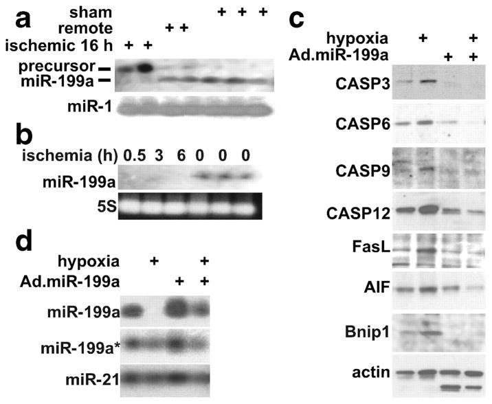
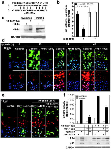
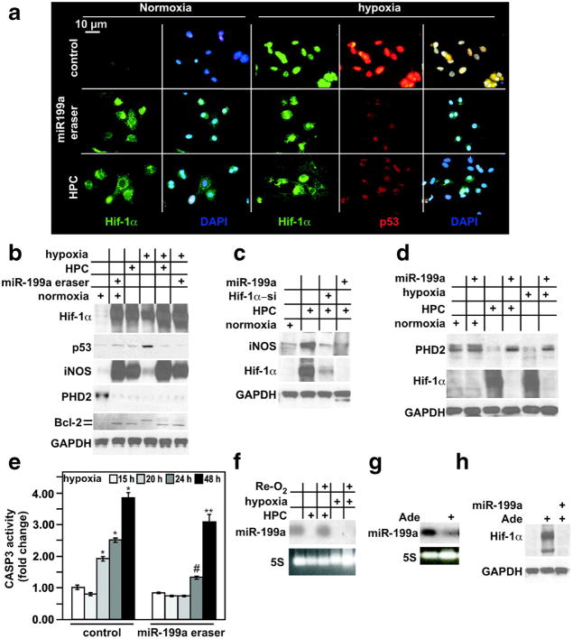
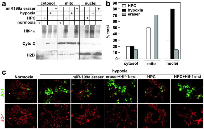
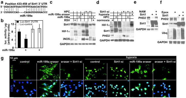
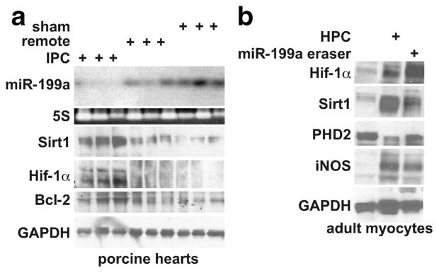
MicroRNAs are posttranscriptional gene regulators that are differentially expressed during various diseases and have been implicated in the underlying pathogenesis. We report here that miR-199a is acutely downregulated in cardiac myocytes on a decline in oxygen tension. This reduction is required for the rapid upregulation of its target, hypoxia-inducible factor (Hif)-1alpha. Replenishing miR-199a during hypoxia inhibits Hif-1alpha expression and its stabilization of p53 and, thus, reduces apoptosis. On the other hand, knockdown of miR-199a during normoxia results in the upregulation of Hif-1alpha and Sirtuin (Sirt)1 and reproduces hypoxia preconditioning. Sirt1 is also a direct target of miR-199a and is responsible for downregulating prolyl hydroxylase 2, required for stabilization of Hif-1alpha. Thus, we conclude that miR-199a is a master regulator of a hypoxia-triggered pathway and can be exploited for preconditioning cells against hypoxic damage. In addition, the data demonstrate a functional link between 2 key molecules that regulate hypoxia preconditioning and longevity.


Figures

Figure 1
Figure 1
Downregulation of miR-199a is required for hypoxia-induced proapoptotic genes. a, C57Bl/6 mice were subjected to left coronary artery occlusion for 16 hour. The ischemic and remote regions of the left ventricle and the sham-operated ventricle were isolated and total RNA was extracted and analyzed by Northern blotting (n = 3). b, Mice were subjected to left coronary artery occlusion for 0.5, 3, and 6 hours and analyzed as in a. c, Myocytes were infected with a control or miR-199a–expressing adenoviruses before exposure to hypoxia for 24 hours in complete culture medium with serum (where marked by +). Protein was extracted and analyzed by Western blotting (n = 3). d, Myocytes were treated as in c. Total RNA was extracted and analyzed by Northern blotting (n = 3).
Figure 2
Figure 2
MiR-199a targets and inhibits Hif-1α. a, The alignment between Mus musculus miR-199a and the 3′UTR of HIF1A, identified by TargetscanS software. b, The miR-199a target region, or a mutant, was cloned into the 3′UTR of a luciferase gene (represented in the graph by black and white bars, respectively). These constructs were introduced into myocytes, in addition to exogenous miR-199a (where marked by +) or a control virus (n = 6). After 24 hours, luciferase activity was measured, averaged, and plotted. The y axis represents arbitrary luciferase activity normalized to micrograms of protein content. Error bars represent SEM. *P<0.01, miR-199a–treated luciferase-Hif-1α 3′UTR target vs control. c, Wild-type Hif-1αcDNA or a mutant lacking miR-199a target site (Hif-1αΔ199a) were delivered to cardiac myocytes or HEK293 cells. After 24 hours, protein was extracted and analyzed by Western blotting (n = 2). d, Myocytes were treated with a control or a miR-199a overexpressing virus for 24 hours before subjecting them to various periods of hypoxia, as indicated. Parallel slides were stained separately with anti–Hif-1α (green) or anti-p53 (red) antibodies and DAPI (blue) (n = 4). e, Myocytes were treated with a control or Hif-1αΔ199a virus, in the absence or presence of a control or miR-199a virus for 24 hours. Cells were then exposed to hypoxia for 24 hours where indicated, before immunostaining with anti-Hif-1α (green), anti-p53 (red), and DAPI (blue) (n = 3). f, Myocytes were treated as in e. Protein was extracted and either assayed for caspase 3 activity (graph, n = 6) or analyzed by Western blotting (n = 3). The treatments are indicated in the grid below the graph by + signs and each aligned with its Western blot results. Results were averaged, normalized to protein content, and plotted as fold change after adjusting basal levels to 1. Error bars represent SEM, *P<0.001, miR-199a–treated vs untreated cells during hypoxia; **P<0.01, miR-199a–treated plus Hif-1aΔ199a vs miR-199a–treated.
Figure 3
Figure 3
Knockdown of miR-199a induces upregulation of Hif-1α, iNOS, and down-regulation of PHD2, mimicking HPC. a, Cardiac myocytes were treated with a control or miR-199a eraser–expressing adenovirus for 24 hours, or HPC, as indicated on the left. A parallel set of myocytes were similarly treated and subsequently subjected to hypoxia for 24 hours, as indicated on the top. Myocytes were then fixed and costained with anti–Hif-1α (green), anti-p53 (red), and DAPI (blue) (n = 5). b, Myocytes were treated as described in a and as indicated in the grid by + signs. Protein was extracted and analyzed by Western blotting (n = 3). c, Myocytes were subjected to HPC before or after pretreatment with a control, miR-199a, and Hif-1α short interfering RNA (Hif-1α-si)-expressing adenoviruses for 24 hours, where indicated by + signs. Protein was extracted and analyzed by Western blotting for the molecules indicated on the left. d, Myocytes were subjected to 24 hours of hypoxia or HPC, before or after treatment with a control or miR-199a–expressing virus for 24 hours where indicated by + signs. Protein was extracted and analyzed by Western blotting for the molecules indicated on the left. e, Myocytes were subjected to 15, 20, 24, or 48 hours of hypoxia before or after treatment with a control or miR-199a eraser for 24 hours, as indicated. Protein was extracted and assayed for caspase 3 activity (n = 6). Results were averaged, normalized to protein content, and plotted as fold change, after adjusting basal levels to 1. Error bars represent SEM. *P<0.01, hypoxia vs normoxia; #P<0.01, miR-199a eraser–pretreated, 24-hour hypoxic, vs control-treated, 24-hour hypoxic, myocytes; **P<0.5, miR-199a eraser–pretreated, 48-hour hypoxic, vs control-treated, 48-hour hypoxic, myocytes. f, Myocytes were subjected to HPC or 24 hours of hypoxia with or without reoxygenation (Re-O2) for 12 hours as indicated with the + sign. Total RNA was then extracted and analyzed by Northern blotting for the miRNA indicated on the left (n = 2). g, Myocytes were stimulated with 100 μmol/L adenosine for 16 hours. Total RNA was then extracted and analyzed by Northern blotting for the miRNA indicated on the left (n = 2). h, Myocytes were treated as in (g). Protein was extracted and analyzed by Western blotting (n = 2).
Figure 4
Figure 4
Hif-1α associates with mitochondria and is required for HPC-mediated protection. a, Cardiac myocytes were subjected to HPC, 24 hours of hypoxia, or treated with a control or the miR-199a eraser–expressing virus for 24 hours, as indicated in the grid by + signs. Cells were fractionated into cytosol, mitochondria, and nuclei and analyzed by Western blotting for the proteins indicated on the left (n = 3). b, The Hif-1α signal shown in a was quantitated in all fractions, for each treatment, and the percentage of total was calculated and plotted (n = 3). c, Cardiac myocytes were plated on gelatin-coated glass chamber slides. Cells were treated with a control or a Hif-1α short interfering RNA (Hif-1α-si)-expressing adenovirus for 48 hours before applying miR-199a eraser or HPC. They were then exposed to hypoxia for 24 hours. Following that, JC-1 dye was applied and the cells were imaged live (n = 4).
Figure 5
Figure 5
Sirt1 is a direct target of miR-199a, is upregulated during HPC, and is required for downregulation of PHD2. a, The alignment between M musculus miR-199a and a 3′UTR region of Sirt1. b, The miR-199a target site, or a mutant, was cloned into the 3′UTR of a luciferase gene (represented in the graph by black and white bars, respectively). These constructs were delivered to myocytes via adenovirus, in addition to exogenous miR-199a (where marked by +) or a control virus (n = 6). After 24 hours, luciferase activity was measured, averaged, and plotted. The y axis represents arbitrary luciferase activity normalized to micrograms of protein content. Error bars represent SEM. *P<0.01, miR-199a–treated, luciferase-Sirt13′UTR target vs control. c, Myocytes were treated with 40 μmol/L resveratrol (RSV) for 24 hours or HPC, with or without exogenous miR-199a for an additional 24 hours, or with miR-199a eraser for 24 hours, where indicted by + signs (n = 3). Protein was then extracted and analyzed by Western blotting. d, Myocytes were treated with Sirt1 short interfering RNA (Sirt1-si) adenovirus for 48 hours. These cells were then exposed to hypoxia for 24 hours or HPC, where indicated by +signs. Protein was then extracted and analyzed by Western blotting (n = 3). e, Myocytes were treated with a control or Sirt1-overexpressing virus in the absence or presence of 20 mmol/L nicotinamide (NAM). Protein was extracted and analyzed by Western blotting (n = 3). f, Myocytes were treated with a control or Sirt1-overexpressing virus in the absence or presence of 0.1 μmol/L epoxomicin (Epox) for 24 hours. Protein was extracted and analyzed by Western blotting (n = 3). g, Myocytes were plated on gelatin-coated glass chamber slides. Cells were treated with a control, miR-199a eraser, or a Sirt1 short interfering RNA (Sirt1-si)-expressing adenovirus for 48 hours, followed miR-199a eraser. A parallel set of similarly treated slides was then exposed to 24 hours of hypoxia, as indicated above. Cells were then fixed and stained with anti-Hif-1α (green) and DAPI (blue) (n = 3).
Figure 6
Figure 6
MiR-199a is downregulated during IPC in porcine hearts and is associated with upregulation of Hif-1α and Sirt1. a, Porcine hearts were preconditioned via 2×10-minute cycles of ischemia/reperfusion of the left ventricle (n = 3). A second set of animals was subjected to a sham operation. The IPC area of the left ventricle, remote zone, and sham-operated ventricles were immediately dissected (early/first window IPC) and analyzed by Northern and Western blotting. The top 2 gels are the results of a Northern blot, and the lower 3 gels are Western blots. b, Cultured adult rat cardiac myocytes were treated with miR-199a eraser for 24 hours or HPC. Protein was extracted and analyzed by Western blotting for the molecules indicated (n = 3).

References

    1. Sayed D, Hong C, Chen IY, Lypowy J, Abdellatif M. MicroRNAs play an essential role in the development of cardiac hypertrophy. Circ Res. 2007;100:416–424. - PubMed
    1. John B, Enright AJ, Aravin A, Tuschl T, Sander C, Marks DS. Human MicroRNA targets. PLoS Biol. 2004;2:1862–1879. - PMC - PubMed
    1. Thomson JM, Newman M, Parker JS, Morin-Kensicki EM, Wright T, Hammond SM. Extensive post-transcriptional regulation of microRNAs and its implications for cancer. Genes Dev. 2006;20:2202–2207. - PMC - PubMed
    1. Newman MA, Thomson JM, Hammond SM. Lin-28 interaction with the Let-7 precursor loop mediates regulated microRNA processing. RNA. 2008;14:1539–1549. - PMC - PubMed
    1. Viswanathan SR, Daley GQ, Gregory RI. Selective blockade of microRNA processing by Lin28. Science. 2008;320:97–100. - PMC - PubMed

Publication types

MeSH terms