. 2009 Apr 10;104(7):860-9.
doi: 10.1161/CIRCRESAHA.108.192765. Epub 2009 Mar 5.

Ciliary polycystin-2 is a mechanosensitive calcium channel involved in nitric oxide signaling cascades

Affiliations

Ciliary polycystin-2 is a mechanosensitive calcium channel involved in nitric oxide signaling cascades

Wissam A AbouAlaiwi et al. Circ Res. .

Abstract

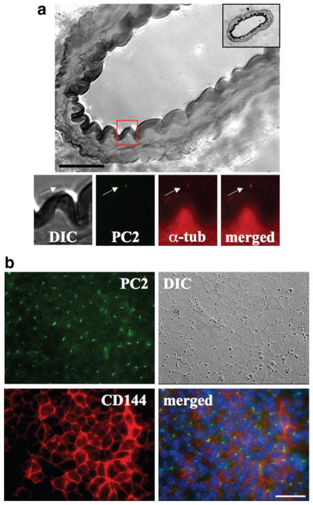
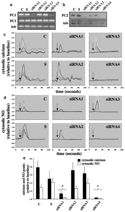
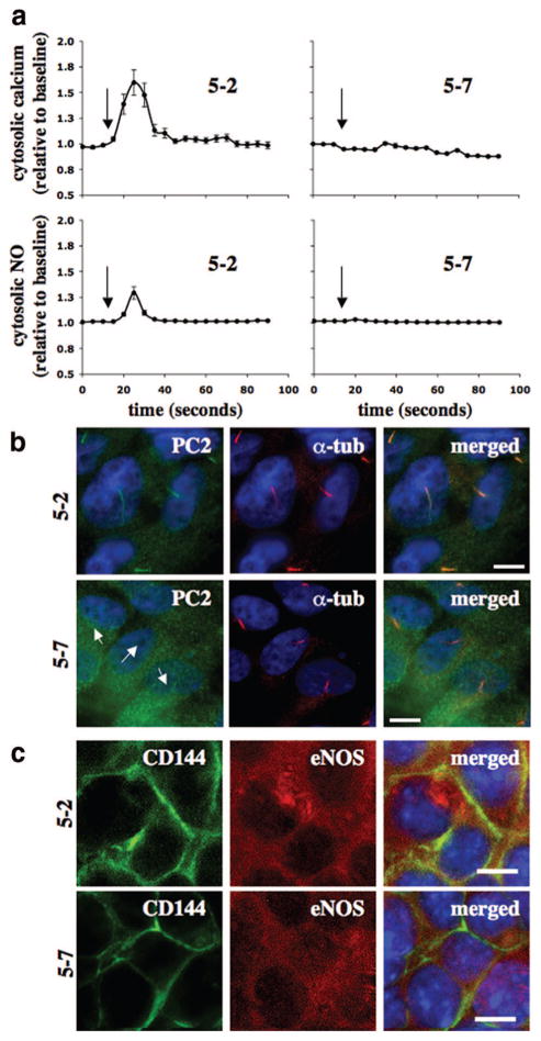
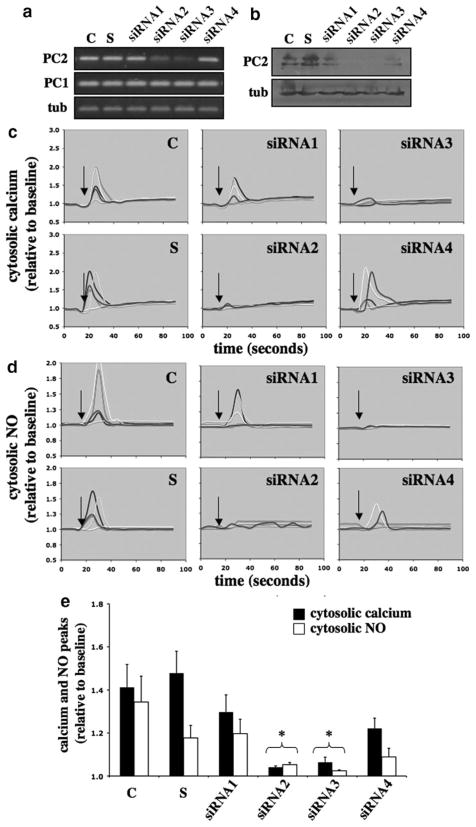
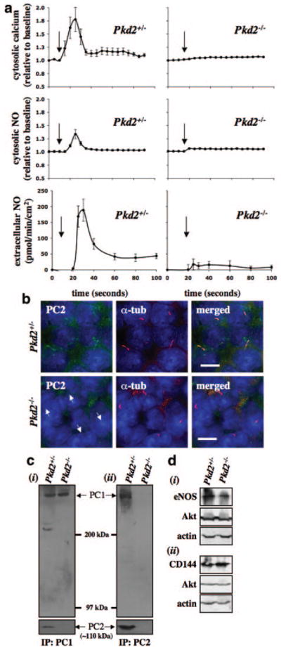
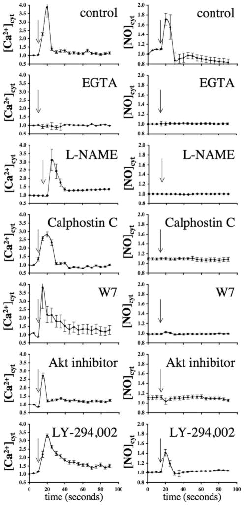
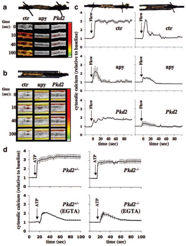
Cardiovascular complications such as hypertension are a continuous concern in patients with autosomal dominant polycystic kidney disease (ADPKD). The PKD2 encoding for polycystin-2 is mutated in approximately 15% of ADPKD patients. Here, we show that polycystin-2 is localized to the cilia of mouse and human vascular endothelial cells. We demonstrate that the normal expression level and localization of polycystin-2 to cilia is required for the endothelial cilia to sense fluid shear stress through a complex biochemical cascade, involving calcium, calmodulin, Akt/PKB, and protein kinase C. In response to fluid shear stress, mouse endothelial cells with knockdown or knockout of Pkd2 lose the ability to generate nitric oxide (NO). Consistent with mouse data, endothelial cells generated from ADPKD patients do not show polycystin-2 in the cilia and are unable to sense fluid flow. In the isolated artery, we further show that ciliary polycystin-2 responds specifically to shear stress and not to mechanical stretch, a pressurized biomechanical force that involves purinergic receptor activation. We propose a new role for polycystin-2 in transmitting extracellular shear stress to intracellular NO biosynthesis. Thus, aberrant expression or localization of polycystin-2 to cilia could promote high blood pressure because of inability to synthesize NO in response to an increase in shear stress (blood flow).


Figures

Figure 1
Figure 1
Polycystin-2 localization in vivo and in vitro. Localization of polycystin-2 (PC2) was examined with immunofluorescence staining. a, High-resolution differential interference contrast (DIC) image shows a section of femoral artery with a thickness of 10 μm. The inset in the top right corner shows the full section of the artery. The red box magnifies an endothelial cell, which shows localization of PC2 to endothelial cilia. Acetylated α-tubulin (α-tub) was used as a ciliary marker. b, Cultured endothelial cells also show the presence of polycystin-2 in cilia, and VE-cadherin (CD144) was used as an endothelial marker. Nuclear marker (DAPI) is shown in the merged images. Scale bar=25 μm.
Figure 2
Figure 2
Effects of polycystin-2 expression in mouse endothelial cells. The expression level of polycystin-2 corresponds to the cytosolic calcium increase and NO biosynthesis in response to fluid shear stress. Mouse endothelial cells were transfected with Lipofectin only (control, C), scramble siRNA (S), siRNA1, siRNA2, siRNA3, or siRNA4 of polycystin-2. a, The transfected cells were collected to examine the transcript levels of polycystin-2 (PC2) and polycystin-1 (PC1), and α-tubulin (tub) was used as a control. b, The transfected cells were analyzed for polycystin-2 expression, and the membrane was reblotted for α-tubulin as a loading control. c, Transfected cells were challenged with fluid shear stress of 7 dyn/cm2 (indicated by arrows), and 5 individual responses were randomly selected and analyzed for changes in cytosolic calcium levels. d, Biosynthesis of NO in response to shear stress was plotted in 5 individual cells. e, Peaks of cytosolic calcium and NO in response to shear stress were averaged. Asterisks indicate statistically significant against control at P<0.05 (N=3).
Figure 3
Figure 3
Effects of fluid shear stress in vascular endothelial cells of an ADPKD patient. Vascular endothelial cells were isolated from several interlobar arteries of an ADPKD kidney. a, Endothelial cells (5-2 and 5-7) from segmental arteries 2 and 7 of patient 5 were cultured and challenged with fluid shear stress, and their cytosolic calcium and NO changes were recorded. Arrows indicate the start of fluid flow. b, The primary culture was then subjected to immunolocalization studies for polycystin-2 (PC2). Acetylated α-tubulin (α-tub) was used as a ciliary marker, and nuclear marker (DAPI) is shown in the merged images. Arrows indicate the absence of polycystin-2. c, Immunofluorescence studies indicated the presence of endothelial markers CD144 and eNOS in both 5-2 and 5-7 cells. N=3 with passages 2, 3, and 4. Scale bar=5 μm.
Figure 4
Figure 4
Effects of polycystin-2 expression in human endothelial cells. The expression level of polycystin-2 corresponds with the cytosolic calcium increase and NO biosynthesis in response to fluid shear stress. Human umbilical vein endothelial cells were transfected with Lipofectin only (control, C), scramble siRNA (S), siRNA1, siRNA2, siRNA3, or siRNA4 of polycystin-2. a, The transfected cells were collected to examine the transcript levels of polycystin-2 (PC2) and polycystin-1 (PC1), and α-tubulin (tub) was used as a control. b, The transfected cells were analyzed for polycystin-2 expression, and the membrane was reblotted for α-tubulin as a loading control. c, Transfected cells were challenged with fluid shear stress of 7 dyn/cm2 (indicated by arrows), and 5 individual responses were randomly selected and analyzed for changes in cytosolic calcium levels. d, Biosynthesis of NO in response to shear stress was plotted in 5 individual cells. e, Peaks of cytosolic calcium and NO in response to shear stress were averaged. Asterisks indicate statistically significant against control at P<0.05 (N=3).
Figure 5
Figure 5
Polycystin-2 required in fluid-flow sensing. Vascular endothelial cells were isolated from Pkd2+/− and Pkd2−/− embryonic aortas. a, The endothelial cells were cultured and challenged with fluid shear stress, and their cytosolic calcium increase, intracellular NO production, and extracellular NO release were measured. Arrows indicate the start of fluid flow. b, The primary culture was then subjected to immunolocalization studies for polycystin-2 (PC2). Acetylated α-tubulin (α-tub) was used as a ciliary marker, and nuclear marker (DAPI) is shown in the merged images. Arrows indicate the absence of polycystin-2. c, Immunoprecipitation studies for polycystin-1 (i) and -2 (ii) confirmed interaction of polycystin-1 and -2 in vascular endothelial cells and absence of polycystin-2 in Pkd2−/− cells. d, The presence of endothelial markers eNOS (i), CD144 (ii), and Akt (i and ii) confirmed the cell type used in our study, and β-actin was used as a loading control. N=3 for calcium and NO measurements with passages 2, 3, and 4. Scale bar=5 μm.
Figure 6
Figure 6
Molecular cascade involved in shear stress–induced calcium and NO signaling. The left and right graphs represent the response to fluid shear stress in mouse endothelial cells for cytosolic calcium ([Ca2+]cyt) and NO ([NO]cyt), respectively. Effects of various inhibitors on response to fluid shear stress, as indicated by arrows, are presented in the line graphs. The extra-cellular calcium chelator EGTA abolished both calcium and NO increases, indicating extracellular calcium influx is required in shear-induced NO biosynthesis. NO synthase inhibitor L-NAME indicated that shear-induced NO production involves a rapid activation of NO synthase. Whereas PKC (calphostin C), calmodulin (W7), and Akt inhibitors played a role in mechanical fluid sensing, PI3K inhibitor (LY-294,002) did not affect shear-induced NO production. N≥4.
Figure 7
Figure 7
Integration of polycystin-2 and purinergic signaling in isolated artery. To differentiate mechanical forces generated by perfusate, an isolated artery was freely placed to provide unrestrained movement, or the artery was capillary-enclosed to limit stretching motion. Artery transfected with scrambled siRNA is denoted as control (ctr), whereas artery transfected with scrambled siRNA and pre-treated and perfused with apyrase is designated as apyrase (apy). Isolated artery was also transfected with Pkd2 siRNA (Pkd2). a, In the freely placed artery, as illustrated by the top image, calcium imaging studies show different cytosolic calcium profiles among control, apyrase, and Pkd2-treated groups. b, In the capillary-enclosed artery, as illustrated by the top image, flow-induced cytosolic calcium increase was diminished in Pkd2, compared to control or apyrase-treated arteries. Changes in cytosolic calcium was pseudocolored, white/green represents a low level, and yellow/red denotes a higher level of cytosolic calcium. In all cases, a similar fluid-flow rate of 164 μL/sec was initiated after time 0 second. c, Five responsive areas within the artery, if any were present, were randomly selected and analyzed for changes in cytosolic calcium levels. d, Both Pkd2+/− and Pkd2−/− endothelial cells were able to respond to 10 μmol/L ATP in the presence or absence of 1 mmol/L EGTA. N=3 arteries for each group and treatment, and N>2 for cells with passage 2, 3, or 4.

References

    1. Boucher C, Sandford R. Autosomal dominant polycystic kidney disease (ADPKD, MIM 173900, PKD1 and PKD2 genes, protein products known as polycystin-1 and polycystin-2) Eur J Hum Genet. 2004;12:347–354. - PubMed
    1. Torres VE, Harris PC, Pirson Y. Autosomal dominant polycystic kidney disease. Lancet. 2007;369:1287–1301. - PubMed
    1. Hateboer N, Veldhuisen B, Peters D, Breuning MH, San-Millan JL, Bogdanova N, Coto E, van Dijk MA, Afzal AR, Jeffery S, Saggar-Malik AK, Torra R, Dimitrakov D, Martinez I, de Castro SS, Krawczak M, Ravine D. Location of mutations within the PKD2 gene influences clinical outcome. Kidney Int. 2000;57:1444–1451. - PubMed
    1. Hateboer N, v Dijk MA, Bogdanova N, Coto E, Saggar-Malik AK, San Millan JL, Torra R, Breuning M, Ravine D. Comparison of phenotypes of polycystic kidney disease types 1 and 2. European PKD1-PKD2 Study Group. Lancet. 1999;353:103–107. - PubMed
    1. Wu G, Markowitz GS, Li L, D’Agati VD, Factor SM, Geng L, Tibara S, Tuchman J, Cai Y, Park JH, van Adelsberg J, Hou H, Jr, Kucherlapati R, Edelmann W, Somlo S. Cardiac defects and renal failure in mice with targeted mutations in Pkd2. Nat Genet. 2000;24:75–78. - PubMed

Publication types

MeSH terms