. 2009 Mar 15;182(6):3398-405.
doi: 10.4049/jimmunol.0803006.

ERK5 knockdown generates mouse leukemia cells with low MHC class I levels that activate NK cells and block tumorigenesis

Affiliations

ERK5 knockdown generates mouse leukemia cells with low MHC class I levels that activate NK cells and block tumorigenesis

Seyma Charni et al. J Immunol. .

Abstract

Tumor cell-based vaccines are currently used in clinical trails, but they are in general poorly immunogenic because they are composed of cell extracts or apoptotic cells. Live tumor cells should be much better Ags provided that they are properly processed by the host immune system. We show herein that stable expression of a small hairpin RNA for ERK5 (shERK5) decreases ERK5 levels in human and mouse leukemic cells and leads to their elimination by NK cells in vivo. The shERK5 cells show down-regulation of MHC class I expression at the plasma membrane. Accordingly, ectopic activation of the ERK5 pathway induces MHC class I gene expression. Coinjection of shERK5-expressing cells into the peritoneum diminishes survival of engrafted wild-type tumor cells. Moreover, s.c. injection of shERK5-expressing cells strongly diminishes tumor development by wild-type cells. Our results show that shERK5 expression in leukemia cells effectively attenuates their tumor activity and allows their use as a tumor cell-based vaccine.


Figures
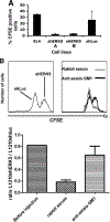
FIGURE 1.
FIGURE 1.
Leukemia cells expressing shERK5 are eliminated within 3 days of peritoneal injection in syngenic mice. A, One million EL4 wt, shERK5 (two independent cell lines: A and B), and shLuc cells labeled with 1 μM CFSE were injected in the peritoneum of syngenic C57BL/6 mice. Peritoneal cells were recovered after 3 days and CFSE+ cells were analyzed by FACS. The percentage positive cells was determined relative to the total number of cells recovered. B, Five million L1210 shLuc or shERK5 cells were labeled with 1 μM (shLuc) or 5 μM (shERK5) CFSE (upper left panel) and injected in syngenic BALB/c mice treated with either anti-asialo GM1, which eliminates NK cells, or control rabbit serum. Two days later, CFSE+ cells in the peritoneal wash, along with the input cells, were analyzed by FACS (upper right panel), and the ratio between shERK5 and shLuc cells was calculated (lower panel).
FIGURE 2.
FIGURE 2.
NK cells target shERK5-expressing leukemia cells in vitro. A, EL4 shERK5 cells loaded with 51Cr were incubated 4 h with total splenocytes, purified NK cells, or splenocytes depleted of NK cells at different ratios. Cytolytic activity was measured using the 51Cr released into the cell supernatants. B, L1210 shERK5 cells (left and middle panels) or shLuc cells (right panel) were labeled with [3H]thymidine and incubated at different ratios with effector cells and either purified NK cells (+NK) or splenocytes depleted of NK cells ( NK) from syngenic (left and right panels) or allogenic (middle panel) mice. C, NK cells use both the perforin/granzyme and the Fas/FasL pathway to kill L1210 shERK5 cells in vitro. Different ratios of purified NK cells (+NKs, E = effector) were incubated with 51Cr-labeled L1210 shERK5 cells (T = target) for 4 h. Two millimolar EGTA and/or 1 μg/ml anti-FasL were added to the media during incubation. D, Fas expression in L1210 cells, either shLuc or shERK5, was analyzed by FACS. The dashed lines represent staining with a nonspecific IgG, and the brackets above the curves indicate the Fas-positive cells.
FIGURE 3.
FIGURE 3.
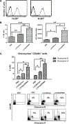
L1210 shERK5 cells recruit and activate NK cells. A, L1210 shLuc (thin line) and L1210 shERK5 cells (thick line) were stained with anti-H-2Dd (left panel) or anti-H-2Kd (right panel) Abs to visualize MHC-I expression and analyzed by FACS. The dash line showed the unspecific binding of a control IgG Ab. B, Five million cells, either L1210 shLuc or L1210 shERK5, or PBS was injected into the peritoneum of syngenic BALB/c mice. Forty-eight hours later, cells were recovered from the site of injection and stained with an Ab to CD49b. The percentage (left panel) and the number (right panel) of NK cells were measured by FACS analysis. The data were evaluated using Student’s t test: *, p < 0.05; **, p < 0.01; ***, p < 0.001. C, Cells from B were stained with anti-granzyme A or anti-granzyme B Abs and analyzed as described in B to evaluate NK cell activation. The bars (upper panel) show the percentage of cells staining positive for both CD49b and granzyme, and they represent the average ± SD of three independent experiments. The lower set of panels shows the data from a typical experiment. The data were evaluated using Student’s t test: *, p < 0.05; **, p < 0.01; ***, p < 0.001.
FIGURE 4.
FIGURE 4.
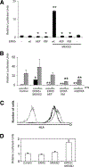
ERK5 catalytic activity is required for the induction of a MHC-I reporter gene in Jurkat cells. A, Ten million Jurkat cells were transfected with the following: 5 μg of expression vectors for either wt ERK5, ERK5 AEF, or ERK5 KM and/or constitutively active MEK5 (MEK5D), 2.5 μg of the reporter gene PD-1-Luc, and 1 μg of the β-galactosidase expression vector. Forty-eight hours later, lysates were prepared and analyzed for luciferase and galactosidase activity. The relative luciferase units represent the ratio of luciferase/galactosidase. B, ERK5 is essential for IFN-induced MHC-I up-regulation. Cells transfected as in A were unstimulated (black bars) or stimulated with 1 nM each IFN-αand IFN-β (gray bars) for 3 days. The data are presented as the mean ± SD of at least three independent experiments, and they were evaluated using Student’s t test: *, p < 0.05 and **, p < 0.005 compared with control cells. C, Fifty million Jurkat Tag cells were transfected with with 3 μg of a GFP expression vector and either 10 μg of pcDNA3 or 5 μg each of expression vectors for ERK5 and MEK5D. Two days later, GFP cells were isolated by FACS and MHC-I expression analyzed in cells cotransfected with empty vector (thick line) or MEKD ERK5 (thin line) using an Ab to human HLA. The dotted line shows background staining in the absence of anti-HLA. D, HeLa cells were transfected with the following: 1 μg of expression vectors for wt ERK5 and/or constituively active MEK5 (MEK5D), 2.5 μg of the reporter gene PD-1-Luc, and 1 μg of a TK-Renilla luciferase reporter gene. For each transfection, a constant amount of DNA was transfected. The data were normalized to the TK-Renilla internal control and expressed as fold activation relative to the empty vector. The bars represent the mean ± SE of two experiments performed in duplicate.
FIGURE 5.
FIGURE 5.
The ERK5 pathway controls transcription of the endogenous β2-m gene. A, RNA was isolated from 107 resting L1210 wt, shLuc, or shERK5 cells and analyzed by quantitative RT-PCR for β2-m and the housekeeping gene S26. The ratio between the two was used to calculate the arbitrary units. The data represent the mean ± SD of at least three independent experiments, which were evaluated using Student’s t test: *, p < 0.05. B, Twenty million Jurkat cells were transfected with 3 μg of a GFP expression vector and either 5 μg each of expression vectors for ERK5 and MEK5D or 10 μg of empty vector. Two days later, GFP+ cells were isolated by FACS, and RNA was isolated and analyzed as described in A. The data represent the mean ± SD of at least three independent experiments, which were evaluated using Student’s t test: *, p < 0.05.
FIGURE 6.
FIGURE 6.
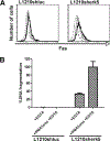
L1210 shERK5 cells are highly sensitive to killing by cytotoxic lymphocytes. A, L1210 shLuc or shERK5 cells (dashed line) were incubated with resting (thin line) or activated (thick line) BM3.3 cells for 1 h and Fas expression in the CD8 population was analyzed by FACS. B, Resting or PMA/ionomycin-activated BM3.3 cells were incubated with L1210 shLuc or shERK5 cells for 2 h in the presence of 2 mM EGTA before measuring DNA fragmentation as described in Materials and Methods.
FIGURE 7.
FIGURE 7.
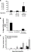
EL4 leukemia cells expressing shERK5 block tumorigenesis by wt cells. A, Five hundred thousand EL4 cells were injected in the peritoneum of syngenic C57BL/6 mice. Three days later, cells were washed from the site of injection and the number of NK cells was determined by FACS using an Ab to NK1.1. The percentage of NK cells was calculated relative to the total number of cells. B, EL4 shLuc loaded with CFSE were injected alone (left bar) or together with nonlabeled EL4 shERK5 cells (right bar, 5 × 10⁵ of each) in the peritoneum of syngenic mice (n = 3 mice/group). The same number of EL4 shERK5 cells loaded with CFSE were injected as a control (middle bar). After 3 days, cells were washed from the site of injection and the number of CFSE+ cells was determined by FACS. The percentage of CFSE+ cells was calculated relative to the total number of cells. C, Syngenic C57BL/6 mice were injected with 2.5 × 10⁵ EL4 shERK5 cells or PBS (n = 4) and then injected s.c. with 2.5 × 10⁵ EL4 wt cells 2 (n = 4) or 6 (n = 5) weeks later. Tumor size was measured at the indicated times afterward.

References

    1. Dunn GP, Koebel CM, and Schreiber RD 2006. Interferons, immunity and cancer immunoediting. Nat. Rev. Immunol 6: 836–848. - PubMed
    1. Nguyen van Binh P, and Duc HT 2006. Analyses and perspectives in cancer immunotherapy. Biomed. Pharmacother 60: 621–628. - PubMed
    1. Berd D, Maguire HC Jr., McCue P, and Mastrangelo MJ 1990. Treatment of metastatic melanoma with an autologous tumor-cell vaccine: clinical and immunologic results in 64 patients. J. Clin. Oncol 8: 1858–1867. - PubMed
    1. Ronchetti A, Rovere P, Iezzi G, Galati G, Heltai S, Protti MP,Garancini MP, Manfredi AA, Rugarli C, and Bellone M 1999. Immunogenicity of apoptotic cells in vivo: role of antigen load, antigen-presenting cells, and cytokines. J. Immunol 163: 130–136. - PubMed
    1. Dzik WH 2003. Apoptosis, TGFβ and transfusion-related immunosuppression: biologic versus clinical effects. Transfus. Apher. Sci 29: 127–129. - PubMed

Publication types

MeSH terms

Substances