. 2009 Mar 15;182(6):3492-3502.
doi: 10.4049/jimmunol.0803052.

Selective targeting of B cells with agonistic anti-CD40 is an efficacious strategy for the generation of induced regulatory T2-like B cells and for the suppression of lupus in MRL/lpr mice

Affiliations

Selective targeting of B cells with agonistic anti-CD40 is an efficacious strategy for the generation of induced regulatory T2-like B cells and for the suppression of lupus in MRL/lpr mice

Paul A Blair et al. J Immunol. .

Abstract

We have previously reported that IL-10(+) regulatory B cells, known to play an important role in controlling autoimmunity and inflammatory disorders, are contained within the transitional 2 immature (T2) B cell pool (T2 Bregs). Therapeutic strategies facilitating their enrichment or enhancing their suppressive activity are highly attractive. In this study, we report that agonistic anti-CD40 specifically targets T2 B cells and enriches Bregs upon short-term in vitro culture. Although transfer of unmanipulated T2 B cells, isolated from mice with established lupus, failed to confer protection to diseased mice, transfer of in vitro anti-CD40-generated T2 B cells (T2-like-Bregs) significantly improved renal disease and survival by an IL-10-dependent mechanism. T2-like-Bregs readily accumulated in the spleen after transfer, suppressed Th1 responses, induced the differentiation of IL-10(+)CD4(+)T cells, and conveyed a regulatory effect to CD4(+)T cells. In addition, in vivo administration of agonistic anti-CD40, currently on trial for the treatment of cancer, halted and reversed established lupus. Taken together, our results suggest a novel cellular approach for the amelioration of experimental lupus.


Figures
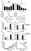
Fig. 1
Fig. 1. T2 and MZ B cells subsets fail to confer protection to MRL/lpr mice
Splenic B cells were labelled with CD19, CD21, CD23, CD24. MZ, FO and T2 B cells were gated as previously described (6, 12). (A) Absolute numbers of MZ and T2 B cells in MRL/lpr mice at different age (B) Scatter plot showing a significant correlation between the proteinuria and the absolute number of T2 or MZ B cells at different age. Splenocytes were harvested from MRL/Mp or C57BL/6 mice at 9 and 24 weeks of age and stained with CD19, CD21, CD23, CD24. C) The percentage of T2 and MZ B cells at different ages. D) Absolute numbers of T2 and MZ B cells calculated as the numbers of cells/live events × the total number of cells in the spleen. 5 × 10⁵ T2 or MZ B cells were transferred i.v. to 9-week old recipient MRL/lpr mice, once a week for three consecutive weeks (n=5/group). (E) Proteinuria was assessed at weekly intervals starting at 9 weeks of age. (F) Survival of recipient mice was recorded weekly until mice died of disease spontaneously or were sacrificed due to general debility. One group of mice was left untreated as control. The data shown are representative of three independent experiments.
Fig. 2
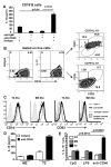
Fig. 2. Anti-CD40 stimulation specifically expands IL10 producing T2-like B cells
Negatively purified splenic B cells from 9-10 week old MRL/lpr mice were cultured for 72h with: isotype controls (10μg/ml), anti-IgM [F(ab’)2] (10μg/ml)/isotype control (10μg/ml), anti-IgM [F(ab’)2] (10μg/ml)/anti-CD40 (5μg/ml) or anti-CD40 alone (5μg/ml)/isotype control (for anti-IgM [F(ab’)2]) (10μg/ml). (A) Supernatants were collected after 72h of culture and IL-10 was measured by ELISA. Data shown mean±se of triplicate wells, and are representative of three independent experiments each with a minimum of five mice. (B) Negatively purified splenic B cells were cultured for 48h with isotype control (5 μg/ml) or anti-CD40 (5 μg/ml). PMA/ionomycin and GolgiStop™ were added for the last 6h of culture. Cells were stained with CD19, CD21, CD23, and IL-10 mAbs. Data shown are representative of 12 mice. (C) The changes in the levels of expression of CD1d and CD93 (AA4) T2-like or mature FO-like and MZ-like B cell subsets, following 48h anti-CD40 stimulation of purified B cells. (D) T2 and MZ B cells were first sorted and then cultured with isotype control (5 μg/ml) or anti-CD40 (5 μg/ml) for 72h. IL-10 production in the supernatants was measured by ELISA. Data shown are representative of three independent experiments. (E) T2, MZ and FO B cells were first sorted and then cultured with isotype control (5 μg/ml), anti-CD40 (5 μg/ml), LPS (10μg/ml) or CpG (25μg/ml) for 48h. PMA/ionomycin and GolgiStop™ were added for the last 6h of culture. IL-10 producing B cells were measured by intracellular staining.
Fig. 3
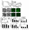
Fig. 3. In vitro, T2-like B cells suppress the production of inflammatory cytokines by CD4+CD25T cells and induce the differentiation of suppressor T cells
Splenic B cells were incubated with anti-CD40 or an irrelevant isotype control for 48h. DAPI-T2, FO, MZ like B cell subsets, or isotype treated B cell subsets, were sorted according to the gates described in figS1. CD4+CD25T cells were MACS sorted from age matched control. CD4+CD25T cells were cultured alone or with T2-, FO-, MZ-like B cells (1:1) and stimulated with anti-CD3 (1μg/ml). A) CD4+TNFα+ T cells, B) CD4+IFNγ+ T cells were measured by intracellular staining. CD4+CD25T cells were cultured either alone, with T2-like B cells (1:1) and with both CD4+CD25+T cells:T2-like B cells (1:1:1) stimulated with anti-CD3. (C) CD4+TNFα+T cell, (D) CD4+IFNγ+T cell, or (E) CD4+IL-10+T cells. CD4+CD25T cells were cultured either alone or with T2-like B cells (1:1) (F). Expression of IL-10 and FoxP3 on CD4+ gated T cells cultured alone (G) or with T2-like B cells. (H) MACS purified CD4+CD25-T cells were cultured either alone or with T2-like B cells and stimulated with anti-CD3 (1μg/ml). 72h later from the first round of stimulation, CD4+ T cells which have been in culture with either T2 B cells or were cultured alone were re-purified by negatively selection (MACS® kit) stained with CFSE (CFSE+CD4+), and cultured 1:1 (1×10⁶/ml total cells) with freshly purified syngeneic CD4+CD25T cells (CD4+) for an additional 72h with anti-CD3 (1μg/ml). TNFα produced by freshly isolated CD4+T cells alone (CD4+CTRL), or by CD4+CTRL cultured with CFSE+CD4+ T cells re-isolated from CD4:T2-likeB cells culture on the 1st round of stimulation, or by CFSE+CD4+CTRL cultured with CD4+T cells cultured alone on the first round, was measured by intracellular cytokine staining. All the data shown are representative of three independent experiments.
Fig. 4
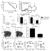
Fig. 4. T2-like B cells control the progression of lupus in MRL/lpr mice
Splenic B cells from 9-10 week old MRL/lpr mice were cultured for 48h with anti-CD40 (5μg/ml). DAPI T2, MZ and FO-like B cells were FACS sorted and 5×10⁵ cells of each subset (purity around or above 90%) were transferred once a week for 3 weeks to 9-10 week old MRL/lpr mice (n=6/group). Control mice received PBS injections. (A) Survival of recipient mice was recorded weekly until mice died of disease spontaneously or were sacrificed due to general debility. (B) Levels of proteinuria in recipient mice were measured once a week. (C) Glomerulonephritis in recipient mice was identified by H&E-staining of kidney sections and, (D) IgG deposition was identified by immunofluorescence with anti-mouse IgG. Kidneys in (C) and (D) were sectioned and stained at 23 weeks of age. (E) Composite renal disease. (F) Sera from 23 weeks old mice were assessed for total anti-dsDNA and anti-dsDNA IgG isotypes by ELISA. Splenocytes were isolated from MRL/lpr mice one week after receiving the final transfer of anti-CD40 stimulated B cell subsets. (G) Splenic cells were stimulated with anti-CD3 for 72 h. Proliferation was determined by 3[H]Tdr incorporation. Reduced proliferative response compared to control group, was also observed when CD4+T cells were purified from T2-like treated and re-stimulated in vitro with anti-CD3 (data not shown). IFNγ and IL-10 production in the supernatants was measured by ELISA. All the data shown are representative of three independent experiments.
Fig. 5
Fig. 5. Neutralization of IL-10, but not TGFβ, abrogated protection conferred by transfer of T2-like B cells
MRL/lpr mice were treated once a week for 3 weeks with T2-like B cells. Mice were treated with anti-IL-10r/IL-10 and with matching irrelevant isotype controls for anti-IL-10R and anti-IL-10 as previously shown (control) (16). (A) Survival rate. (B) Levels of proteinuria was assessed at weekly intervals. (C, D) antibody levels. Sera from 20 week old mice were assessed for total anti-dsDNA IgG and anti-dsDNA IgG isotype by ELISA. The data shown are representative of two independent experiments. MRL/lpr mice were treated once a week for 3 weeks with T2-like B cells. Mice were treated i.p. with anti-TGF-β (2 mg/mouse) or with matching irrelevant isotype controls at day 1 of cell transfer and twice weekly for the duration of the experiment. (E) Survival of recipient mice was recorded weekly until mice died of disease spontaneously or were sacrificed due to general debility. (F) Levels of proteinuria in recipient mice were measured once a week. The data are representative of two independent experiments.
Fig. 6
Fig. 6. Differentiation of transferred hCD20+T2-like B cells
Splenic B cells from 9-10 week old hCD20Tg MRL/lpr mice were cultured for 48h with isotype or anti-CD40. Approximately 2×10⁷ hCD20Tg MRL/lpr B cells were transferred to age matched MRL/lpr mice. Host spleens and inguinal LN were harvested after transfer at the indicated time points, and re-stained with CD19, CD21, CD23, CD24 and anti-human CD20. Representative FACS plots of hCD20+ B cell subsets recovered from spleen and lymph node (A) 1 day post transfer (B) 7 days post transfer. 1-2 ×10⁶ sorted hCD20Tg (C) T2 isotype control (D) or anti-CD40 treated T2-like B cells were transferred to 9-10 week old MRL/lpr mice. Recipient spleens were harvested 7 days after transfer, and re-stained with CD19, CD21, CD23, anti-humanCD20. Gates were pre-set on endogenous B cells subset. Representative FACS plots of one out of 4 independent transfers using a minimum of 5 wild type mice as recipients.
Fig. 7
Fig. 7. Therapeutic effect of agonistic anti-CD40 in MRL/lpr mice
A group of nine 10-week-old MRL/lpr mice were treated daily with 300μg/day anti-CD40 (FGK-45) for four weeks. A control group of nine 10-week-old MRL/lpr mice received isotype control injections (AFRC MAC-1) over the same time periods. Disease progression was measured by survival, proteinuria and skin disease. (A) Survival of recipient mice was recorded weekly until mice died of disease spontaneously or were scarified due to general debility. (B) Levels of proteinuria in recipient mice, were measured, once a week, by Albustix. Groups of mice were compared by ANOVA or Mann-Whitney U statistical test. All mice had detectable level of proteinuria at the start of treatment. C) Skin disease 10 weeks after the start of treatment (20 weeks of age). All mice had a skin disease score of zero at the start of treatment. Scoring system: 0 – no rash, 1 – beginning of rash but little or no hair loss, 2 – discrete hairless rash < ~ 10mm in length, 3 – extensive rash > ~15mm in length, 4 – extensive rash that includes the ears. The p values were calculated by 2 way ANOVA. The data shown are representative of two independent experiments. (D) Sera from 23 week old CD40 and isotype treated mice were assessed for total anti-dsDNA and anti-dsDNA IgG isotypes by ELISA. (E) Representative FACS plots showing an increase in T2 B cells after anti-CD40 treatment compared to isotype treated mice. (F) Bar chart showing the significant difference in the absolute B cell numbers between anti-CD40 and isotype treated mice. Mice were treated for two weeks with either 300μg/mouse/day of anti-CD40 or with an irrelevant isotype control. Splenocytes were isolated from treated mice at different time point, stimulated for 4 hrs with PMA, ionomycin and Golgi Stop. Frequencies of (G) IL-10+CD19+ B cells and (H) IFNγ+, TNFα+ and IL-10+ CD40+CD19 non-B cells were assessed by intracellular staining and flow cytometry.

References

    1. Mauri C, Gray D, Mushtaq N, Londei M. Prevention of arthritis by interleukin 10-producing B cells. J Exp Med. 2003;197:489–501. - PMC - PubMed
    1. Mizoguchi A, Mizoguchi E, Takedatsu H, Blumberg RS, Bhan AK. Chronic intestinal inflammatory condition generates IL-10-producing regulatory B cell subset characterized by CD1d upregulation. Immunity. 2002;16:219–230. - PubMed
    1. Fillatreau S, Sweenie CH, McGeachy MJ, Gray D, Anderton SM. B cells regulate autoimmunity by provision of IL-10. Nat Immunol. 2002;3:944–950. - PubMed
    1. Mauri C, Ehrenstein MR. The ‘short’ history of regulatory B cells. Trends Immunol. 2008;29:34–40. - PubMed
    1. Yanaba K, Bouaziz JD, Haas KM, Poe JC, Fujimoto M, Tedder TF. A regulatory B cell subset with a unique CD1dhiCD5+ phenotype controls T cell-dependent inflammatory responses. Immunity. 2008;28:639–650. - PubMed

Publication types

MeSH terms