. 2009 Mar 15;182(6):3668-77.
doi: 10.4049/jimmunol.0803579.

ESAT-6 inhibits production of IFN-gamma by Mycobacterium tuberculosis-responsive human T cells

Affiliations

ESAT-6 inhibits production of IFN-gamma by Mycobacterium tuberculosis-responsive human T cells

Xisheng Wang et al. J Immunol. .

Abstract

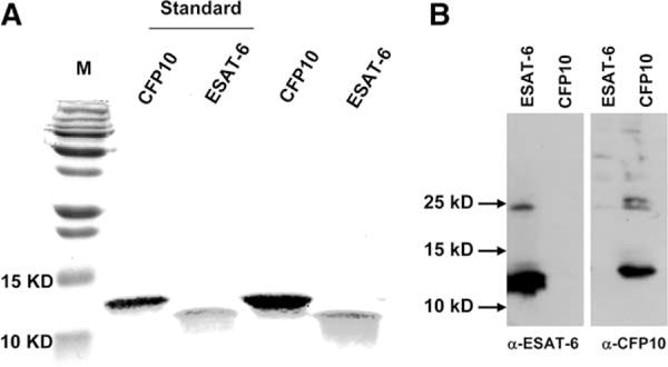
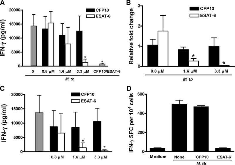
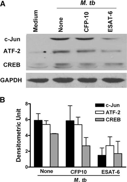
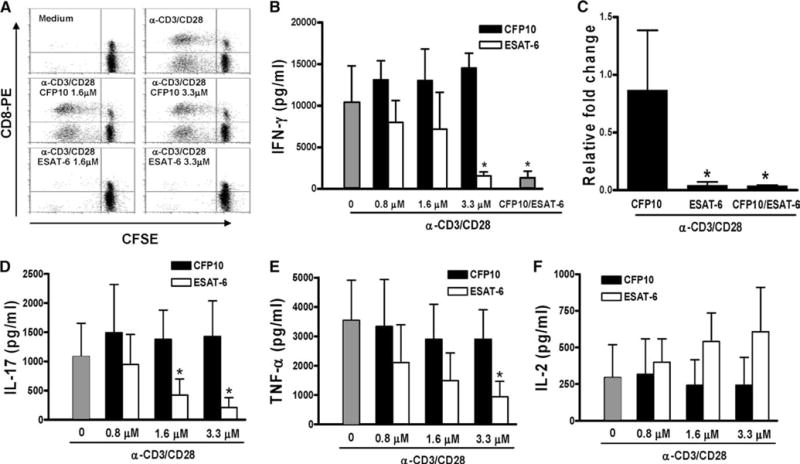
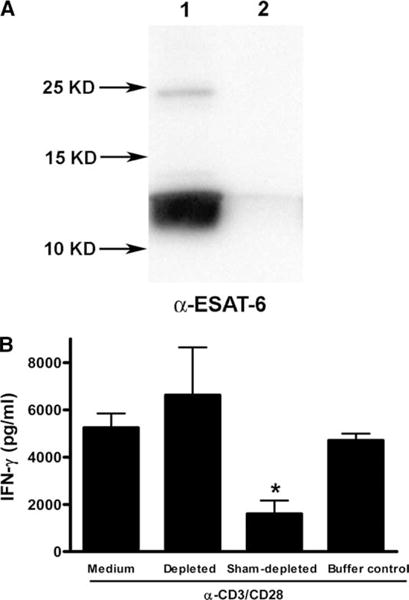
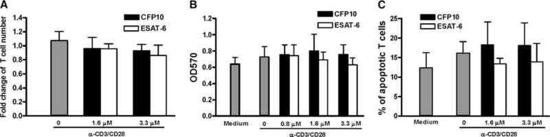
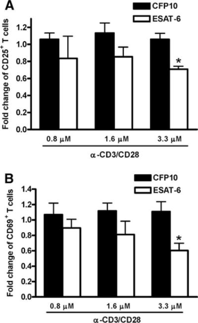
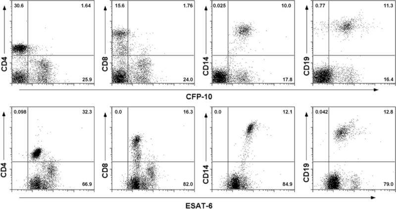
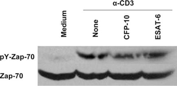
The Mycobacterium tuberculosis early secreted Ag of 6 kDa (ESAT-6) is a potent Ag for human T cells and is a putative vaccine candidate. However, ESAT-6 also contributes to virulence in animal models, mediates cellular cytolysis, and inhibits IL-12 production by mononuclear phagocytes. We evaluated the effects of ESAT-6 and its molecular chaperone, culture filtrate protein of 10 kDa (CFP10), on the capacity of human T cells to produce IFN-gamma and proliferate in response to TCR activation. Recombinant ESAT-6, but not CFP10, markedly inhibited IFN-gamma production by T cells stimulated with M. tuberculosis or with the combination of anti-CD3 and anti-CD28, in a dose-dependent manner. ESAT-6 also inhibited T cell production of IL-17 and TNF-alpha but not IL-2. Preincubation of ESAT-6 with CFP10 under conditions that favor dimer formation did not affect inhibition of IFN-gamma. ESAT-6 decreased IFN-gamma transcription and reduced expression of the transcription factors, ATF-2 and c-Jun, which normally bind to the IFN-gamma proximal promoter and stimulate mRNA expression. ESAT-6 inhibited T cell IFN-gamma secretion through mechanisms that did not involve cellular cytotoxicity or apoptosis. ESAT-6, but not CFP10, bound to T cells and inhibited expression of early activation markers without reducing activation of ZAP70. We conclude that ESAT-6 directly inhibits human T cell responses to mycobacterial Ags by affecting TCR signaling pathways downstream of ZAP70.


Conflict of interest statement

Disclosures

The authors have no financial conflict of interest.

Figures

FIGURE 1
FIGURE 1
Preparation of recombinant ESAT-6 and CFP10. Plasmids containing inserts encoding histidine-tagged ESAT-6 or CFP10 were expressed in E. coli, and proteins were purified, as described in Materials and Methods. Purity of the recombinant proteins was analyzed by 15% SDS- PAGE, and visualized by staining with Coomassie Blue, followed by destaining the gel (A), or by Western blot (B), after electroblotting to a nitrocellulose membrane and blotting with Abs as indicated (bottom). M: m.w. markers. Standards are recombinant proteins from the TB Vaccine Testing and Research Materials Contract, the two far right lanes show the recombinant proteins we prepared. A representative result from three independent preparations is shown.
FIGURE 2
FIGURE 2
ESAT-6 inhibits M. tuberculosis-induced IFN-γ secretion by PBMC. A, PBMC from nine healthy tuberculin rectors were incubated with different concentrations of ESAT-6, CFP10, or ESAT-6/CFP10 heterodimers (ESAT-6 and CFp10 at 3.3 μM, far right gray bar) for 1 h, before adding 2 μg/ml heat-killed M. tuberculosis. Some wells were cultured with medium alone (far left gray bar). After 48 h of incubation, supernatants were collected and IFN-γ levels were measured by ELISA. For 0.8 μM, ESAT-6 = 5 and CFP10 = 8.3 μg/ml. IFN-γ levels with medium alone were <50 pg/ml. Means and SD are shown. *, p < 0.05, compared with the cells treated with M. tuberculosis and 3.3 μM CFP10. B, PBMC from three healthy tuberculin reactors were cultured, as in A. After 48 h, total RNA from cells was isolated and reverse transcribed to cDNA, and IFN-γ mRNA expression was measured by real-time PCR, after normalization for 18S rRNA content. The fold-change of IFN-γ mRNA in M. tuberculosis-stimulated cells, compared with cells cultured in medium alone, was given an arbitrary value of 1.0. Due to significant person-to-person variability in IFN-γ mRNA level, the expression of IFN-γ mRNA in cells with ESAT-6 or CFP10 was expressed as relative fold change, compared with that expressed in the cells without CFP10 or ESAT-6. *, p < 0.05, compared with the cells treated with M. tuberculosis and equimolar concentrations of CFP10. C, PBMC from six healthy tuberculin reactors were incubated with increasing concentrations of ESAT-6 or CFP10 for 48 h. Stimulation with heat-killed M. tuberculosis Erdman is shown for comparison (far left gray bar). IFN-γ levels in culture supernatants were measured by ELISA. Means and SD are shown. *, p < 0.005, compared with the cells stimulated with M. tuberculosis and equimolar concentrations of CFP10. D, PBMC were stimulated, as in A. After 48 h, CD3+ cells were purified and cultured in a 96-well ELISPOT plate, precoated with anti-IFN-γ capture Ab. After 16 h further incubation, the number of IFN-γ+ cells was determined by incubation with IFN-γ detection Ab, followed by development of the substrate. A representative result from experiments with three donors is shown.
FIGURE 3
FIGURE 3
ESAT-6 inhibits expression of transcription factors that positively regulate IFN-γ gene expression in human T cells. PBMC from three healthy tuberculin reactors were treated with medium alone, or with 3.3 μM ESAT-6 or CFP10 for 1 h, before adding heat-killed M. tuberculosis. After 48 h, cells were collected, total protein extracts were prepared, and the proteins were separated by SDS-PAGE (30 μg per lane). A, Western blotting was performed to evaluate the expression of CREB, ATF-2, and c-Jun. GAPDH was used as a loading control. A representative result is shown. B, Means and SD of densitometry analysis from three experiments for each transcription factor is shown.
FIGURE 4
FIGURE 4
Effect of ESAT-6 on proliferation and cytokine production by CD3+ cells stimulated with anti-CD3 and anti-CD28. A, Purified CD3+ cells were labeled with CFSE and cultured in medium alone (top right panel), CFP10 (middle panels), or ESAT-6 (lower panels) for 1 h, before addition of anti-CD3 (10 μg/ml) and anti-CD28 (1 μg/ml). The top left panel shows cells cultured in medium alone, without anti-CD3 or anti-CD28. After 72 h, the cells were stained with anti-CD8-PE, and CFSE content was analyzed by flow cytometry. One representative result of experiments performed for three donors is shown. B, Purified CD3+ cells from five healthy donors were cultured with medium alone, ESAT-6, CFP10, or ESAT-6/CFP10 heterodimer (3.3 μM each, far right bar) for 1 h, before addition of anti-CD3 and anti-CD28, as in A. After 48 h, IFN-γ levels in the culture supernatants were measured by ELISA. Mean values and SD are shown. *, p < 0.001, compared with the cells treated with anti-CD3 plus anti-CD28 mAbs and equimolar concentration of CFP10. C, Purified CD3+ cells from three healthy donors were cultured as described in B for 1 h, before addition of anti-CD3 and anti-CD28. After 48 h, IFN-γ mRNA expression was measured by real-time PCR. *, p < 0.005, compared with the cells stimulated with anti-CD3 plus anti-CD28 in the presence of CFP10. D–F, Production of IL-17 (D), TNF-α (E) and IL-2 (F) by purified CD3+ cells, cultured as described in B. Mean values and SD from five subjects (IL-17), and eight individuals (TNF-α and IL-2), respectively, are shown. *, p < 0.005 (both D and E), compared with cells stimulated with anti-CD3 plus anti-CD28 in the presence of CFP10.
FIGURE 5
FIGURE 5
Depletion of ESAT-6 abrogates the inhibition of IFN-γ production by T cells. A, Preparations of recombinant histidine-tagged ESAT-6 were sham-depleted (lane 1) or depleted of ESAT-6 by incubation with a nickel resin (lane 2), and the efficiency of depletion was determined by Western blotting for ESAT-6 with mAb HYB76-8, after resolution by 15% SDS-PAGE and electroblotting to a nitrocellulose membrane. A representative result of two experiments is shown. B, Purified CD3+ cells from three healthy tuberculin-negative individuals were cultured for 1 h in medium alone, 3.3 μM of the sham-depleted recombinant ESAT-6, an equal volume of the ESAT-6-depleted preparation, or the buffer used for the depletion process, before stimulation with anti-CD3 and anti-CD28. After 48 h, IFN-γ levels in the culture supernatants were measured by ELISA. Mean values and SD are shown. *, p < 0.05, compared with the cells treated with the ESAT-6-depleted protein preparation.
FIGURE 6
FIGURE 6
Effect of ESAT-6 and CFP10 on the viability and apoptosis of T cells. CD3+ cells from three healthy donors were cultured with medium alone, ESAT-6, or CFP10 for 1 h, then stimulated with anti-CD3 and anti-CD28. After 48 h, cell viability was analyzed by trypan blue exclusion (A) and MTT assay (B). C, Apoptosis was analyzed by annexin V staining, followed by flow cytometry analysis. In A, the number of viable cells in the wells treated with medium alone was arbitrarily given a value of 1.0. Mean values and SD are shown.
FIGURE 7
FIGURE 7
Effect of ESAT-6 and CFP10 on expression of early T cell activation markers. Purified CD3+ cells from PBMC of three healthy donors were incubated with medium alone, ESAT-6, or CFP10 for 1 h, then cultured with anti-CD3 and anti-CD28. After 48 h, the expression levels of CD25 (A) and CD69 (B) on CD3 + cells were analyzed by flow cytometry. Due to significant variability from donor to donor, the percentage of CD25+ or CD69+ cells obtained after stimulation with anti-CD3 and anti- CD28 was arbitrarily given a value of 1.0. Mean values and SD are shown. *, p < 0.05, compared with the cells treated with an equimolar concentration of CFP10, anti-CD3, and anti-CD28.
FIGURE 8
FIGURE 8
Binding of ESAT-6 to T cells and other cell populations. PBMC from a healthy donor were incubated with 3.3 μM biotinylated ESAT-6 or CFP10 in combination with PE-conjugated mAbs to CD4, CD8, CD14, and CD19. After washing, the cells were incubated with FITC-streptavidin and analyzed by flow cytometry. The numbers indicate the percentage of cells in the corresponding quadrants. One representative result of experiments with cells from three donors is shown.
FIGURE 9
FIGURE 9
Effect of ESAT-6 on phosphorylation of ZAP70. CD3+ cells purified by negative selection from four healthy donors were incubated on ice with medium alone, or with 3.3 μM of CFP10 or ESAT-6 for one hour, and the cells were treated with anti-CD3 mAb and cross-linked with goat anti-mouse IgG as described in methods. Total cell protein extracts of cells were prepared and the level of phosphorylated and total ZAP70 was evaluated by Western blotting. A representative result is shown.

Similar articles

Cited by

References

    1. Raviglione MC, Snider DE, Jr, Kochi A. Global epidemiology of tuberculosis. Morbidity and mortality of a worldwide epidemic. J Am Med Assoc. 1995;273:220–226. - PubMed
    1. Andersen P, Andersen AB, Sorensen AL, Nagai S. Recall of long-lived immunity to Mycobacterium tuberculosis infection in mice. J Immunol. 1995;154:3359–3372. - PubMed
    1. Sorensen AL, Nagai S, Houen G, Andersen P, Andersen AB. Purification and characterization of a low-molecular-mass T-cell antigen secreted by Mycobacterium tuberculosis. Infect Immun. 1995;63:1710–1717. - PMC - PubMed
    1. Dietrich J, Billeskov R, Doherty TM, Andersen P. Synergistic effect of bacillus calmette guerin and a tuberculosis subunit vaccine in cationic liposomes: increased immunogenicity and protection. J Immunol. 2007;178:3721–3730. - PubMed
    1. Pym AS, Brodin P, Majlessi L, Brosch R, Demangel C, Williams A, Griffiths KE, Marchal G, Leclerc C, Cole ST. Recombinant BCG exporting ESAT-6 confers enhanced protection against tuberculosis. Nat Med. 2003;9:533–539. - PubMed

Publication types

MeSH terms