. 2009 Mar 15;182(6):3855-65.
doi: 10.4049/jimmunol.0803330.

Neuromodulatory activities of CD4+CD25+ regulatory T cells in a murine model of HIV-1-associated neurodegeneration

Affiliations

Neuromodulatory activities of CD4+CD25+ regulatory T cells in a murine model of HIV-1-associated neurodegeneration

Jianuo Liu et al. J Immunol. .

Abstract

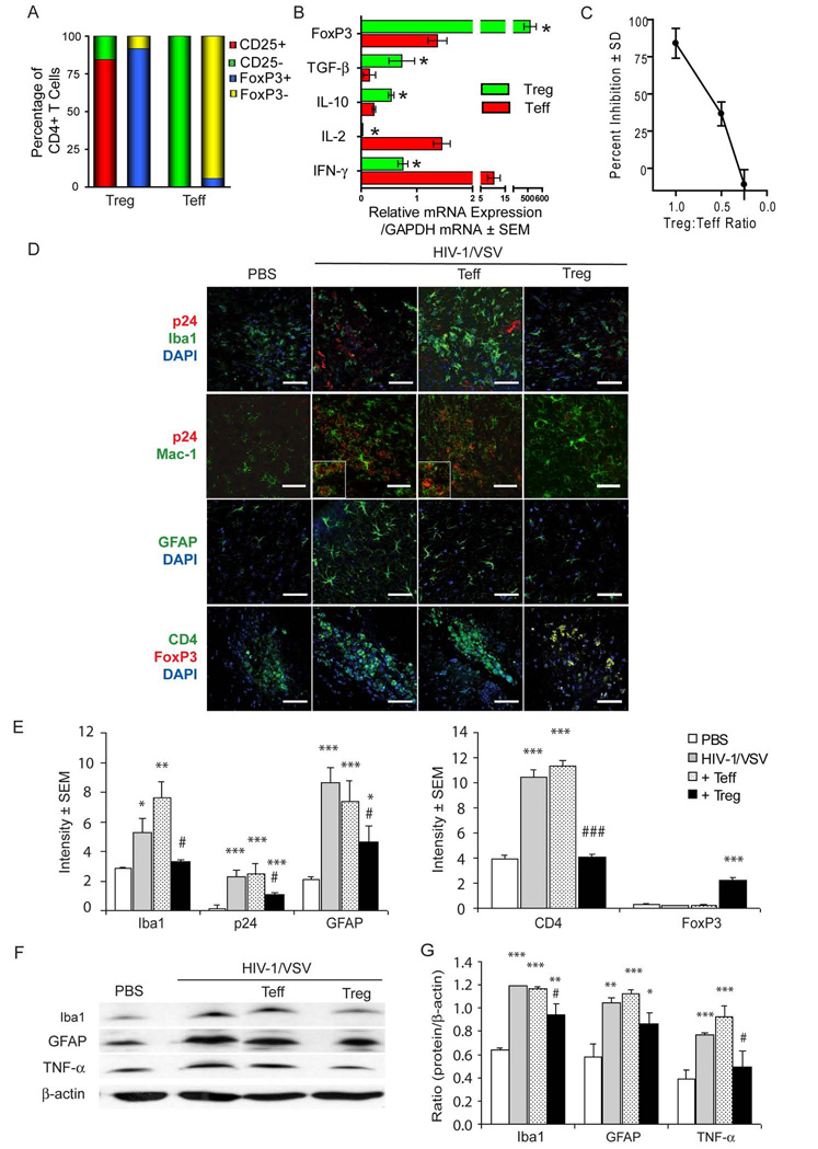
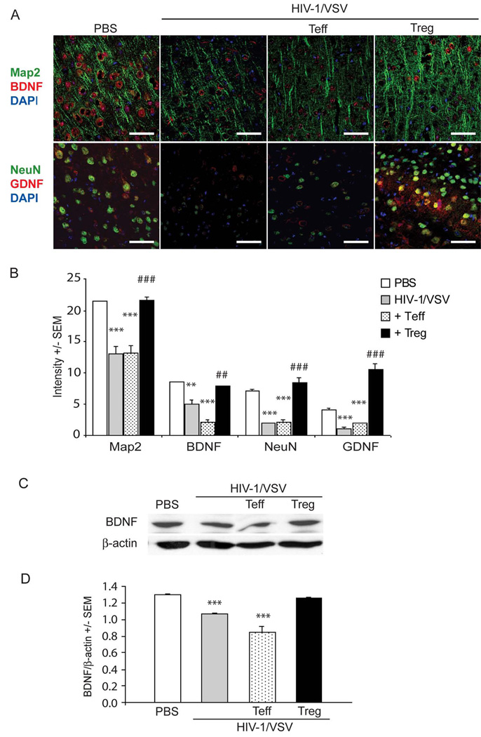
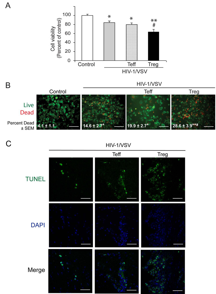
HIV-1-associated neurocognitive impairments are intrinsically linked to microglial immune activation, persistent viral infection, and inflammation. In the era of antiretroviral therapy, more subtle cognitive impairments occur without adaptive immune compromise. We posit that adaptive immunity is neuroprotective, serving in both the elimination of infected cells through CD8(+) cytotoxic T cell activities and the regulation of neuroinflammatory responses of activated microglia. For the latter, little is known. Thus, we studied the neuromodulatory effects of CD4(+) regulatory T cells (Treg; CD4(+)CD25(+)) or effector T cells in HIV-1-associated neurodegeneration. A newly developed HIV-1 encephalitis mouse model was used wherein murine bone marrow-derived macrophages are infected with a full-length HIV-1(YU2)/vesicular stomatitis viral pseudotype and injected into basal ganglia of syngeneic immunocompetent mice. Adoptive transfer of CD3-activated Treg attenuated astrogliosis and microglia inflammation with concomitant neuroprotection. Moreover, Treg-mediated anti-inflammatory activities and neuroprotection were associated with up-regulation of brain-derived neurotrophic factor and glial cell-derived neurotrophic factor expression and down-regulation of proinflammatory cytokines, oxidative stress, and viral replication. Effector T cells showed contrary effects. These results, taken together, demonstrate the importance of Treg in disease control and raise the possibility of their utility for therapeutic strategies.


Conflict of interest statement

Disclosures

The authors have no financial conflict of interest.

Figures

FIGURE 1
FIGURE 1
HIV-1/VSV-infected BMM induce HIVE pathology. Inflammatory pathological effects of viral-infected BMM in brains of immune competent mice were evaluated. BMM infected with HIV-1/VSV were stereotactically injected via the intracranial (i.c.) route into the basal ganglia of syngeneic C57BL/6J mice. Brain tissues were dissected on day 7 and HIVE pathology was analyzed after immunostaining for expression of HIV-1p24, anti-CD3, GFAP and Iba1 antigens. Representative micrographs are shown at the original magnification of x100 for HIV-1p24, CD3, GFAP and Iba1, x400 for bone marrow cell and HIV-1p24 positive BMM. Flow cytometric analysis demonstrates > 95% CD11b+ BMM.
FIGURE 2
FIGURE 2
Treg attenuate chronic neuroinflammatory responses in HIVE mice. (A) Flow cytometric analysis of Treg and Teff subsets from naïve C57BL/6 mice showing percentage distribution of CD4+ T cell phenotypes: CD4+CD25+ (red), CD4+CD25- (green), CD4+FoxP3+ (blue), or CD4+FoxP3- (yellow). (B) Quantitative PCR of mRNA encoding FoxP3, TGF-β, IL-10, IL-2, and IFN-γ from CD3-activated Treg (green bars) and Teff (red bars). Mean ± SEM of mRNA levels was determined for triplicate cell samples and normalized to GAPDH. Significant differences in relative expression of mRNA from Treg compared to Teff were determined by Student’s t test, p < 0.05. (C) Treg inhibition of anti-CD3 mediated proliferation of 1×10⁴ Teff was assessed. Cells were co-cultured for 72 hours and pulsed with 3H-thymidine for the final 18 hours of culture, harvested onto filters, and counted by β-scintillation spectrometry. (D) To induce HIVE, HIV-1/VSV infected BMM were stereotactically injected i.c. into the basal ganglia of syngeneic C57BL/6J mice. Sham controls were injected i.c. with PBS. Treg or Teff (1 ×10⁶) were adoptively transferred into HIVE mice one day post-infection. Serial sections of brain tissue that comprise the injection area, were obtained on day 7 post infection and analyzed by immunohistochemical and Western blot assays for p24, Iba1, Mac-1, GFAP, CD4 and FoxP3 antigens. Brains were collected at day 7 after intravenous injections. Representative brain sections from PBS, HIV-1/VSV, HIV-1/VSV/Teff, and HIV-1/VSV/Treg group showing expression of HIV-1p24 (red) Iba1 (green), Mac-1 (green), GFAP (green), CD4 (green) and FoxP3 (red), Where indicated nuclei were stained with DAPI. (Scale bars: 50 μm, magnification x400). Cellular co-localization of intracellular HIV-1p24 and membrane Mac-1 expression are shown in magnified inserts. (E) Digital image quantification of fluorescence intensity in the stained area was analyzed under 400x magnification using Image J software (NIH, Bethesda, MD). Eight fields in 4 sections in each experiment were subjected to quantitative analysis. Bar graphs reviewed mean of area stained positive intensities in a field of view (PBS: open bars, HIV-1/VSV: gray bars, HIV-1/VSV/Teff: speckled bars, or HIV-1/VSV/Treg: black bars). (F) Representative Western blot analysis of Iba1, GFAP and TNF-α levels from brains of mice treated with PBS, HIV-1/VSV (HIV), HIV-1/VSV/Teff (Teff), or HIV-1/VSV/Treg (Treg). (G) Densitometric quantification of Western blots for Iba1, GFAP, and TNF-α levels in mice treated with PBS (open bars), HIV-1/VSV (gray bars), HIV-1/VSV/Teff (speckled bars), or HIV-1/VSV/Treg (black bars). Levels were normalized to β-actin levels and mean densities ± SEM were determined from four mice per group. (E and G) Compared to PBS, *p < 0.05; **p < 0.01; ***p < 0.001, and compared to HIV/VSV group, #p < 0.05 and ###p < 0.001.
FIGURE 3
FIGURE 3
Treg are neuroprotective in HIVE mice. Mice were stereotactically injected into the basal ganglia with HIV-1/VSV-infected syngeneic BMM or with PBS alone as sham controls. After 1 day, Treg or Teff (1 ×10⁶) were adoptively transferred into HIVE mice. Serial sections of brain tissue that encompassed the injection area were obtained on day 7 post infection and analyzed by immunohistochemistry. (A) Serial sections of brains from PBS control, HIV-1/VSV, HIV-1/VSV/Teff, and HIV-1/VSV/Treg were stained for Map2 (green), NeuN (green), BDNF (red) and GDNF (red) and visualized by confocal laser-scanning microscopy. Images are shown at x400 and scale bar equals 50 μm. (B) Stained sections from mice (eight fields in 4 section in each mouse) treated with PBS (open bars), HIV-1/VSV (gray bars), HIV-1/VSV/Teff (speckled bars), or HIV-1/VSV/Treg (black bars) were digitally analyzed using Image J software (NIH, Bethesda, MD). Bar graphs showed mean of fluorescence intensity in a field of view. (C) Western blot analysis of BDNF expression and (D) densitometric quantification of BDNF Western blots from lysates of brain tissues from mice treated with PBS, HIV-1/VSV, HIV-1/VSV/Teff, or HIV-1/VSV/Treg. BDNF levels were normalized to β-actin expression. Values are expressed as mean ± SEM for four mice per group and were significant compared to PBS group, ***p < 0.001; and compared to HIV/VSV group, ##p < 0.01; and ###p < 0.001.
FIGURE 4
FIGURE 4
Treg induce apoptosis in HIV-1/VSV-infected BMM. HIV-1/VSV-infected BMM were exposed to Teff or Treg for 3 days in the absence of M-CSF at a ratio of 3:1 (BMM:Treg or Teff). Uninfected BMM and HIV-1/VSV-infected BMM served as negative and infection controls. (A) After 3 days, cell viabilities were determined by MTT assay. (B) Cell survival/cytotoxicity was assayed by live/dead cytotoxicity immunostaining and visualized by fluorescent microscopy at x400 magnification. Scale bar equals 50 μm. Mean percentages of dead cells ± SEM were determined for 3 cultures/group and significant differences in means were determined by Student’s t test where *p < 0.05 and **p < 0.001 compared to uninfected control group; and #p < 0.001 compared with HIV-1/VSV group. (C) HIV-1/VSV infected BMM were stereotactically injected i.c. into the basal ganglia of syngeneic C57BL/6J mice. Treg or Teff (1 ×10⁶) were adoptively transferred into HIVE mice one day post-infection. Serial sections of brain tissue that comprise the injection area, were obtained on day 7 post infection and analyzed by immunohistochemistry for TUNEL (green) and nuclei by DAPI stain (blue). (Scale bars: 50 μm, magnification x400).
FIGURE 5
FIGURE 5
Treg reduce HIV-1 viral replication in BMM. HIV-1/VSV-infected BMM were cocultured with Teff or Treg at a ratio of 3:1 (BMM vs Treg or Teff). (A) Supernatants from cultures of uninfected BMM (Control, closed triangles), HIV-1/VSV-infected BMM (closed squares), HIV-1/VSV/Teff (open circles), and HIV-1/VSV/Treg (open diamonds) were collected from day 1 to day 6 after addition of T cells and assessed for RT activity. (B) Viral-infected BMM cultured in the absence or presence Teff or Treg or uninfected BMM (Control) were harvested at the end of day 6 and stained for expression of HIV-1p24 and visualized by light microscopy at x400 magnification. Scale bar equals 50 μm. (C) HIV-1p24-positive BMM were counted and percentages of HIV-p24 positive BMM were determined. Means ± SEM were determined from 3 independent experiments. Compared to HIV-1/VSV BMM group by Student's t-test, #p < 0.01, ##p < 0.001.
FIGURE 6
FIGURE 6
Treg inhibit ROS production and cytotoxicity in BMM. Anti-CD3-activated Tregs or Teffs were co-cultured in the absence or presence of HIV-1/VSV infected BMM for 24 hours. After removal of T cells, ROS production was measured as a function of H2O2 accumulation using an Amplex Red assay. Uninfected BMM cultured alone served as ROS baseline controls. (A) Compared to uninfected BMM controls (open bar), percentage increase of ROS as a function of H2O2 levels were determined for HIV-1/VSV-infected BMM cultured alone (gray bar) or co-cultured in the presence of Teff (HIV-1/VSV/Teff, speckled bar), or Treg (HIV-1/VSV/Treg, black bar). (B) Uninfected BMM cultured in the absence (left set) or presence of PMA (right set) were co-cultured without TNF-α or T cells (white bars), in the presence of TNF-α (gray bars), with TNF-α and Teff (speckled bars), or with TNF-α and Treg (black bars). (A and B) Mean ± SEM were determined for 3 experimental determinations and significant differences were determined by Student’s t test wherein **p < 0.01 and ***p < 0.001 compared to control group, and ##p < 0.01 and ###p < 0.001 compared with HIV-1/VSV group. (C) Uninfected BMM (Control) and HIV-1/VSV-infected BMM were cultured in the absence or presence of Teff or Treg for 24 hours and supernatants were harvested. Collected supernatants were then subjected to cytokine array blots (RayBiotech). Red ovals encompass replicate blots to detect expression of (1) IL-2, (2) IL-12p40/p70, (3) MCP-1, and (4) MCP-5. (D) Densitometric analysis of cytokine array blots were achieved by digital image analysis with Image J software, and mean densities ± SD were determined for replicate determinations. Mean densities ± SD of positive controls for each array of control BMM, HIV-1/VSV-infected BMM cultured in the absence or presence of Teff or Treg were 204.4 ± 9.6, 206.3 ± 6.0, 201.7 ± 14.8, and 202.5 ± 11.9.
FIGURE 7
FIGURE 7
Treg induce neuroprotective responses from HIV-1/VSV-infected BMM. Mouse primary neurons were exposed for 24 hours to 10% of conditioned media (CM) collected from uninfected BMM (Control), HIV-1/VSV-infected BMM, or HIV-1/VSV infected BMM co-cultured with Treg or Teff. (A) Treated primary neuronal isolates were immunostained for expression of Map-2 (green) and NeuN (red). Images are at x400 magnification and scale bar equals 50 μm (top row). Apoptotic neurons were determined by TUNEL staining showing apoptotic cells (green) and DAPI nuclear staining (blue). Micrographs are shown at x200 magnification and scale bars equal 100 μm (middle and lower rows). (B) Percentages of apoptotic neurons treated with CM from control BMM, HIV-1/VSV infected BMM, HIV-1/VSV infected BMM treated with Teff, or HIV-1/VSV infected BMM treated with Treg. Mean percentages ± SEM were determined from three experiments. Compared to control group, ***p < 0.001; and compared to HIV-1/VSV infected BMM group, ###p < 0.001.

Similar articles

Cited by

References

    1. Antinori A, Arendt G, Becker JT, Brew BJ, Byrd DA, Cherner M, Clifford DB, Cinque P, Epstein LG, Goodkin K, Gisslen M, Grant I, Heaton RK, Joseph J, Marder K, Marra CM, McArthur JC, Nunn M, Price RW, Pulliam L, Robertson KR, Sacktor N, Valcour V, Wojna VE. Updated research nosology for HIV-associated neurocognitive disorders. Neurology. 2007;69:1789–1799. - PMC - PubMed
    1. Hult B, Chana G, Masliah E, Everall I. Neurobiology of HIV. Int Rev Psychiatry. 2008;20:3–13. - PubMed
    1. Cherner M, Masliah E, Ellis RJ, Marcotte TD, Moore DJ, Grant I, Heaton RK. Neurocognitive dysfunction predicts postmortem findings of HIV encephalitis. Neurology. 2002;59:1563–1567. - PubMed
    1. Masliah E. In vivo modeling of HIV-1 mediated neurodegeneration. Am J Pathol. 1996;149:745–750. - PMC - PubMed
    1. Gisslen M, Hagberg L, Rosengren L, Brew BJ, Cinque P, Spudich S, Price RW. Defining and evaluating HIV-related neurodegenerative disease and its treatment targets: a combinatorial approach to use of cerebrospinal fluid molecular biomarkers. J Neuroimmune Pharmacol. 2007;2:112–119. - PubMed

Publication types

MeSH terms

Substances