. 2009 Mar 15;182(6):3866-76.
doi: 10.4049/jimmunol.0713949.

Epilysin (MMP-28) restrains early macrophage recruitment in Pseudomonas aeruginosa pneumonia

Affiliations

Epilysin (MMP-28) restrains early macrophage recruitment in Pseudomonas aeruginosa pneumonia

Anne M Manicone et al. J Immunol. .

Abstract

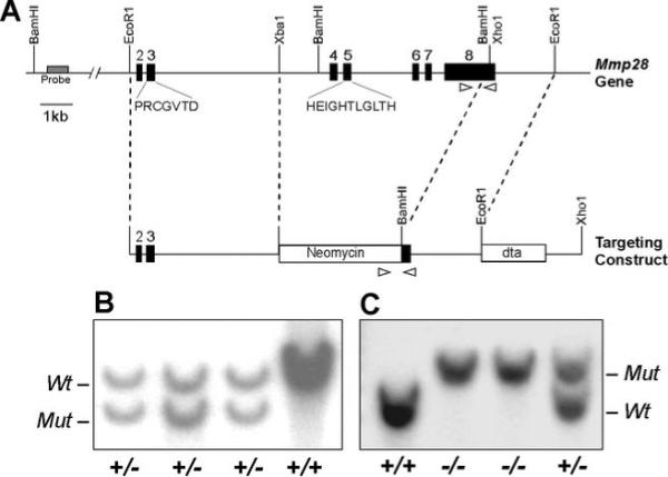
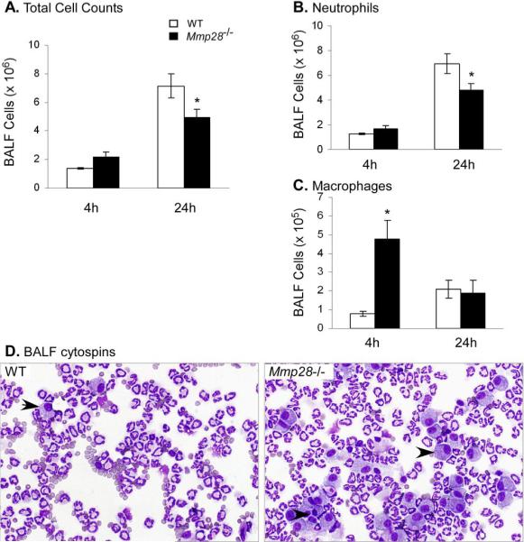
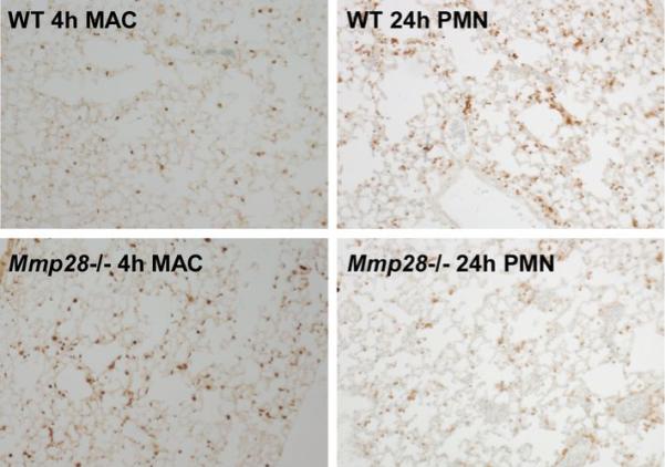
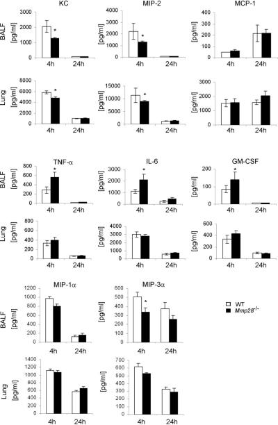
Several members of the matrix metalloproteinase (MMP) family function in various processes of innate immunity, particularly in controlling leukocyte influx. Epilysin (MMP-28) is expressed in numerous tissues and, in adult mice, it has the highest expression in lung, where it is detected in bronchial epithelial cells (Clara cells). Epilysin is also expressed by bone marrow-derived macrophages, but not by alveolar macrophages, suggesting that its expression by macrophages is dependent on localization and differentiation. To assess the role of this MMP, we generated epilysin-null (Mmp28(-/-)) mice. Although epilysin is constitutively expressed in normal tissues, Mmp28(-/-) mice have no overt phenotype. However, using a murine model of Pseudomonas aeruginosa pneumonia, we found that Mmp28(-/-) mice had an early increase in macrophage recruitment into the lungs, as well as enhanced bacterial clearance and reduced pulmonary neutrophilia, which we predicted were due to accelerated macrophage influx. Macrophage depletion in WT and Mmp28(-/-) mice confirmed a role for macrophages in clearing P. aeruginosa and regulating neutrophil recruitment. Furthermore, we observed that macrophages derived from Mmp28(-/-) mice migrated faster than did wild-type cells to bronchoalveolar lavage fluid from P. aeruginosa-treated mice of either genotype. These observations indicate that epilysin functions as an intrinsic negative regulator of macrophage recruitment by retarding the chemotaxis of these cells.


Figures

Fig. 1
Fig. 1. Generation of Epilysin-null (Mmp28−/−) Mice
A, The targeting construct was designed to replace most of the exons, including exon 5, which codes for the catalytic domain. A diphtheria-toxin cassette was included to selected against ES cells with mistargeted recombination. The targeting construct was linearized with XhoI and injected in C57Bl/6 blastocyst. After recombination, exons 4 to 7 and the full coding portion of the exon 8 are removed, and the epilysin transcript only contains exons 1 to 3 (coding for the pro-region only). B, ES clones (144 total) were screened by Southern hybridization using a 694 bp probe in the 3'terminal end of intron 1 just outside of the targeting construct. With Bam HI digestion, wildtype epilysin gene gives a ∼9 kb band while targeted gene gives a ∼7.5 kb band. C, F1 offspring of chimeric mice were screened by Southern and PCR assays. For the PCR assay, the recombined locus produces a larger DNA fragment (324 nt) than that from the wildtype gene (259 nt) and both bands from heterozygotes. Three primers were used, the position and orientation of which are indicated by arrowheads: a wildtype forward primer, a neomycin cassette forward primer, and a common reverse primer.
Fig. 2
Fig. 2. MMP-28 is expressed by Clara cells in the lung
(A) Immunofluorescence staining for epilysin in the airway epithelium from naïve WT mice (top panel) compared to MMP-28 mice (bottom panel). Nuclei were labeled with 4',6-diamidino-2-phenylindole (DAPI), demonstrating epilysin expression in the cytoplasm of airway epithelial cells. (B) Co-immunostaining of epilysin and CCSP (Clara cell marker). (C) MMP-28 mRNA from whole lung and LCM-retrieved bronchiolar epithelium. MMP-28 expression is enriched in LCM-retrieved bronchiolar epithelium compared to whole lung. Samples were normalized to β2-microglobulin. (D) Immunofluorescence staining for epilysin in the airway epithelium from naïve WT mice (0 h), and P. aeruginosa-treated mice harvested at 4 and 24 h after infection. Immunostaining demonstrates decreased epilysin levels in the airway epithelium from infected mice.
Fig. 3
Fig. 3. Lung MMP Expression in response to P. aeruginosa Pneumonia
Whole lung RNA was collected at serial times after exposure to P. aeruginosa and levels of specific transcripts were determined by qRT-PCR and normalized to HPRT. Reported are fold change in mRNA levels relative to time 0. (A) MMP-28 expression was rapidly downregulated and returned to baseline after resolution of infection and inflammation. (B-E) In contrast to MMP-28, other MMPs, (MMP-7,−10,−12, and −14) were induced during infection (*p<0.05 compared with timepoint 0). n=4−5 mice/timepoint.
Fig. 4
Fig. 4. Upregulation of MMP-28 Expression in Macrophages
(A) BMDM were incubated in medium alone, E. Coli LPS, or P. aeruginosa, and mRNA was collected for qRT-PCR analysis at 24, 48 and 72 h post-exposure. MMP-28 expression by BMDM was stimulated by both LPS and P. aeruginosa at 24 and 48 h. Samples were normalized to HPRT. *p value < 0.05 when compared to time-controlled medium alone samples. (B) Immunofluorescence staining for epilysin and MAC-2 48 h after P. aeruginosa-exposure in WT mice. Images demonstrate co-expression of epilysin and MAC-2 in selected macrophages.
Fig. 5
Fig. 5. Bronchoalveolar fluid (BALF) cell counts following P. aeruginosa challenge
Wildtype (white bar) and Mmp28−/− mice (black bar) mice were exposed to aerosolized P. aeruginosa and harvested at 4 and 24 h for BALF cell counts and differential. (A) Total cell counts, demonstrating significantly reduced cell counts at 24 h in Mmp28−/− mice. (B) Total neutrophils (PMNs), demonstrating reduced neutrophil recruitment at 24 h in the Mmp28−/− mice, and (C) Total macrophages (MACs), demonstrating increased early macrophage recruitment at 4 h in Mmp28−/− mice. (*p<0.05) (D) Representative cytospins of BALF collected 4 h after P. aeruginosa infection in.wildtype and Mmp28−/− mice. Macrophages (arrow heads). Data are mean ± SEM, results represent 10 mice/timepoint.
Fig. 6
Fig. 6. Lung histology after P. aeruginosa inhalation
These are representative images demonstrating enhanced macrophage influx into the lung of Mmp28−/− mice at 4 h post-infection with P. aeruginosa (Bottom Left), and reduced neutrophil influx into the lung of Mmp28−/− mice 24 h post-infection with P. aeruginosa (Bottom Right) compared to WT controls (Top). Lungs are fixed in 10% formalin, paraffin-embedded and stained with anti-mouse MAC-2 antibody (left) or anti-neutrophil antibody (right). Secondary antibody was HRP-conjugated donkey anti-rat. Secondary antibodies were labeled with the Vectastain ABC kit and colorimetric detection was done with diaminobenzidine staining.
Fig. 7
Fig. 7. P. aeruginosa clearance from the lung at 4 and 24 h following bacterial challenge in wildtype and Mmp28−/− mice
Mice were exposed to aerosolized P. aeruginosa, and lungs and spleens (a surrogate marker for bacteremia) were harvested at 0, 4 and 24 h for bacterial counts. Results are expressed as colony forming units (CFU) per lung or spleen. (A) At 4 h, CFU in the lung are significantly decreased in Mmp28−/− mice versus wildtype control. (*p<0.05). (B) There is no difference in CFU from the spleen between genotypes. Data are mean ± SEM, results n=10 mice/timepoint.
Fig. 8
Fig. 8. Macrophage Depletion using Clodronate
Alveolar macrophages and circulating monocytes were depleted using IN and IP coldronate, respectively. Control mice received IN and IP liposome only. Forty-eight hours after treatment, mice were harvested for BALF, lung histology and circulating leukocyte cell counts and differential. Lungs were inflated with 10% formalin and embedded in paraffin. Sections were stained with MAC-2 to analyze pulmonary macrophage content. (A) BALF cell count and differential demonstrating a 10 fold reduction in alveolar macrophages with no significant difference in neutrophil recruitment in clodronate-treated mice. Immunostaining confirmed a significant reduction of pulmonary macrophages in the clodronate-treated group. (B) Flow cytometry of circulating leukocytes. Leukocytes were isolated from liposome-treated (left) and clodronate-treated (right) mice using gradient separation with Histopaque 1077. Cells were stained with FITC-conjugated anti-mouse CD11b and analyzed by flow cytometry. The dot plots represent the CD11b+ population of leukocytes, with CD11b staining intensity on the X axis. The CD11b+ cells are grouped into 3 subgroups, a-c, based on level of expression. The percentages represent number of CD11b+ cells in area/total CD11b+ cells. Clodronate-treated mice demonstrated depletion of a CD11bhigh population of circulating monocytes (area c) from 21% to 8% of all CD11b+ cells (areas a-c). (C) Total circulating monocyte cell counts as determined by manual differential and cell counts from control and clodronate-treated wildtype mice. There was a significant reduction in the numbers of circulating monocytes in clodronate-treated mice (*p value <0.05).
Fig. 9
Fig. 9. Macrophage-Deficient Mice have impaired bacterial clearance and increased pulmonary neutrophilia
(A-C) Results from control WT (white bar) and Mmp28−/− (grey bar) mice vs. clodronate-treated WT (white striped bar) and Mmp28−/− (grey striped bar) mice. All groups have been treated with intranasal instillation of P. aeruginosa (1 × 10⁷ organisms/lung) and harvested at 4 and 24 h for BALF cell counts, differential and bacterial clearance. (A) BALF neutrophil cell counts at 4 and 24 h post-infection, (B) BALF macrophage cell counts at 4 and 24 h post-infection, (C) Whole lung bacterial counts (CFU) 4 h after infection. *p value < 0.05 when compared to same genotype mice.
Fig. 10
Fig. 10. BALF and Lung Homogenate Cytokines
Wildtype (white bar) and Mmp28−/− mice (black bar) were exposed to aerosolized P. aeruginosa and harvested at 4 and 24 h for whole lung homogenates of the right lung, and BALF from the left lung. The levels of several chemokines and cytokines were assessed from these two compartments using luminex-based assays and ELISAs. KC and MIP-2 are significantly decreased in the BALF and LH of Mmp28−/− mice, 4 h post-infection. There is no difference in MCP-1 or MIP-1α levels. TNF-alpha, IL-6 and GM-CSF are significantly increased in the BALF, but not LH of Mmp28−/− mice, 4 h post-infection. Data are means ± SEM, results represent 10 mice/timepoint. (*p value <0.05)
Fig. 11
Fig. 11. Mmp28−/− Macrophages Migrate Faster than WT cells
(A) Migration of WT and Mmp28−/− BMDM to BALF obtained from P. aeruginosa-treated WT or Mmp28−/− mice. At 50 minutes, Mmp28−/− BMDM migrated faster to all BALF samples compared to WT BMDM. There was a slight decrease in chemoattractant activity in BALF from Mmp28−/− compared to WT. (B) Mmp28−/− vs. WT BMDM migrated faster to WT BALF. LPS-stimulated BMDM (black bar) did not affect chemotaxis of BMDM compared to unstimulated cells (white bar).

Similar articles

Cited by

References

    1. Suzuki T, Chow CW, Downey GP. Role of innate immune cells and their products in lung immunopathology. Int J Biochem Cell Biol. 2008;40:1348–1361. - PubMed
    1. Bartlett JA, Fischer AJ, McCray PB., Jr. Innate immune functions of the airway epithelium. Contrib Microbiol. 2008;15:147–163. - PubMed
    1. Kantari C, Pederzoli-Ribeil M, Witko-Sarsat V. The role of neutrophils and monocytes in innate immunity. Contrib Microbiol. 2008;15:118–146. - PubMed
    1. Marriott HM, Dockrell DH. The role of the macrophage in lung disease mediated by bacteria. Exp Lung Res. 2007;33:493–505. - PubMed
    1. Parks WC, Wilson CL, Lopez-Boado YS. Matrix metalloproteinases as modulators of inflammation and innate immunity. Nat Rev Immunol. 2004;4:617–629. - PubMed

Publication types

MeSH terms

Substances