. 2009 Mar 15;182(6):3919-27.
doi: 10.4049/jimmunol.0803834.

Villitis of unknown etiology is associated with a distinct pattern of chemokine up-regulation in the feto-maternal and placental compartments: implications for conjoint maternal allograft rejection and maternal anti-fetal graft-versus-host disease

Affiliations

Villitis of unknown etiology is associated with a distinct pattern of chemokine up-regulation in the feto-maternal and placental compartments: implications for conjoint maternal allograft rejection and maternal anti-fetal graft-versus-host disease

Mi Jeong Kim et al. J Immunol. .

Abstract

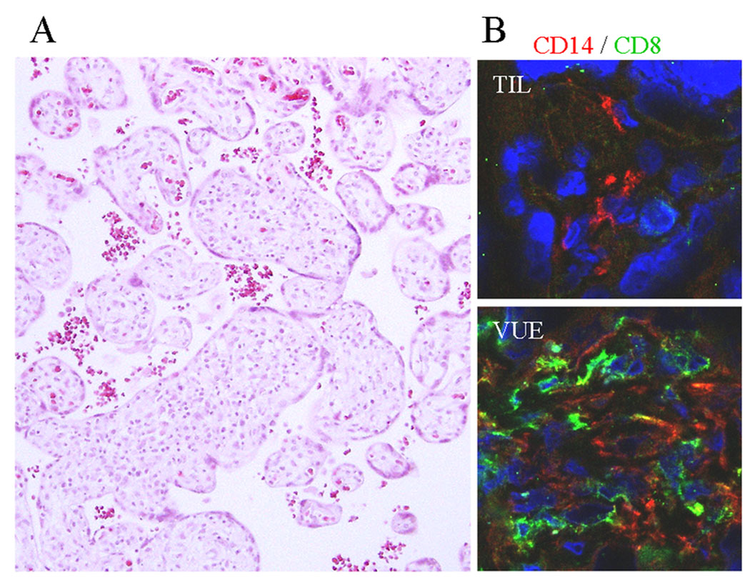
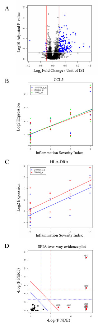
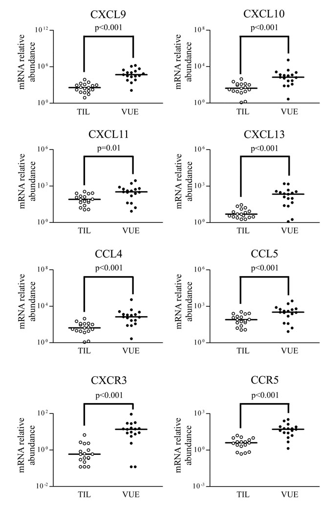
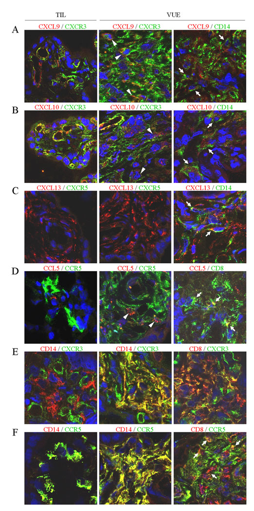
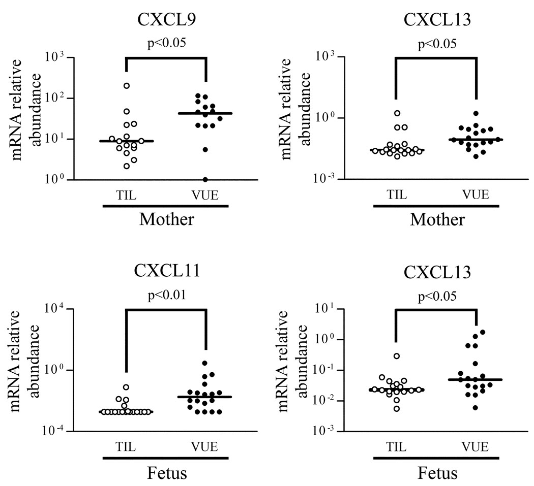
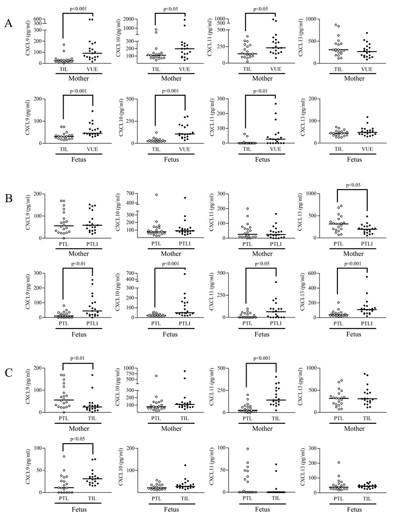
The co-presence of histoincompatible fetal and maternal cells is a characteristic of human placental inflammation. Villitis of unknown etiology (VUE), a destructive inflammatory lesion of villous placenta, is characterized by participation of Hofbauer cells (placental macrophages) and maternal T cells. In contrast to acute chorioamnionitis of infection-related origin, the fundamental immunopathology of VUE is unknown. This study was performed to investigate the placental transcriptome of VUE and to determine whether VUE is associated with systemic maternal and/or fetal inflammatory response(s). Comparison of the transcriptome between term placentas without and with VUE revealed differential expression of 206 genes associated with pathways related to immune response. The mRNA expression of a subset of chemokines and their receptors (CXCL9, CXCL10, CXCL11, CXCL13, CCL4, CCL5, CXCR3, CCR5) was higher in VUE placentas than in normal placentas (p < 0.05). Analysis of blood cell mRNA showed a higher expression of CXCL9 and CXCL13 in the mother, and CXCL11 and CXCL13 in the fetus of VUE cases (p < 0.05). The median concentrations of CXCL9, CXCL10, and CXCL11 in maternal and fetal plasma were higher in VUE (p < 0.05). Comparison of preterm cases without and with acute chorioamnionitis revealed elevated CXCL9, CXCL10, CXCL11, and CXCL13 concentrations in fetal plasma (p < 0.05), but not in maternal plasma with chorioamnionitis. We report for the first time the placental transcriptome of VUE. A systemic derangement of CXC chemokines in maternal and fetal circulation distinguishes VUE from acute chorioamnionitis. We propose that VUE be a unique state combining maternal allograft rejection and maternal antifetal graft-vs-host disease mechanisms.


Figures

Figure 1
Figure 1
Microscopic findings of VUE. A, VUE is characterized by patchy lymphocytic infiltrates in the affected chorionic villi. B, Hofbauer cells (placental tissue macrophages) in the chorionic villi of a term control placenta (TIL) are immunopositive for CD14. Chorionic villi in the placenta with VUE show infiltration of CD8+ T cells in addition to CD14+ Hofbauer cells. (CD14, red; CD8, green).
Figure 2
Figure 2
Transcriptome analysis of VUE placentas using microarray. A, A volcano plot showing the relationship between the FDR-corrected p values of the genes and their fold changes (log2 of). Blue dots outside the two red lines and above the gray line have fold changes > 2 and FDR-corrected p values < 0.05. Positive values on the x-axis indicate genes whose expression increased with inflammation severity, while negative values indicate genes down-regulated with inflammation severity. A gene located at 0.25 on the x-axis increases in average 4 × 0.25 = 1 U of log2 fold change (2-fold) from the group without disease (ISI = 0) to the most severe inflammation group (ISI = 3). B and C, The expression levels of the Affymetrix probe sets for the CCL5 (B) and HLA-DRA (C) genes were plotted according to inflammation severity. Gene expression increased along with inflammation severity. D, The list of significant genes and their log-fold changes were analyzed using the SPIA algorithm to identify signaling pathways for which there is high enrichment evidence (high x-axis) and high perturbation evidence (high y-axis). Each KEGG signaling pathway analyzed is shown as a bullet point. Pathways above the oblique red line are significant at 5% after Bonferroni correction based on the combined evidence, while those above the oblique blue line are significant at 5% after FDR correction. The vertical and horizontal thresholds represent the same corrections for each type of evidence. Ag processing and presentation (KEGG ID: 4612) and cytokine-cytokine receptor interaction (KEGG ID: 4060) are the most relevant according to the signaling perturbation-based evidence (y-axis).
Figure 3
Figure 3
Changes in mRNA expression of a subset of chemokines and receptors in VUE placentas. mRNA expressions of CXCL9, CXCL10, CXCL11, CXCL13, CCL4, and CCL5 are higher in VUE placentas compared with those in term control placentas. mRNA of CXCR3 and CCR5 is also expressed at higher levels in VUE placentas than in term control placentas.
Figure 4
Figure 4
Immunofluorescent staining for selected chemokines and their receptors in VUE placentas. Immunofluorescent signals for corresponding chemokines, receptors, and CD Ags are indicated in green (Alexa 488) and red (Alexa 568 or Alexa 594). AD, Increased immunoreactivity of CXCR9, CXCR10, CXCR13, and CCL5 in VUE placentas. CXCR3+ and CCR5+ leukocytes infiltrate the chorionic villi, expressing their chemokine ligands in VUE placentas. CXCR5 (C) is not found in either VUE placentas or normal term (TIL) placentas. Immunoreactivity of CXCL9 (A), CXCL10 (B), and CXCL13 (C) is localized in CD14+ Hofbauer cells (arrows) and endothelial cells of VUE placentas. CCL5 (D) is positive in some CD14+ Hofbauer cells (not shown) and CD8+ T cells (arrows). Some infiltrating leukocytes were doubly positive for both a chemokine and its receptor (arrowheads). E and F, CD14+ Hofbauer cells in TIL placentas are negative for CXCR3 (E) while being immunoreactive for CCR5 (F). Infiltrating CD8+ T cells and CD14+ Hofbauer cells in VUE foci are positive for CXCR3 and CCR5. The endothelial cells of villous capillaries are also positive for CXCR3.
Figure 5
Figure 5
Differential mRNA expression of chemokines and receptors in maternal and fetal blood. Maternal blood shows higher mRNA expression of CXCL9 and CXCL13 in VUE cases than in control cases (TIL). CXCL11 and CXCL13 mRNA expression is higher in fetal blood of VUE cases than in control cases.
Figure 6
Figure 6
Chemokine concentrations in maternal and fetal plasma of VUE and acute chorioamnionitis. A, The median concentrations of chemokines CXCL9, CXCL10, and CXCL11 in both maternal and fetal plasma are higher in VUE cases than in term control cases (TIL), respectively. On the other hand, the median concentration of CXCL13 is not different between VUE and control. B, The median fetal plasma concentrations of CXCL9, CXCL10, CXCL11, and CXCL13 are also higher in PTLI cases than in PTL cases. In contrast, maternal plasma concentrations of CXCL9, CXCL10, and CXCL11 are not different between PTL and PTLI cases. CXCL13 concentration is lower in PTLI cases than in PTL cases. C, Comparison between PTL and TIL cases showed that maternal plasma CXCL9 concentration is higher in PTL cases than in TIL cases, whereas CXCL11 concentration is lower in PTL than in TIL cases.

References

    1. Kraus FT, Redline RW, Gersell DJ, Nelson DM, Diche JM. Inflammation and infection. In: Kraus FT, Redline RW, Gersell DJ, Nelson DM, Diche JM, editors. Placental Pathology Atlas of Non Tumor Pathology 3. 1st ed. Washington, DC: AFIP; 2004. pp. 75–116.
    1. Gomez R, Romero R, Ghezzi F, Yoon BH, Mazor M, Berry SM. The fetal inflammatory response syndrome. Am. J. Obstet. Gynecol. 1998;179:194–202. - PubMed
    1. Murtha AP, Greig PC, Jimmerson CE, Roitman-Johnson B, Allen J, Herbert WN. Maternal serum interleukin-6 concentrations in patients with preterm premature rupture of membranes and evidence of infection. Am. J. Obstet. Gynecol. 1996;175:966–969. - PubMed
    1. Shimoya K, Matsuzaki N, Taniguchi T, Okada T, Saji F, Murata Y. Interleukin-8 level in maternal serum as a marker for screening of histological chorioamnionitis at term. Int. J. Gynaecol. Obstet. 1997;57:153–159. - PubMed
    1. Brito H, Juliano P, Altemani C, Altemani A. Is the immunohistochemical study of the inflammatory infiltrate helpful in distinguishing villitis of unknown etiology from non-specific infection villitis? Placenta. 2005;26:839–841. - PubMed

Publication types

MeSH terms