. 2009 Dec;41(6):661-70.
doi: 10.1165/rcmb.2008-0182OC. Epub 2009 Mar 5.

Role of proinflammatory cytokines IL-18 and IL-1beta in bleomycin-induced lung injury in humans and mice

Affiliations

Role of proinflammatory cytokines IL-18 and IL-1beta in bleomycin-induced lung injury in humans and mice

Tomoaki Hoshino et al. Am J Respir Cell Mol Biol. 2009 Dec.

Abstract

Administration of several chemotherapeutic drugs, such as bleomycin, busulfan, and gefitinib, often induces lethal lung injury. However, the precise mechanisms responsible for this drug-induced lung injury are still unclear. In the present study, we examined the role of the proinflammatory cytokines IL-18 and IL-1beta in the mechanism of bleomycin-induced lung injury. We performed immunohistochemical analysis of IL-18 and IL-18 receptor (R) alpha chain expression in the lungs of five patients with bleomycin-induced lethal lung injury. Enhanced expression of both IL-18 and IL-18Ralpha was observed in the lungs of all five patients with bleomycin-induced lung injury. To support the data obtained from patient samples, the levels of IL-1beta and IL-18 mRNA and protein, pulmonary inflammation, and lung fibrosis were examined in mouse models of bleomycin-induced lung injury. Intravenous administration of bleomycin induced the expression of IL-1beta and IL-18 in the serum and lungs of wild-type C57BL/6 mice. IL-18-producing F4/80(+) neutrophils, but not CD3(+) T cells, were greatly increased in the lungs of treated mice. Moreover, bleomycin-induced lung injury was significantly attenuated in caspase-1(-/-), IL-18(-/-), and IL-18Ralpha(-/-) mice in comparison with control mice. Thus, our results provide evidence for an important role of IL-1beta and IL-18 in chemotherapy-induced lung injury.


Figures

Figure 1.
Figure 1.
Enhanced IL-18 and IL-18 receptor (R) α expression in the lungs of patients with bleomycin-induced lethal lung injury. Lung tissues were obtained from two patients with bleomycin-induced lethal lung injury at autopsy. Hematoxylin and eosin (H&E) staining, immunostaining with anti–IL-18 monoclonal antibodies (mAbs), and immunostaining with anti–IL-18Rα mAb were performed as reported previously (21). (A) Patient 1 (67-yr-old female treated with 300 mg of bleomycin) demonstrated alveolar interstitial edema and diffused fibrinous exudates with hyaline membrane formation. Patient 2 was a 66-year-old male treated with 190 mg bleomycin. Original magnification, ×200. (B) Lung tissues obtained from a 66-year-old male control subject. Original magnification, ×200.
Figure 1.
Figure 1.
Enhanced IL-18 and IL-18 receptor (R) α expression in the lungs of patients with bleomycin-induced lethal lung injury. Lung tissues were obtained from two patients with bleomycin-induced lethal lung injury at autopsy. Hematoxylin and eosin (H&E) staining, immunostaining with anti–IL-18 monoclonal antibodies (mAbs), and immunostaining with anti–IL-18Rα mAb were performed as reported previously (21). (A) Patient 1 (67-yr-old female treated with 300 mg of bleomycin) demonstrated alveolar interstitial edema and diffused fibrinous exudates with hyaline membrane formation. Patient 2 was a 66-year-old male treated with 190 mg bleomycin. Original magnification, ×200. (B) Lung tissues obtained from a 66-year-old male control subject. Original magnification, ×200.
Figure 2.
Figure 2.
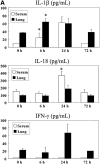
Induction of expression of IL-1β and IL-18 by intravenous administration of bleomycin in mice. (A) Wild-type (WT) C57BL/6 (B6) mice received an intravenous tail vein injection of 2 mg bleomycin suspended in 200 μl of sterile PBS. Mice (n = 4 per group) were killed 0, 6, 24, and 72 hours after the treatment, and serum samples were obtained and subjected to ELISA assay. The whole lung tissues were homogenized in 2 ml of lysis buffer (1% Triton X-100, 10 mM Tris-HCL, 5 mM EDTA, pH 7.6) containing a protease inhibitor cocktail (Complete Mini; Boehringer Mannheim GmbH) and centrifuged at 20,000 × g for 15 minutes, and the supernatants were collected and stored at −80°C until ELISA assay. The sandwich ELISA kits were used for mouse IL-1β, mouse IFN-γ (R&D Systems) and mouse IL-18 (MBL). *P < 0.05 versus 0 hours. (B) B6 mice received an intravenous tail vein injection of 2 mg bleomycin suspended in 200 μl of sterile PBS, and were then killed 6 and 24 hours after the treatment. As a control, the lung tissue was immediately harvested, and total RNA (1 μg) was used for mRNA analysis using a multiprobe RNAse protection assay. Lanes 1–3, 24 hours after PBS treatment (control); lanes 4–7, 6 hours after bleomycin treatment; lanes 8–10, 24 hours after bleomycin treatment. (C) Quantitative RNase protection analysis (n = 3 or 4 per group) was performed, and mRNA levels were quantitated using a Typhoon 8600 densitometer. Mouse glyceraldehyde 3-phosphate dehydrogenase was used as the control. *P < 0.05 versus 0 hours.
Figure 2.
Figure 2.
Induction of expression of IL-1β and IL-18 by intravenous administration of bleomycin in mice. (A) Wild-type (WT) C57BL/6 (B6) mice received an intravenous tail vein injection of 2 mg bleomycin suspended in 200 μl of sterile PBS. Mice (n = 4 per group) were killed 0, 6, 24, and 72 hours after the treatment, and serum samples were obtained and subjected to ELISA assay. The whole lung tissues were homogenized in 2 ml of lysis buffer (1% Triton X-100, 10 mM Tris-HCL, 5 mM EDTA, pH 7.6) containing a protease inhibitor cocktail (Complete Mini; Boehringer Mannheim GmbH) and centrifuged at 20,000 × g for 15 minutes, and the supernatants were collected and stored at −80°C until ELISA assay. The sandwich ELISA kits were used for mouse IL-1β, mouse IFN-γ (R&D Systems) and mouse IL-18 (MBL). *P < 0.05 versus 0 hours. (B) B6 mice received an intravenous tail vein injection of 2 mg bleomycin suspended in 200 μl of sterile PBS, and were then killed 6 and 24 hours after the treatment. As a control, the lung tissue was immediately harvested, and total RNA (1 μg) was used for mRNA analysis using a multiprobe RNAse protection assay. Lanes 1–3, 24 hours after PBS treatment (control); lanes 4–7, 6 hours after bleomycin treatment; lanes 8–10, 24 hours after bleomycin treatment. (C) Quantitative RNase protection analysis (n = 3 or 4 per group) was performed, and mRNA levels were quantitated using a Typhoon 8600 densitometer. Mouse glyceraldehyde 3-phosphate dehydrogenase was used as the control. *P < 0.05 versus 0 hours.
Figure 3.
Figure 3.
Increase of IL-18–producing F4/80-positive macrophages, but not CD3+ T cells, in bronchoalveolar lavage fluid (BALF) from bleomycin-treated, WT B6 mice. Recovered BALF cells were isolated from WT B6 mice treated intraperitoneally with bleomycin or control PBS on Day 4. Isolated BALF cells were stained with PE-Cy7–conjugated anti-mCD3 and PE-F4/80 mAb in the presence of anti-mCD16/CD32 mAb. The cells were then fixed, permeabilized, and stained with FITC anti–mIL-18 (M5). Three-color analysis was performed for analysis of cytoplasmic IL-18 expression in F4/80+ neutrophils and CD3+ T cells.
Figure 4.
Figure 4.
Prevention of bleomycin-induced lung injury in IL-18–deficient (−/−), IL-18Rα−/−, and caspase-1−/− mice. Juvenile (<10 wk old) female IL-18−/− mice and control WT B6 mice were intraperitoneally injected twice with bleomycin (2 mg) on Days 0 and 7, then killed on Day 28. Juvenile female caspase-1−/−, IL-18Rα−/−, and control WT B6 × 129 mice were intraperitoneally injected three times with bleomycin (2 mg) on Days 0, 7, and 14, then killed on Day 28. (A) The lung tissue was examined microscopically after H&E staining. Original magnification, ×40 and ×200. (B) Semiquantitative histopathological analysis was performed as previously reported (29). Briefly, H&E sections were observed at 400× and the lesions were defined as follows: Score 0, no lesions; Score 1, occasional small localized subpleural fibrotic foci; Score 2, thickening of intra-alveolar septa and subpleural fibrotic foci; and Score 3, thickened continuous subpleural fibrous foci and intraalveolar septa. IL-18−/− mice and control WT B6 mice were intraperitoneally injected three times with bleomycin (2 mg) on Days 0, 7, and 14, then killed on Day 28. Caspase-1−/−, IL-18Rα−/−, and control WT B6 × 129 mice were intraperitoneally injected four times with bleomycin (2 mg) on Days 0, 7, 14, and 21, then killed on Day 28. (C) Wet lung weights and (D) lung hydroxyproline content were measured at Day 28 (n = 5 in each group) as described in Materials and Methods. Results are expressed as the mean (±SD) for five mice per group. *P < 0.05 versus bleomycin-treated control WT mice.
Figure 4.
Figure 4.
Prevention of bleomycin-induced lung injury in IL-18–deficient (−/−), IL-18Rα−/−, and caspase-1−/− mice. Juvenile (<10 wk old) female IL-18−/− mice and control WT B6 mice were intraperitoneally injected twice with bleomycin (2 mg) on Days 0 and 7, then killed on Day 28. Juvenile female caspase-1−/−, IL-18Rα−/−, and control WT B6 × 129 mice were intraperitoneally injected three times with bleomycin (2 mg) on Days 0, 7, and 14, then killed on Day 28. (A) The lung tissue was examined microscopically after H&E staining. Original magnification, ×40 and ×200. (B) Semiquantitative histopathological analysis was performed as previously reported (29). Briefly, H&E sections were observed at 400× and the lesions were defined as follows: Score 0, no lesions; Score 1, occasional small localized subpleural fibrotic foci; Score 2, thickening of intra-alveolar septa and subpleural fibrotic foci; and Score 3, thickened continuous subpleural fibrous foci and intraalveolar septa. IL-18−/− mice and control WT B6 mice were intraperitoneally injected three times with bleomycin (2 mg) on Days 0, 7, and 14, then killed on Day 28. Caspase-1−/−, IL-18Rα−/−, and control WT B6 × 129 mice were intraperitoneally injected four times with bleomycin (2 mg) on Days 0, 7, 14, and 21, then killed on Day 28. (C) Wet lung weights and (D) lung hydroxyproline content were measured at Day 28 (n = 5 in each group) as described in Materials and Methods. Results are expressed as the mean (±SD) for five mice per group. *P < 0.05 versus bleomycin-treated control WT mice.
Figure 5.
Figure 5.
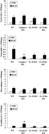
Prevention of neutrophil accumulation in bleomycin-treated caspase-1−/−, IL-18Rα−/−, and IL-18−/− mice. Juvenile (< 10 wk old) female B6 background caspase-1−/−, B6 IL-18−/−, B6 IL-18Rα−/−, and control WT B6 mice were injected intraperitoneally twice with bleomycin (2 mg) or PBS (vehicle) on Days 0 and 7. Recovered BALF cells were isolated on Day 28. BALF cells were centrifuged onto glass slides at 800 rpm for 10 minutes, air dried, and stained with Wright-Giemsa. Cell populations were calculated as described in Materials and Methods. *P < 0.05 versus bleomycin-treated control WT B6 mice.

Similar articles

Cited by

References

    1. Sleijfer S. Bleomycin-induced pneumonitis. Chest 2001;120:617–624. - PubMed
    1. Piguet PF, Collart MA, Grau GE, Kapanci Y, Vassalli P. Tumor necrosis factor/cachectin plays a key role in bleomycin-induced pneumopathy and fibrosis. J Exp Med 1989;170:655–663. - PMC - PubMed
    1. Nakao A, Fujii M, Matsumura R, Kumano K, Saito Y, Miyazono K, Iwamoto I. Transient gene transfer and expression of Smad7 prevents bleomycin-induced lung fibrosis in mice. J Clin Invest 1999;104:5–11. - PMC - PubMed
    1. Scheule RK, Perkins RC, Hamilton R, Holian A. Bleomycin stimulation of cytokine secretion by the human alveolar macrophage. Am J Physiol 1992;262:L386–L391. - PubMed
    1. Smith RE, Strieter RM, Phan SH, Lukacs NW, Huffnagle GB, Wilke CA, Burdick MD, Lincoln P, Evanoff H, Kunkel SL. Production and function of murine macrophage inflammatory protein-1 alpha in bleomycin-induced lung injury. J Immunol 1994;153:4704–4712. - PubMed

MeSH terms