. 2009 May 1;284(18):11971-81.
doi: 10.1074/jbc.M808761200. Epub 2009 Mar 5.

Differential behavior of missense mutations in the intersubunit contact domain of the human pyruvate kinase M2 isozyme

Affiliations

Differential behavior of missense mutations in the intersubunit contact domain of the human pyruvate kinase M2 isozyme

Kamal Akhtar et al. J Biol Chem. .

Abstract

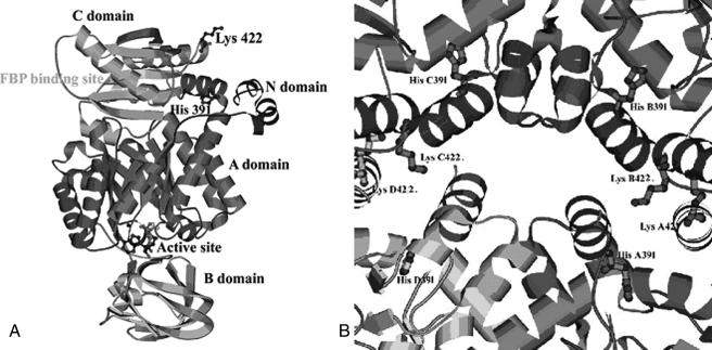
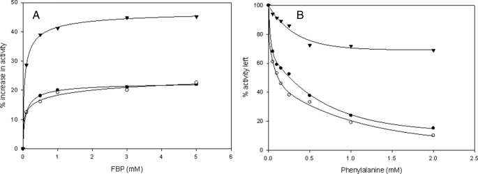
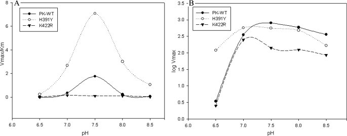
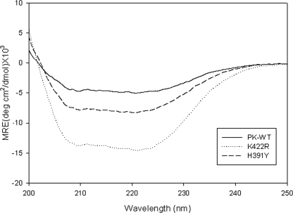
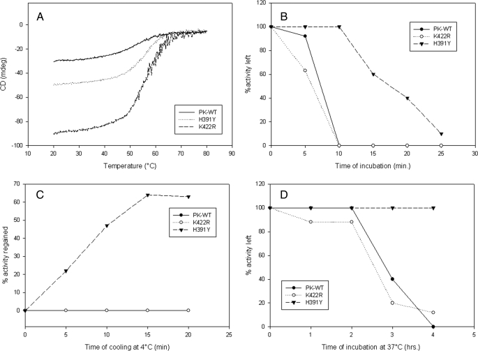
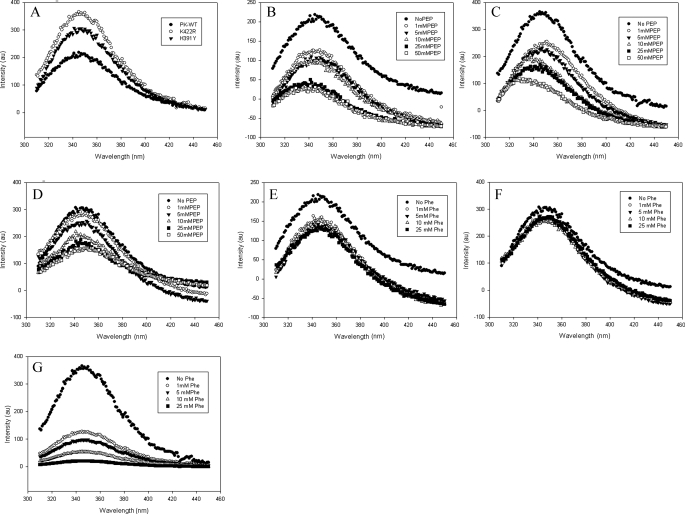
In this study, we attempted to understand the mechanism of regulation of the activity and allosteric behavior of the pyruvate kinase M(2) enzyme and two of its missense mutations, H391Y and K422R, found in cells from Bloom syndrome patients, prone to develop cancer. Results show that despite the presence of mutations in the intersubunit contact domain, the K422R and H391Y mutant proteins maintained their homotetrameric structure, similar to the wild-type protein, but showed a loss of activity of 75 and 20%, respectively. Interestingly, H391Y showed a 6-fold increase in affinity for its substrate phosphoenolpyruvate and behaved like a non-allosteric protein with compromised cooperative binding. However, the affinity for phosphoenolpyruvate was lost significantly in K422R. Unlike K422R, H391Y showed enhanced thermal stability, stability over a range of pH values, a lesser effect of the allosteric inhibitor Phe, and resistance toward structural alteration upon binding of the activator (fructose 1,6-bisphosphate) and inhibitor (Phe). Both mutants showed a slight shift in the pH optimum from 7.4 to 7.0. Although this study signifies the importance of conserved amino acid residues in long-range communications between the subunits of multimeric proteins, the altered behavior of mutants is suggestive of their probable role in tumor-promoting growth and metabolism in Bloom syndrome patients with defective pyruvate kinase M(2).


Figures

FIGURE 1.
FIGURE 1.
A, ribbon diagram of the overall structure of PK showing the positions of the two mutations, H391Y and K422R, along with the active site and Fru-1,6-P2-binding site. B, intersubunit contact domain of PK. The major amino acid residues and side chains at the tetramer interface region are shown.
FIGURE 2.
FIGURE 2.
A, Fru-1,6-P2 titration curves. B, phenylalanine titration curves for PK-WT (•) and the K422R (○) and H391Y (▾) mutant proteins. Enzyme activity was assayed at 25 °C and pH 7.5 as described under “Experimental Procedures” with 2.5 mm P-enolpyruvate and 2.5 mm ADP.
FIGURE 3.
FIGURE 3.
A, PK-WT and the mutant enzymes were incubated at different pH values in 50 mm Tris-Cl buffer and assayed under standard conditions as described under “Experimental Procedures.” A graph of log Vmax and pH was plotted to check the pH optimum of the P-enolpyruvate-bound state of the enzymes. B, P-enolpyruvate titration study was done at different pH values in Tris-Cl buffer, and Km values at each individual pH were calculated. The Vmax/Km ratios obtained at different pH values were plotted against pH as shown.
FIGURE 4.
FIGURE 4.
The CD spectra of PK-WT and the mutant proteins were recorded in the far-UV region (from 200 to 250 nm) at 25 °C at a concentration of 0.3 mg/ml in 10 mm phosphate buffer (pH 7.5), 3 mm MgCl2, 100 mm KCl, and 5% glycerol. The mean residual ellipticity (MRE) was plotted for a wavelength from 200 to 250 nm. deg, degrees.
FIGURE 5.
FIGURE 5.
A, loss of ellipticity upon increasing temperature analyzed by CD spectroscopy for PK-WT and the mutant proteins. B, loss of biological activity at 60 °C. C, regain of activity after cooling the denatured proteins on ice. D, activity assay at physiological temperature (37 °C) over a given time period. All assays were done under standard reaction conditions as described under “Experimental Procedures.” mdeg, millidegrees.
FIGURE 6.
FIGURE 6.
The intrinsic tryptophan fluorescence of the proteins (50 μg/ml) was measured upon excitation at 295 nm and by scanning emission in the range of 310–450 nm. A, emission spectra of PK-WT and the K422R and H391Y mutant proteins. B–D, P-enolpyruvate titration of PK-WT, K422R, and H391Y, respectively, showing quenching with increasing concentrations of P-enolpyruvate (PEP). E–G, PK-WT, H391Y, and K422R, respectively, showing quenching with increasing concentrations of the inhibitor phenylalanine. au, absorbance units.
FIGURE 7.
FIGURE 7.
A, the wild-type human PKM2 structure shows His391 (red) of the A-chain and Glu386 (blue) of the C-chain. B, a unique H-bond 2.98 Å in length was predicted between the replaced tyrosine residue (red) and the backbone of Glu386 (blue) connecting the A- and C-domains of the protein. It is hypothesized that formation of a new hydrogen bond connecting the A- and C-domains could restrict the movements along the hinges of the A- and C-chains and cause compromised dynamics in the molecule.
FIGURE 8.
FIGURE 8.
A, His391 in human PKM2 is surrounded by hydrophobic residues such as Leu392, Pro446, Tyr390, and Phe395, which make a local hydrophobic cluster. B, Lys422 (blue) with its protruding side chain is found to interact with Ala419 (turquoise) to align the position of Glu418 (orange) of the same monomer to make a stable H-bond with Arg399 (red) of the neighboring monomer.

Similar articles

Cited by

References

    1. Kayne, F. J., and Price, N. C. (1973) Arch. Biochem. Biophys. 159 292–296 - PubMed
    1. Kuo, D. J., O'Connell, E. L., and Rose, I. A. (1979) J. Am. Chem. Soc. 101 5025–5030
    1. Valentini, G., Chiarelli, L., Fortin, R., Speranza, M. L., Galizzi, A., and Mattevi, A. (2000) J. Biol. Chem. 275 18145–18152 - PubMed
    1. Mazurek, S., Boschek, C. B., and Eigenbrodt, E. (1997) J. Bioenerg. Biomembr. 29 315–330 - PubMed
    1. Imamura, K., and Tanaka, T. (1982) Methods Enzymol. 90 150–165 - PubMed

Publication types

Substances

LinkOut - more resources