. 2009 May 1;284(18):12541-9.
doi: 10.1074/jbc.M808376200. Epub 2009 Mar 5.

Heat shock protein gp96 interacts with protein phosphatase 5 and controls toll-like receptor 2 (TLR2)-mediated activation of extracellular signal-regulated kinase (ERK) 1/2 in post-hypoxic kidney cells

Affiliations

Heat shock protein gp96 interacts with protein phosphatase 5 and controls toll-like receptor 2 (TLR2)-mediated activation of extracellular signal-regulated kinase (ERK) 1/2 in post-hypoxic kidney cells

Sanae Ben Mkaddem et al. J Biol Chem. .

Abstract

Ischemia/reperfusion injury (IRI) induces an innate immune response, leading to an inflammatory reaction and tissue damage that have been attributed to engagement of the Toll-like receptor (TLR) 2 and 4. However, the respective roles of TLR2 and/or TLR4 in mediating downstream activation of mitogen-activated protein kinase (MAPK) pathways during IRI have not been fully elucidated. Here we show that extracellular signal-regulated kinase (ERK)1/2 is activated in both intact kidneys and cultured renal tubule epithelial cells (RTECs) from wildtype and Tlr4 knockout mice, but not those from Tlr2 knockout mice subjected to transient ischemia. Geldanamycin (GA), an inhibitor of heat shock protein 90 and reticulum endoplasmic-resident gp96, and gp96 mRNA silencing (siRNA), did not affect ERK1/2 activation in either post-hypoxic wild-type or Tlr4-deficient RTECs, but did restore its activation in post-hypoxic Tlr2-deficient RTECs. Immunoprecipitation studies revealed that gp96 co-immunoprecipitates with the serine-threonine protein phosphatase 5 (PP5), identified as a negative modulator of the mitogen extracellular kinase (MEK)-ERK pathway, in unstressed wild-type and post-hypoxic Tlr2-deficient RTECs. In contrast, PP5 co-immunoprecipitation with gp96 was strikingly reduced in post-hypoxic wild-type RTECs, suggesting that the inactivation of PP5 resulting from the dissociation of PP5 from gp96 allows the activation of ERK1/2 to occur. Inhibition of PP5 by okadaic acid, and Pp5 siRNA also restored TLR2-mediated phosphorylation of ERK1/2, and apoptosis signal-regulating kinase 1 (ASK1)/c-Jun N-terminal kinase (JNK)-mediated apoptosis in post-hypoxic Tlr2-deficient RTECs. These findings indicate that gp96 interacts with PP5 and controls TLR2-mediated induction of ERK1/2 in post-hypoxic renal tubule cells.


Figures

FIGURE 1.
FIGURE 1.
Differential expression of ERK1/2 in ischemic-reperfused wild-type, Tlr2-, and Tlr4-deficient mice. A, levels of serum creatinine in WT, Tlr2-/-, and Tlr4-/- sham-operated control (C) mice, and one (D-1), two (D-2), and seven (D-7) days after bilateral clamping of the renal pedicles. B, detection of TUNEL-positive tubule cells (arrowheads) in kidney sections from control (Control) and day-2 post-ischemic (I/R (D-2)) mice. Scale bar, 20 μm. C, quantification of the number of active caspase-3 positive cells per tissue section (200 μm2) in post-ischemic kidneys. Data are means ± S.E. (A, B: 6-9 kidneys). *, p < 0.05 between groups. D, immunoblot analysis of phospho (p-) and total ASK1-, JNK-, and ERK1/2-labeled bands detected in kidney homogenates from control (C), and D-1, D-2, and D-7 post-ischemic mice kidneys. E, immunoblot analysis of p- and total ERK1/2-labeled bands in D-2 post-ischemic WT, Tlr2-/-, Tlr4-/-, and Tlr2,4-/- DKO mice kidneys.
FIGURE 2.
FIGURE 2.
Inhibition of ASK1 and JNK activation impairs the induction of apoptosis in post-hypoxic wild-type renal tubule cells. A, immunoblot analysis of p- and total ASK1 and JNK in non-hypoxic (Control) and 24-h post-hypoxic (Hypoxia) wild-type RTECs preincubated without or with thioredoxin (200 ng/ml). B, immunoblot analysis of p- and total c-Jun and ASK1 in nonhypoxic and 24-h post-hypoxic wild-type RTECs preincubated without or with SP600125 (2 μm). C and D, percentage of apoptotic cells measured in non-hypoxic (Control) and 24 h post-hypoxic (Hypoxia) wild-type RTECs pre-incubated without or with thioredoxin (C) or SP600125 (D). Data are means ± S.E. from four different cell cultures from two different kidneys for each set of experimental conditions. *, p < 0.05 between groups.
FIGURE 3.
FIGURE 3.
Geldanamycin restores the phosphorylation of ERK1/2 in post-hypoxic Tlr2-/- renal tubule cells. A, immunoblot analyses of p- and total ERK1/2 and JNK in non-hypoxic (C) and 24-h post-hypoxic (H) WT, Tlr2-/-, Tlr4-/- RTECs preincubated without or with GA (+GA). B, immunoblot analyses of p- and total AKT in non-hypoxic (C) and day-1 post-hypoxic (H) WT, Tlr2-/-, Tlr4-/- RTECs preincubated without or with geldanamycin (+GA, 10 nm). C, immunoblot analyses of p- and total AKT and ERK1/2 in day-1 post-hypoxic (H) WT, and Tlr4-/- RTECs preincubated (+) or not (-) with LY294002 (20 μm).
FIGURE 4.
FIGURE 4.
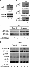
gp96 requirement for the induction of TLR2-mediated activation of ERK1/2. A, immunoblot analyses of gp96 and the correspondingβ-actin in untreated (C) and day-1 post-hypoxic (H) WT or Tlr2-/- RTECs. B and C, immunoblot analysis of gp96 and the corresponding β-actin (B), and phospho (p-) and total ERK1/2 (C) in non-hypoxic (C or Control) and day-1 post-hypoxic (H or Hypoxia) WT RTECs transfected or not with a specific gp96 siRNA or negative control siRNA. D, immunoblot analyses of p- and total ERK1/2, ASK1, and JNK in non-hypoxic and day-1 post-hypoxic Tlr2-/- RTECs transfected or not with gp96 siRNA or negative control siRNA.
FIGURE 5.
FIGURE 5.
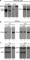
PP5 interacts with gp96. Lysates from non-hypoxic PKSV-PR cells (A) and wild-type or Tlr2-/- RTECs (B, C) were subjected to IP using an antibody against gp96. The IP material was then subjected to Western blot analysis, and proteins were detected with anti-PP5 or anti-gp96 antibodies, then revealed with the ECL anti-rabbit IgG, horseradish peroxidase-linked species-specific whole antibody (GE Healthcare) (A, lanes 1-4 and C) or the Rabbit IgG TrueBlot™ (eBioscience) (A, lanes 5 and 6, and B). A, untreated PKSV-PR cells lysates were immunoprecipitated (IP) with the anti-gp96 antibody, then blotted for PP5 (lane 1). As controls, immunoprecipitated cell lysates were blotted for gp96 (lane 2) and non-immunoprecipitated PKSV-PR cell lysates (None) were blotted for PP5 (lane 3) or gp96 (lane 4). Western blot identification of PP5 in non-hypoxic PKSV-PR cell lysates from co-IP (lane 5) and non-immunoprecipitated (None, lane 6) cell lysates using the secondary IgG TrueBlot™ antibody. B and C, untreated control (C) and post-hypoxic (H) WT or Tlr2-/- RTECs were immunoprecipitated with the anti-gp96 antibody, then blotted for PP5 (B) or gp96 (C). As controls, non-immunoprecipitated cell lysates were blotted either for PP5 (B) or gp96 (C).
FIGURE 6.
FIGURE 6.
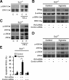
PP5 controls the phosphorylation of ERK1/2, ASK1, and JNK in post-hypoxic Tlr2-/- RTECs. A, immunoblot analyses of p- and total ERK1/2 in non-hypoxic (C) and 24-h post-hypoxic (H) Tlr2-/- RTECs preincubated or not with 5 nm okadaic acid (+Ok.a). B, immunoblot analyses of PP5 and corresponding β-actin, and p- and total ERK1/2 in non-hypoxic (Control) and day-1 post-hypoxic (Hypoxia) Tlr2-/- RTECs transfected or not with a Pp5 siRNA or negative control siRNA. C and D, immunoblot analyses of p- and total ASK1 and JNK in non-hypoxic (C) and day-1 post-hypoxic (H) Tlr2-/- RTECs preincubated or not with 10 nm okadaic acid (+Ok.a)(C), and non-hypoxic (Control) or day-1 post-hypoxic (Hypoxia) Tlr2-/- RTECs transfected or not with a Pp5 siRNA or negative control siRNA (D). E, percentage of apoptotic cells measured in non-hypoxic (Control) or day-1 post-hypoxic (Hypoxia) Tlr2-/- RTECs transfected or not with a Pp5 siRNA or negative control siRNA. Data are means ± S.E. from 4 different cell cultures from 3 different kidneys for each set of experimental conditions. *, p < 0.05 between groups.
FIGURE 7.
FIGURE 7.
Diagrammatic representation of the mechanism of TLR2-mediated ERK activation in post-hypoxic renal tubule cells. A, in non-hypoxic renal tubule cells, PP5 is associated with gp96, while transient hypoxia (Hypoxia) stimulates the expression of gp96, and induces the dissociation of gp96 bound to PP5, resulting in the inhibition of PP5 activity and downstream ERK1/2 phosphorylation. B, in Tlr2-/--deficient RTECs, hypoxia stimulates gp96, but does not trigger the dissociation of gp96 bound to PP5. In this case, PP5 can be expected to remain active, and inhibit raf-1 activity and downstream ERK1/2 phosphorylation. Inhibition of gp96 activity by GA, or mRNA extinction of gp96 induces the reactivation of ERK1/2, but not of JNK or ASK1 in Tlr2-/--deficient RTECs, and extinction of Pp5 mRNA expression also induces the reactivation of ERK1/2 phosphorylation and reactivation of p-JNK and ASK-1 (not shown in the diagram).

Similar articles

Cited by

References

    1. Bonventre, J. V., and Weinberg, J. M. (2003) J. Am. Soc. Nephrol. 14 2199-2210 - PubMed
    1. Beg, A. A. (2002) Trends Immunol. (2002) 23 509-512 - PubMed
    1. Akira, S., and Takeda, K. (2004) Nat. Rev. Immunol. 4 499-511 - PubMed
    1. Marshak-Rothstein, A. (2006) Nat. Rev. Immunol. 6 823-835 - PMC - PubMed
    1. Tsuboi, N., Yoshikai, Y., Matsuo, S., Kikuchi, T., Iwami, K., Nagai, Y., Takeuchi, O., Akira, S., and Matsuguchi, T. (2002) J. Immunol. 169 2026-2033 - PubMed

Publication types

MeSH terms