. 2009 Apr 24;284(17):11039-47.
doi: 10.1074/jbc.M808058200. Epub 2009 Mar 5.

The D816V mutation of c-Kit circumvents a requirement for Src family kinases in c-Kit signal transduction

Affiliations

The D816V mutation of c-Kit circumvents a requirement for Src family kinases in c-Kit signal transduction

Jianmin Sun et al. J Biol Chem. .

Abstract

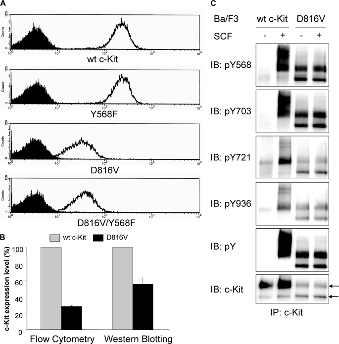
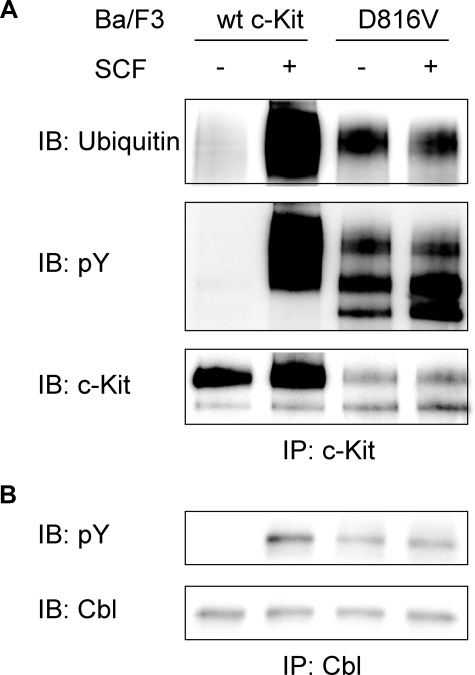
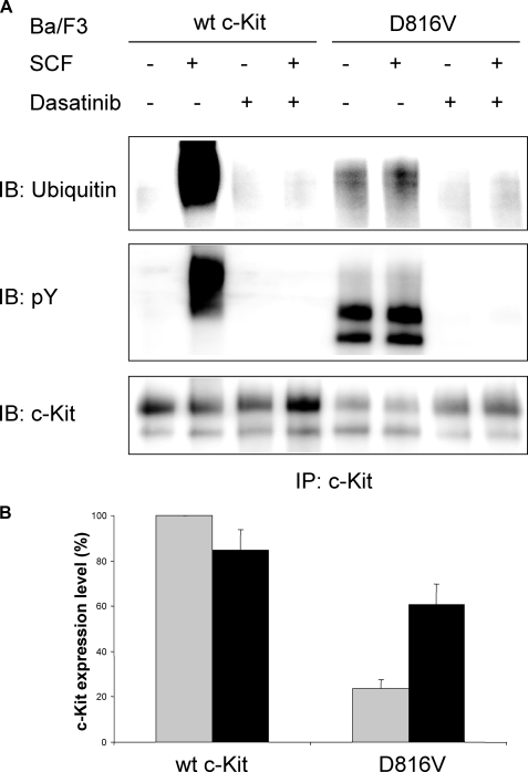
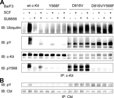
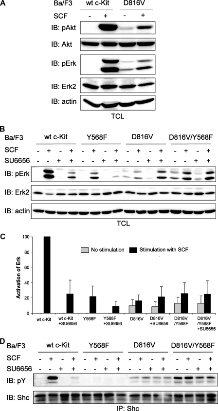
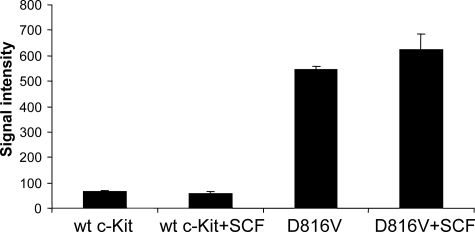
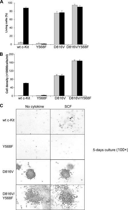
The receptor tyrosine kinase c-Kit plays a critical role in hematopoiesis, and gain-of-function mutations of the receptor are frequently seen in several malignancies, including acute myeloid leukemia, gastrointestinal stromal tumors, and testicular carcinoma. The most common mutation of c-Kit in these disorders is a substitution of the aspartic acid residue in position 816 to a valine (D816V), leading to constitutive activation of the receptor. In this study, we aimed to investigate the role of Src family kinases in c-Kit/D816V signaling. Src family kinases are necessary for the phosphorylation of wild-type c-Kit as well as of activation of downstream signaling pathways including receptor ubiquitination and the Ras/Mek/Erk pathway. Our data demonstrate that, unlike wild-type c-Kit, the phosphorylation of c-Kit/D816V is not dependent on Src family kinases. In addition, we found that neither receptor ubiquitination nor Erk activation by c-Kit/D816V required activation of Src family kinases. In vitro kinase assay using synthetic peptides revealed that c-Kit/D816V had an altered substrate specificity resembling Src and Abl tyrosine kinases. We further present evidence that, in contrast to wild-type c-Kit, Src family kinases are dispensable for c-Kit/D816V cell survival, proliferation, and colony formation. Taken together, we demonstrate that the signal transduction pathways mediated by c-Kit/D816V are markedly different from those activated by wild-type c-Kit and that altered substrate specificity of c-Kit circumvents a need for Src family kinases in signaling of growth and survival, thereby contributing to the transforming potential of c-Kit/D816V.


Figures

FIGURE 1.
FIGURE 1.
Expression and activation of wild-type (wt) c-Kit and c-Kit/D816V in Ba/F3 cells. A, Ba/F3 cells expressing wild-type c-Kit, c-Kit/Y568F, c-Kit/D816V, or c-Kit/D816V/Y568F were stained with PE-conjugated anti-c-Kit antibody or isotype control followed by examination of receptor cell surface expression using flow cytometry (open curve, anti-c-Kit; filled curve, isotype control). B, expression of c-Kit was quantified based on both flow cytometry and Western blotting. Error bars indicate standard deviation. C, Ba/F3 cells expressing wild-type c-Kit or c-Kit/D816V were starved, SCF-stimulated (100 ng/ml) and subjected to immunoprecipitation (IP) using a c-Kit antibody. Phosphospecific antibodies toward pTyr-568 (pY568), pTyr-703 (pY703), pTyr-721 (pY721), and pTyr-936 (pY936) of c-Kit were used to detect the activation of individual tyrosine residues. As controls, phosphotyrosine (pY) and c-Kit expression levels were assessed. Arrows indicate the position of the 130-kDa immature band of c-Kit and the 145-kDa mature band of c-Kit, respectively. IB, immunoblot.
FIGURE 2.
FIGURE 2.
Constitutive activation of Cbl and ubiquitination of c-Kit in Ba/F3 cells expressing c-Kit/D816V. Ba/F3 cells expressing c-Kit/D816V were starved and stimulated with SCF (100 ng/ml). A, lysates were subjected to immunoprecipitation (IP) using a c-Kit antibody followed by Western blot (IB) analysis using antibodies against ubiquitin, phosphotyrosine(pY), and c-Kit. wt, wild type. B, lysates were subjected to immunoprecipitation using a Cbl antibody followed by Western blot analysis using antibodies against phosphotyrosine and Cbl. Ba/F3 cells expressing wild-type c-Kit were used as control.
FIGURE 3.
FIGURE 3.
Dasatinib inhibits the constitutive ubiquitination of c-Kit/D816V and increases the cell surface expression of the receptor. Ba/F3 cells expressing wild-type (wt) c-Kit or c-Kit/D816V mutant were incubated with or without Dasatinib (2 nm) for 24 h. A, cells were starved and stimulated with SCF (100 ng/ml) and subjected to immunoprecipitation using a c-Kit antibody. Levels of ubiquitination, tyrosine phosphorylation, and c-Kit expression were analyzed by Western blot (IB). pY, phosphotyrosine. B, cells were stained with PE-conjugated anti-c-Kit antibody or isotype control followed by examination of expression using flow cytometry. Cell surface expression levels of c-Kit were quantified, and the expression level of wild-type c-Kit without incubation with Dasatinib was considered as 100% and compared with other cells (gray column: no incubation; black column: incubation with Dasatinib). The data shown are the average of three independent experiments with error bars defining the standard deviation.
FIGURE 4.
FIGURE 4.
Src family kinases are not required for the phosphorylation and ubiquitination of c-Kit/D816V. Ba/F3 cells expressing wild-type (wt) c-Kit, c-Kit/Y568F, c-Kit/D816V or c-Kit/D816V/Y568F were starved. A, thereafter cells were preincubated with the Src inhibitor SU6656 (2 μm) for 30 min before SCF stimulation (100 ng/ml) followed by immunoprecipitation (IP) using a c-Kit antibody. After SDS-PAGE and electrotransfer, membranes were probed with ubiquitin antibody and reprobed with phosphotyrosine (pY) and c-Kit antibodies. IB, immunoblot; pY568, pTyr-568. B, cells were preincubated with the Src inhibitor SU6656 (2 μm) for 30 min before SCF stimulation (100 ng/ml) and immunoprecipitation using a Cbl antibody. After SDS-PAGE and electrotransfer, membranes were probed with phosphotyrosine and Cbl antibodies to show the activation of Cbl.
FIGURE 5.
FIGURE 5.
Inhibition of Src family kinases has different effects on Erk activation in Ba/F3 cells expressing wild-type (wt) c-Kit and c-Kit/D816V. A, Ba/F3 cells expressing wild-type c-Kit or c-Kit/D816V were starved and thereafter stimulated with SCF (100 ng/ml). Whole cell lysates were investigated by Western blot (IB) analysis using antibodies against phospho-Erk (pErk) and phospho-Akt (pAkt). Filters were reprobed with Erk2, Akt, and β-actin antibodies to verify equal loading. B, Ba/F3 cells expressing wild-type c-Kit, c-Kit/Y568F, c-Kit/D816V, or c-Kit/D816V/Y568F were starved followed by preincubation with the Src inhibitor SU6656 (2 μm) for 30 min before SCF stimulation (100 ng/ml). Total cell lysates (TCL) were subjected to Western blot analysis using an antibody against phospho-Erk. Erk2 and β-actin were used as loading control. C, quantitation of data obtained in panel B. The level of Erk phosphorylation in Ba/F3 cells expressing wild-type c-Kit was set to 100. The data represent the average of triplicate determinations with the error bars showing standard deviation. D, cell lysates were immunoprecipitated (IP) using a Shc antibody, and samples were analyzed by Western blot using antibodies against phosphotyrosine and Shc. pY, phosphotyrosine.
FIGURE 6.
FIGURE 6.
c-Kit/D816V gains Src activity. Ba/F3 cells expressing wild-type (wt) or c-Kit/D816V were starved, stimulated with SCF (100 ng/ml), and subsequently immunoprecipitated using a c-Kit antibody. The immunoprecipitates were used in a kinase assay together with Src optimal peptide as substrate. The phosphorylation of the substrate by wild-type c-Kit and c-Kit/D816V was detected by Fuji FLA 3000, and signal intensity was quantified by the Multigauge software. Error bars indicate standard deviation.
FIGURE 7.
FIGURE 7.
Phosphorylation of Tyr-568 is not required in c-Kit/D816V-induced cell survival, proliferation, and growth in methylcellulose. Ba/F3 cells expressing wild-type (wt) c-Kit, c-Kit/Y568F, c-Kit/D816V, or c-Kit/D816V/Y568F were washed and thereafter seeded in 6-well plates in 100,000 cells/ml with SCF (100 ng/ml, black column) or without cytokine (gray column) as control followed by a 48-h incubation. A, cells were stained with the annexin V-PE apoptosis detection kit and subsequently subjected to flow cytometry to examine the proportion of living cells, apoptotic cells, and dead cells. Error bars indicate standard deviation. B, living cells were counted using trypan blue exclusion. Error bars indicate standard deviation. C, colony formation in methylcellulose.

Similar articles

Cited by

References

    1. Lennartsson, J., and Rönnstrand, L. (2006) Curr. Cancer Drug Targets 6, 65–75 - PubMed
    1. Rönnstrand, L. (2004) CMLS Cell. Mol. Life Sci. 61, 2535–2548 - PMC - PubMed
    1. Ning, Z. Q., Li, J., and Arceci, R. J. (2001) Leuk. Lymphoma 41, 513–522 - PubMed
    1. Beghini, A., Peterlongo, P., Ripamonti, C. B., Larizza, L., Cairoli, R., Morra, E., and Mecucci, C. (2000) Blood 95, 726–727 - PubMed
    1. Tian, Q., Frierson, H. F., Jr., Krystal, G. W., and Moskaluk, C. A. (1999) Am. J. Pathol. 154, 1643–1647 - PMC - PubMed

Publication types

LinkOut - more resources