

The outcome of Cryptococcus neoformans intracellular pathogenesis in human monocytes
- PMID: 19265539
- PMCID: PMC2670303
- DOI: 10.1186/1471-2180-9-51
The outcome of Cryptococcus neoformans intracellular pathogenesis in human monocytes
Abstract
Background: Cryptococcus neoformans is an encapsulated yeast that is a facultative intracellular pathogen. The interaction between macrophages and C. neoformans is critical for extrapulmonary dissemination of this pathogenic yeast. C. neoformans can either lyse macrophages or escape from within them through a process known as phagosomal extrusion. However, most studies of intracellular pathogenesis have been made with mouse cells and their relevance to human infection is uncertain. In this study we extended studies of C. neoformans-macrophage cellular interaction/s to human peripheral blood monocytes.
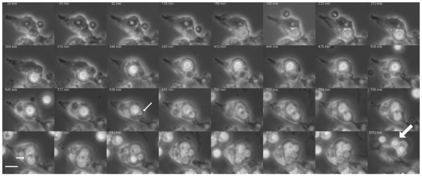
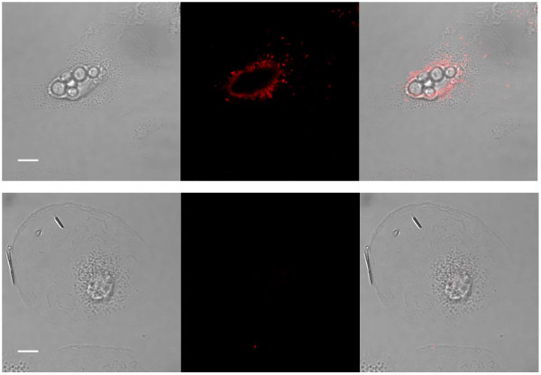
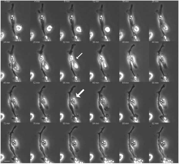
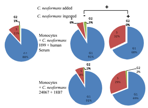
Results: This study demonstrated that C. neoformans can shed polysaccharide within human monocytes, spread from cell to cell, and be extruded from them. Furthermore, human monocytes responded to ingestion of C. neoformans with cell cycle progression from G1 to S.
Conclusion: Similarities between mouse and human cells support the suitability of mouse cells for the study of intracellular pathogenesis mechanisms. Given that these hosts diverged over 70 million years ago, the similar pathogenic strategies for C. neoformans in murine and human cells supports the hypothesis that the mechanism that underlies the mammalian intracellular pathogenesis of C. neoformans originated from interactions with a third host, possibly soil amoeboid predators, before the mammalian radiation.
Figures
Similar articles
-
Phagosome extrusion and host-cell survival after Cryptococcus neoformans phagocytosis by macrophages.Curr Biol. 2006 Nov 7;16(21):2161-5. doi: 10.1016/j.cub.2006.09.061. Curr Biol. 2006. PMID: 17084702
-
Assessing Phagocytosis of Cryptococcus neoformans Cells in Human Monocytes or the J774 Murine Macrophage Cell Line.Methods Mol Biol. 2024;2775:157-169. doi: 10.1007/978-1-0716-3722-7_11. Methods Mol Biol. 2024. PMID: 38758317
-
Cryptococcus neoformans is a facultative intracellular pathogen in murine pulmonary infection.Infect Immun. 2000 Jul;68(7):4225-37. doi: 10.1128/IAI.68.7.4225-4237.2000. Infect Immun. 2000. PMID: 10858240 Free PMC article.
-
Catch me if you can: phagocytosis and killing avoidance by Cryptococcus neoformans.FEMS Immunol Med Microbiol. 2012 Mar;64(2):147-61. doi: 10.1111/j.1574-695X.2011.00871.x. FEMS Immunol Med Microbiol. 2012. PMID: 22029633 Review.
-
The origin and maintenance of virulence for the human pathogenic fungus Cryptococcus neoformans.Microbes Infect. 2003 Jun;5(7):667-75. doi: 10.1016/s1286-4579(03)00092-3. Microbes Infect. 2003. PMID: 12787743 Review.
Cited by
-
The Diverse Roles of Monocytes in Cryptococcosis.J Fungi (Basel). 2020 Jul 16;6(3):111. doi: 10.3390/jof6030111. J Fungi (Basel). 2020. PMID: 32708673 Free PMC article. Review.
-
Conservation of Intracellular Pathogenic Strategy among Distantly Related Cryptococcal Species.Infect Immun. 2018 Jun 21;86(7):e00946-17. doi: 10.1128/IAI.00946-17. Print 2018 Jul. Infect Immun. 2018. PMID: 29712729 Free PMC article.
-
The role of host gender in the pathogenesis of Cryptococcus neoformans infections.PLoS One. 2013 May 31;8(5):e63632. doi: 10.1371/journal.pone.0063632. Print 2013. PLoS One. 2013. PMID: 23741297 Free PMC article.
-
Cryptococcus gattii, no longer an accidental pathogen?Curr Fungal Infect Rep. 2012 Dec;6(4):245-256. doi: 10.1007/s12281-012-0111-0. Curr Fungal Infect Rep. 2012. PMID: 23243480 Free PMC article.
-
Fc gamma receptor 3A polymorphism and risk for HIV-associated cryptococcal disease.mBio. 2013 Aug 27;4(5):e00573-13. doi: 10.1128/mBio.00573-13. mBio. 2013. PMID: 23982074 Free PMC article.
References
-
- Casadevall A, Perfect J. Cryptococccus neoformans. Washington, DC: American Society for Microbiology Press; 1998.
-
- Shao X, Mednick A, Alvarez M, van Rooijen N, Casadevall A, Goldman DL. An innate immune system cell is a major determinant of species-related susceptibility differences to fungal pneumonia. J Immunol. 2005;175(5):3244–3251. - PubMed
-
- Lee SC, Kress Y, Zhao ML, Dickson DW, Casadevall A. Cryptococcus neoformans survive and replicate in human microglia. Lab Invest. 1995;73(6):871–879. - PubMed
Publication types
MeSH terms
Substances
Grants and funding
LinkOut - more resources
Full Text Sources
