. 2009 Mar 5:6:29.
doi: 10.1186/1743-422X-6-29.

Epstein-Barr Nuclear Antigen 1 modulates replication of oriP-plasmids by impeding replication and transcription fork migration through the family of repeats

Affiliations

Epstein-Barr Nuclear Antigen 1 modulates replication of oriP-plasmids by impeding replication and transcription fork migration through the family of repeats

Ashok Aiyar et al. Virol J. .

Abstract

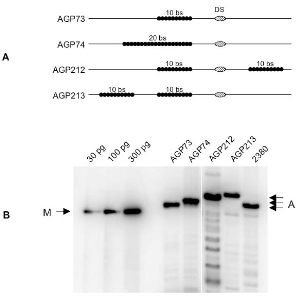
Background: Epstein-Barr virus is replicated once per cell-cycle, and partitioned equally in latently infected cells. Both these processes require a single viral cis-element, termed oriP, and a single viral protein, EBNA1. EBNA1 binds two clusters of binding sites in oriP, termed the dyad symmetry element (DS) and the family of repeats (FR), which function as a replication element and partitioning element respectively. Wild-type FR contains 20 binding sites for EBNA1.

Results: We, and others, have determined previously that decreasing the number of EBNA1-binding sites in FR increases the efficiency with which oriP-plasmids are replicated. Here we demonstrate that the wild-type number of binding sites in FR impedes the migration of replication and transcription forks. Further, splitting FR into two widely separated sets of ten binding sites causes a ten-fold increase in the efficiency with which oriP-plasmids are established in cells expressing EBNA1. We have also determined that EBNA1 bound to FR impairs the migration of transcription forks in a manner dependent on the number of EBNA1-binding sites in FR.

Conclusion: We conclude that EBNA1 bound to FR regulates the replication of oriP-plasmids by impeding the migration of replication forks. Upon binding FR, EBNA1 also blocks the migration of transcription forks. Thus, in addition to regulating oriP replication, EBNA1 bound to FR also decreases the probability of detrimental collisions between two opposing replication forks, or between a transcription fork and a replication fork.


Figures

Figure 1
Figure 1
Plasmids with an FR containing more than 20 EBNA1 binding sites form minute colonies under selection. The indicated plasmids were transfected into 293/EBNA1 cells, which were subjected to puromycin selection for 18 days in colony formation assays as described in the Materials and Methods section. Representative images of methylene blue stained colonies are shown. The identity of the transfected plasmid is indicated above each image, and the number of EBNA1 binding sites present in the FR of each plasmid is indicated below each image. As a negative control, an assay was also performed with AGP83, a DS-only plasmid that replicates transiently, but is not partitioned, and forms colonies with very low efficiency. The colonies formed from cells transfected with AGP81 and AGP82 did not increase in size even after several weeks of growth in selective media. The number of colonies formed in such assays that were 2 mm in size and larger is indicated in Table 1.
Figure 2
Figure 2
Two models to explain decreases in copy number when oriP plasmids contain an FR with 20 or more EBNA1 binding sites. DS is represented as a striped oval, and EBNA1-binding sites in FR are represented as black filled circles. For simplicity, only ten binding sites are shown. EBNA1 dimers bound to DS or FR are represented as gray ovals. (A) Replication factor titration model. EBNA1 bound to FR is proposed to non-functionally titrate cellular replication factors, such as ORC proteins, away from EBNA1 bound to DS, thus decreasing replication initiation events at DS. The titration efficiency is proportional to the amount of EBNA1 at FR, which in turn is dependent on the number of EBNA1 binding sites in FR. (B) The replication fork barrier model in which EBNA1 bound to FR is proposed to act post-initiation to impede the progression of replication forks initiated at DS. A decreased efficiency of progression is indicated by the gradation in line color from black to light gray. The strength of this barrier is proportional to the amount of EBNA1 present at FR, which is also dependent on the number of EBNA1 binding sites in FR.
Figure 3
Figure 3
Split FRs distinguish between the replication factor titration and replication fork barrier models. (A) Schematic representation of the oriP region from plasmids designed to distinguish between the replication factor titration and the replication fork barrier models. The identity of the plasmid is indicated to the left of each schematic. DS is represented as a striped oval, and the EBNA1-binding sites in FR as filled black circles. The number of EBNA1 binding sites within each FR is indicated above each FR. FRs are separated from each other (plasmid AGP213) or from DS (plasmid AGP212) by the EBV sequences normally present between FR and DS. (B) Stable replication of oriP replication reporters under selection in 293/EBNA1 cells. 293/EBNA1 cells were transfected with the indicated plasmid, placed under puromycin selection for 18 days, at which time replicated DpnI-resistant episomal DNAs were recovered and quantified as described in the Methods. "M" indicates the migration position of standards used for quantitation, and the amounts of standards loaded are indicated above each lane. The identity of the transfected plasmid is indicated above each lane. "A" indicates the migration position of DpnI-resistant, linearized plasmid DNAs.
Figure 4
Figure 4
EBNA1 bound to FR blocks the progression of replication forks in transfected cells. A) Plasmids containing the SV40 replication origin, and FR regions with ten or 20 EBNA1-binding sites were linearized, and co-transfected into 293/EBNA1 cells with large T-antigen expression plasmid. A schematic representation of bidirectional replication fork movement from the SV40 origin is indicated above and below the linear transfected DNA, with the position of FR and the SV40 origin indicated. The leading strands from the SV40 origin are indicated as long arrows, and Okazaki fragments as the short arrows. Dark lines indicate unimpeded fork progression, while light gray lines indicated segments where diminished DNA synthesis is predicted. The positions and identities of restriction enzyme recognition sites to liberate fragments "ONE" and "TWO" from replicated DNA are shown. (B) Hirt extraction was sued to recover DNAs from transfected 293/EBNA1 cells that were subsequently digested with DpnI and the specified restriction endonucleases to release fragments ONE and TWO, which were separated by electrophoresis, and quantified by Southern blot. Two independent experiments are shown with the migration of fragments ONE and TWO, and the number of EBNA1-binding sites in FR indicated. The TWO:ONE ratio is also shown.
Figure 5
Figure 5
EBNA1 bound to FR impedes transcription fork progression. (A) Representation of transcription reporter plasmids used here. In pRSVL, the RSV LTR drives transcription of the luciferase gene, and the SV40 late polyadenylation signal is used for polyadenylation. Derivatives of pRSVL with ten, 20 or 40 EBNA1-binding sites (filled black circles) between the luciferase gene and the polyadenylation signal were constructed. Primary transcripts, and prematurely terminated transcripts present in total RNA preparations are indicated, as are mature luciferase mRNAs present in total and polyA+ RNA preparations. Primers used for RT-PCR are indicated as arrows. (B) Luciferase expression from the reporter plasmids described above. Plasmids were co-transfected with either pcDNA3 (stippled bars), or a EBNA1 DNA binding domain expression plasmid (black bars) into 293 cells. The number of EBNA1 binding sites in the reporter plasmid is indicated below each pair of bars. Luciferase activity is reported relative to the activity observed when pRSVL was co-transfected with pcDNA3. (C) RT-PCRs to detect luciferase and GAPDH transcripts in total or polyadenylated RNAs recovered from the transfected cells described in B. PCR products were visualized with ethidium bromide and the identity of the transfected plasmid is indicated above each lane.

References

    1. Adams A. Replication of latent Epstein-Barr virus genomes in Raji cells. J Virol. 1987;61:1743–1746. - PMC - PubMed
    1. Yates JL, Guan N. Epstein-Barr virus-derived plasmids replicate only once per cell cycle and are not amplified after entry into cells. J Virol. 1991;65:483–488. - PMC - PubMed
    1. Lupton S, Levine AJ. Mapping genetic elements of Epstein-Barr virus that facilitate extrachromosomal persistence of Epstein-Barr virus-derived plasmids in human cells. Mol Cell Biol. 1985;5:2533–2542. - PMC - PubMed
    1. Yates JL, Warren N, Sugden B. Stable replication of plasmids derived from Epstein-Barr virus in various mammalian cells. Nature. 1985;313:812–815. doi: 10.1038/313812a0. - DOI - PubMed
    1. Ambinder RF, Shah WA, Rawlins DR, Hayward GS, Hayward SD. Definition of the sequence requirements for binding of the EBNA-1 protein to its palindromic target sites in Epstein-Barr virus DNA. J Virol. 1990;64:2369–2379. - PMC - PubMed

Publication types

MeSH terms

Substances

LinkOut - more resources