

Acetaminophen protects brain endothelial cells against oxidative stress
- PMID: 19265712
- PMCID: PMC2818055
- DOI: 10.1016/j.mvr.2009.02.002
Acetaminophen protects brain endothelial cells against oxidative stress
Abstract
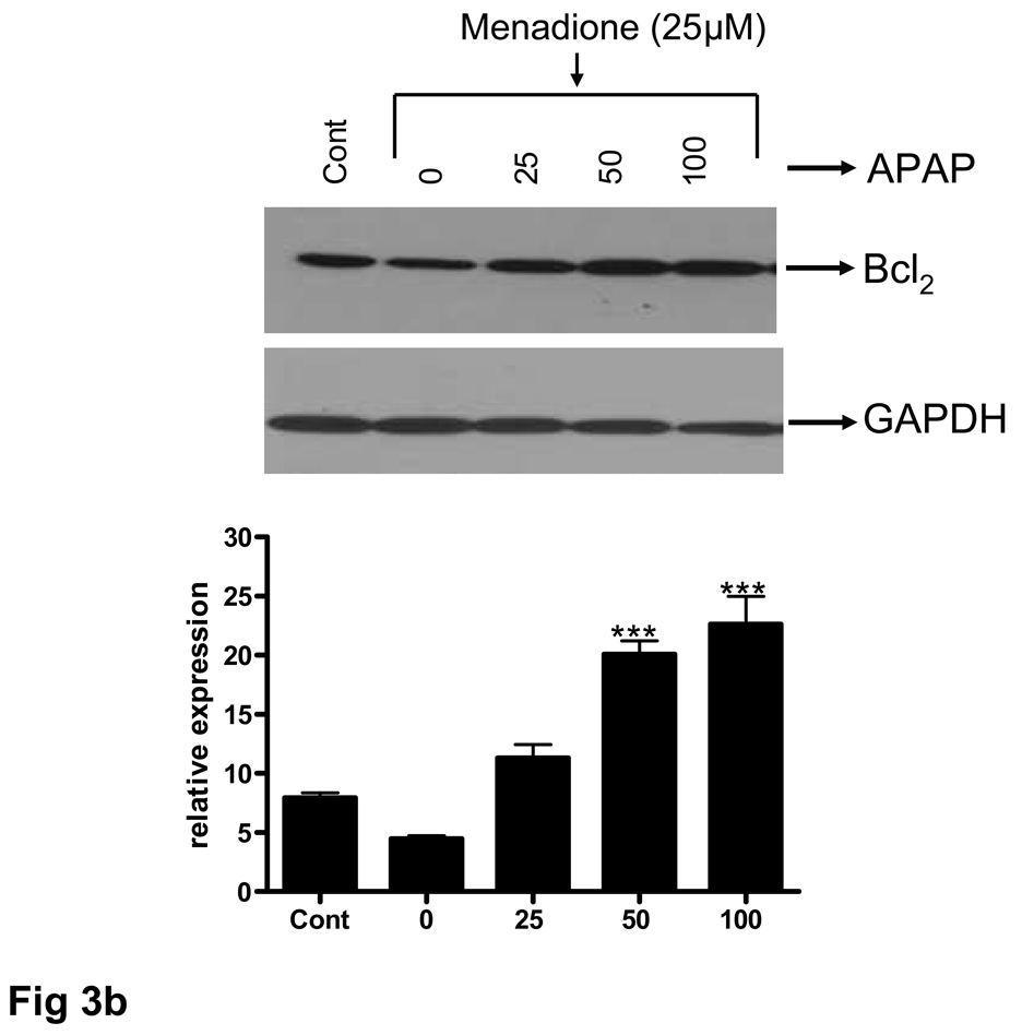
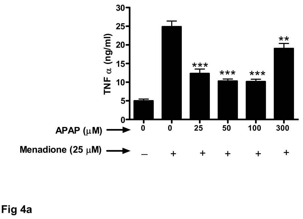
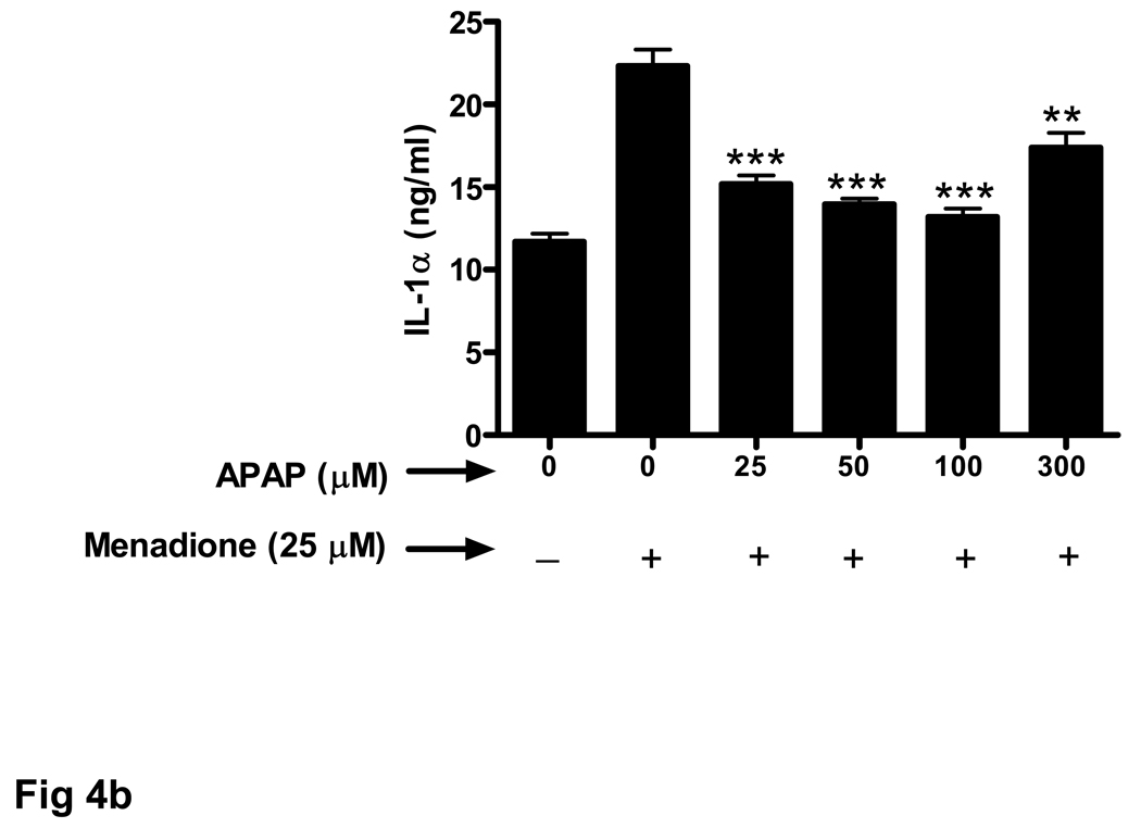
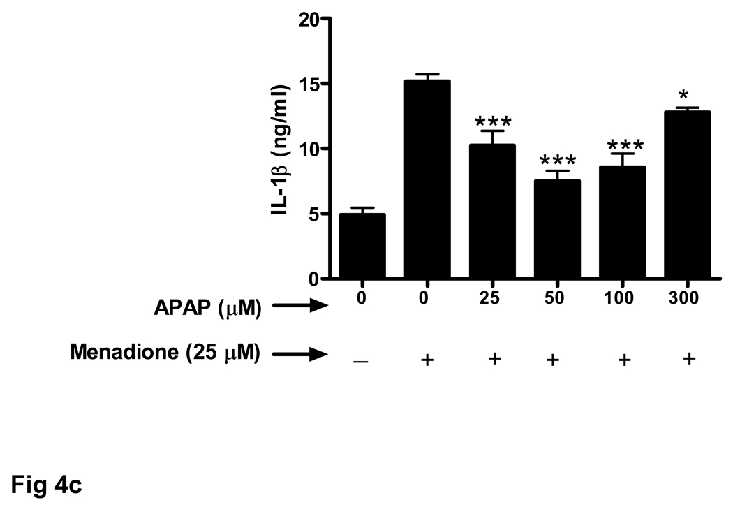
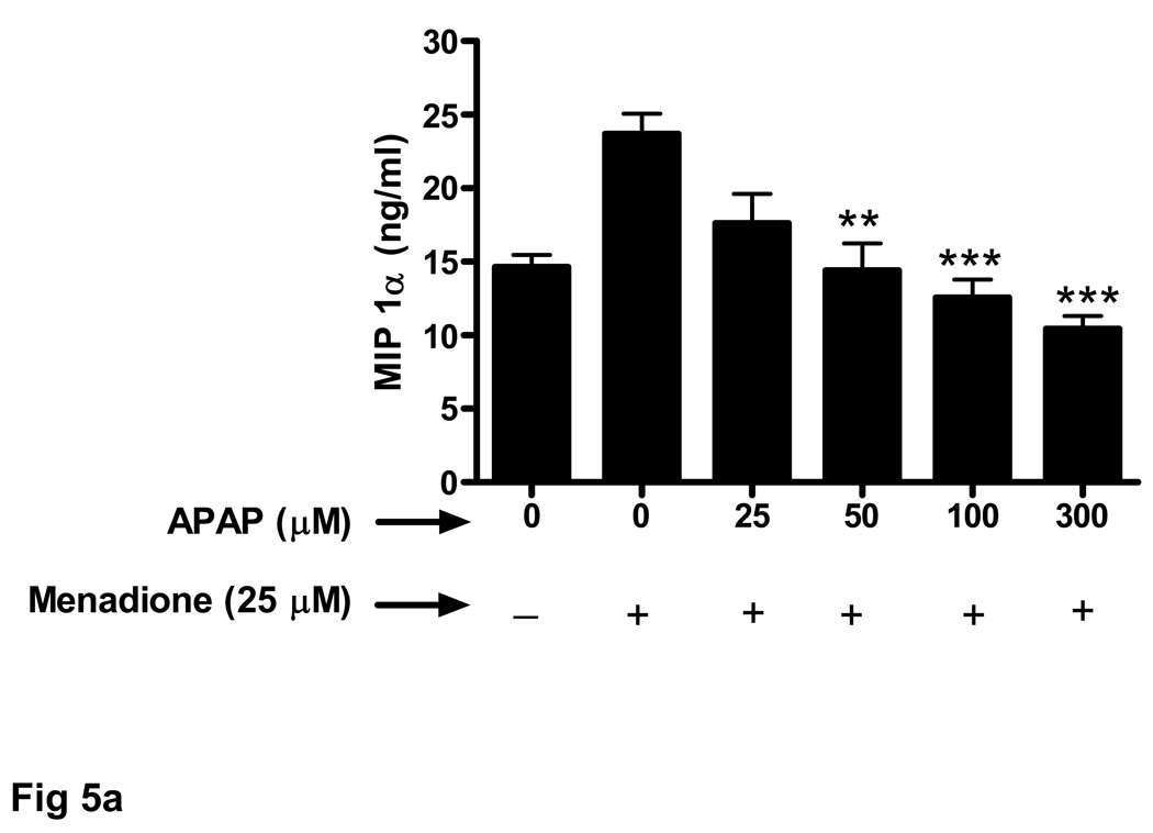
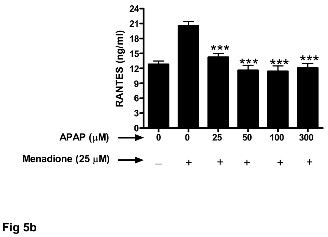
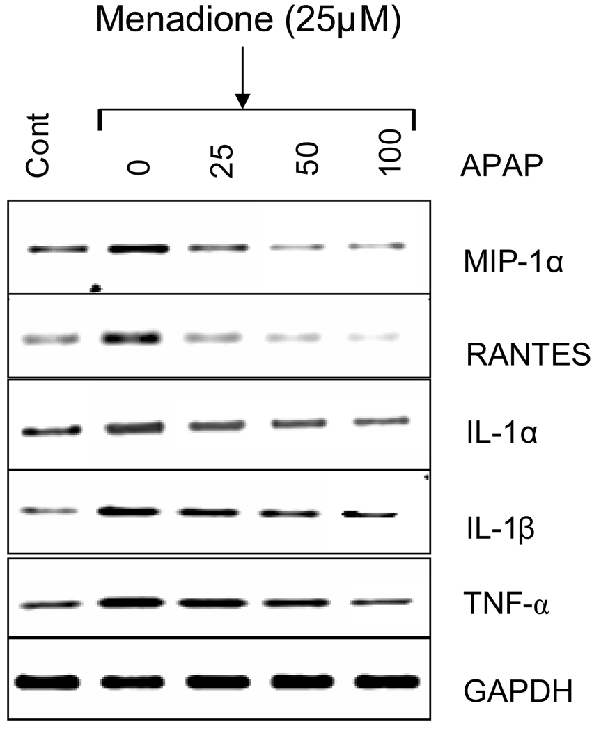
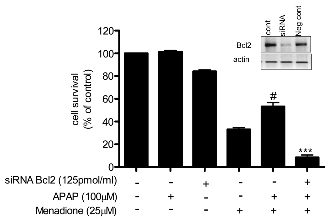
Increasing evidence suggests that acetaminophen has unappreciated anti-oxidant and anti-inflammatory properties. Drugs that affect oxidant and inflammatory stress in the brain are of interest because both processes are thought to contribute to the pathogenesis of neurodegenerative disease. The objective of this study is to determine whether acetaminophen affects the response of brain endothelial cells to oxidative stress. Cultured brain endothelial cells are pre-treated with acetaminophen and then exposed to the superoxide-generating compound menadione (25 microM). Cell survival, inflammatory protein expression, and anti-oxidant enzyme activity are measured. Menadione causes a significant (p<0.001) increase in endothelial cell death as well as an increase in RNA and protein levels of tumor necrosis factor alpha, interleukin-1, macrophage inflammatory protein alpha, and RANTES. Menadione also evokes a significant (p<0.001) increase in the activity of the anti-oxidant enzyme superoxide dismutase (SOD). Pre-treatment of endothelial cell cultures with acetaminophen (25-100 microM) increases endothelial cell survival and inhibits menadione-induced expression of inflammatory proteins and SOD activity. In addition, we document, for the first time, that acetaminophen increases expression of the anti-apoptotic protein Bcl2. Suppressing Bcl2 with siRNA blocks the pro-survival effect of acetaminophen. These data show that acetaminophen has anti-oxidant and anti-inflammatory effects on the cerebrovasculature and suggest a heretofore unappreciated therapeutic potential for this drug in neurodegenerative diseases such as Alzheimer's disease that are characterized by oxidant and inflammatory stress.
Figures
 ) with acetaminophen (APAP) (0–300 µM) and then exposed to menadione or co-incubated (
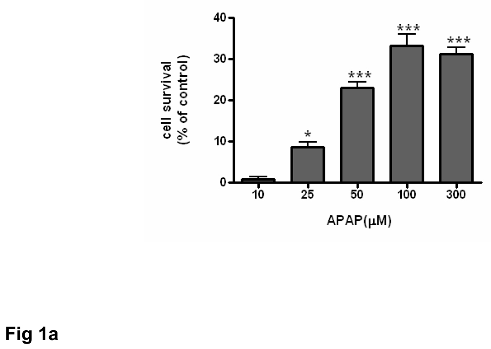
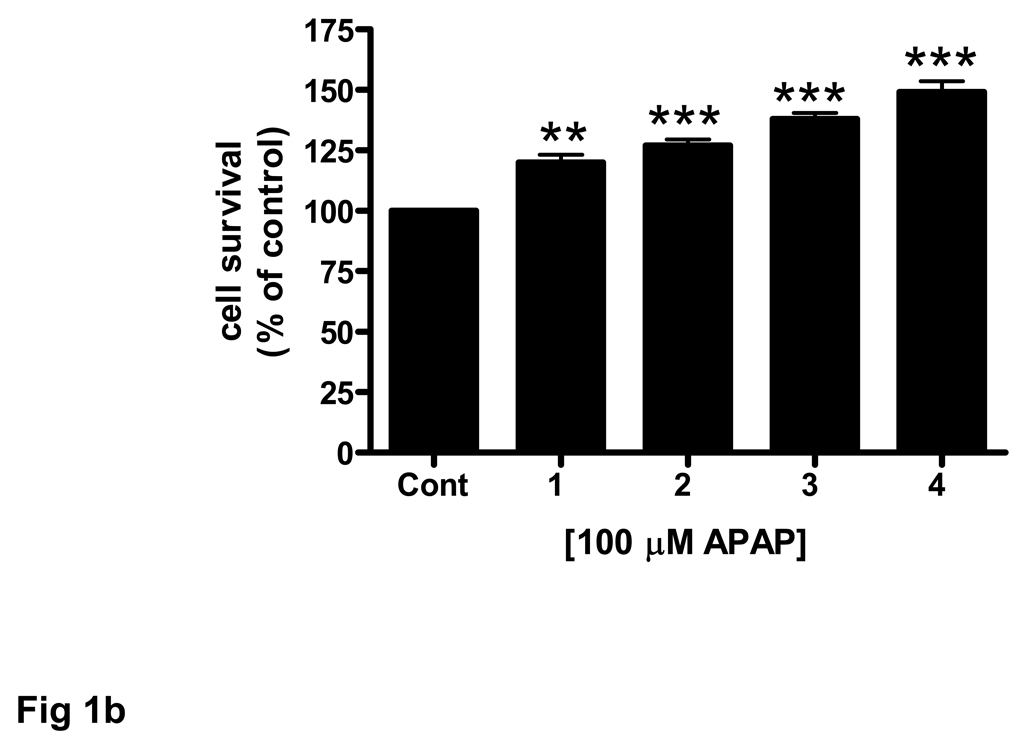
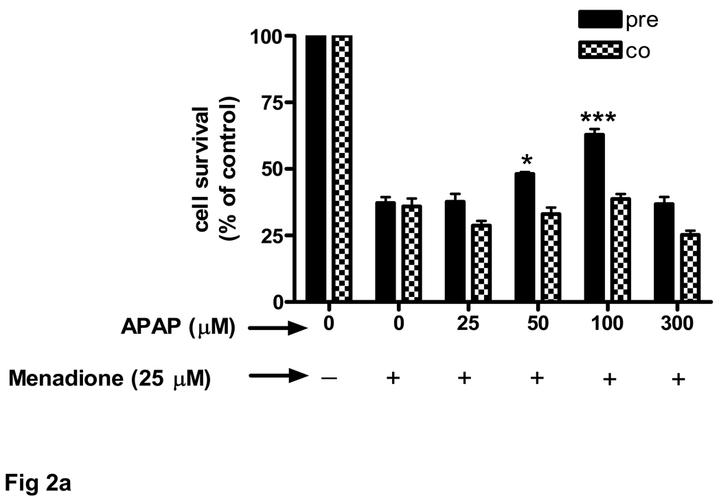
 ) with APAP and menadione. *p<0.05 vs. menadione alone; ***p<0.001 vs. menadione alone. (b) Brain endothelial cells received 1 to 4 pretreatments of 100 µM acetaminophen (APAP) prior to 3 h exposure to menadione (25 µM). Each treatment period was 8 h. *p<0.05 vs. menadione alone; **p<0.01 vs. menadione alone; ***p<0.001 vs. menadione alone. (c) Rat brain endothelial cells were exposed to 4 pretreatments (32 h) of varying doses (10–100 µM) of acetaminophen (APAP) and then incubated with 25 µM of menadione for 3 h. ***p<0.001 vs. menadione alone. (d) Rat brain endothelial cells were exposed to 4 pretreatments (32 h) of varying doses (10–100 µM) of acetaminophen and then incubated with 1 mM H2O2 for 3 h. ***p<0.001 vs. H2O2 alone. Data are mean ± SD from 3 separate experiments.
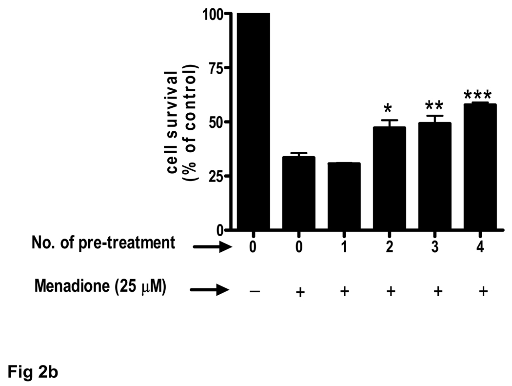 ) with acetaminophen (APAP) (0–300 µM) and then exposed to menadione or co-incubated () with APAP and menadione. *p<0.05 vs. menadione alone; ***p<0.001 vs. menadione alone. (b) Brain endothelial cells received 1 to 4 pretreatments of 100 µM acetaminophen (APAP) prior to 3 h exposure to menadione (25 µM). Each treatment period was 8 h. *p<0.05 vs. menadione alone; **p<0.01 vs. menadione alone; ***p<0.001 vs. menadione alone. (c) Rat brain endothelial cells were exposed to 4 pretreatments (32 h) of varying doses (10–100 µM) of acetaminophen (APAP) and then incubated with 25 µM of menadione for 3 h. ***p<0.001 vs. menadione alone. (d) Rat brain endothelial cells were exposed to 4 pretreatments (32 h) of varying doses (10–100 µM) of acetaminophen and then incubated with 1 mM H2O2 for 3 h. ***p<0.001 vs. H2O2 alone. Data are mean ± SD from 3 separate experiments.
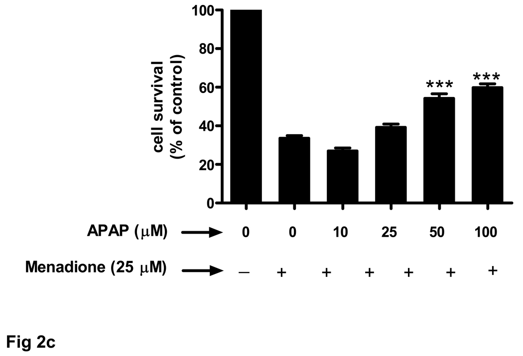 ) with acetaminophen (APAP) (0–300 µM) and then exposed to menadione or co-incubated () with APAP and menadione. *p<0.05 vs. menadione alone; ***p<0.001 vs. menadione alone. (b) Brain endothelial cells received 1 to 4 pretreatments of 100 µM acetaminophen (APAP) prior to 3 h exposure to menadione (25 µM). Each treatment period was 8 h. *p<0.05 vs. menadione alone; **p<0.01 vs. menadione alone; ***p<0.001 vs. menadione alone. (c) Rat brain endothelial cells were exposed to 4 pretreatments (32 h) of varying doses (10–100 µM) of acetaminophen (APAP) and then incubated with 25 µM of menadione for 3 h. ***p<0.001 vs. menadione alone. (d) Rat brain endothelial cells were exposed to 4 pretreatments (32 h) of varying doses (10–100 µM) of acetaminophen and then incubated with 1 mM H2O2 for 3 h. ***p<0.001 vs. H2O2 alone. Data are mean ± SD from 3 separate experiments.
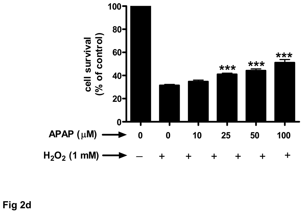 ) with acetaminophen (APAP) (0–300 µM) and then exposed to menadione or co-incubated () with APAP and menadione. *p<0.05 vs. menadione alone; ***p<0.001 vs. menadione alone. (b) Brain endothelial cells received 1 to 4 pretreatments of 100 µM acetaminophen (APAP) prior to 3 h exposure to menadione (25 µM). Each treatment period was 8 h. *p<0.05 vs. menadione alone; **p<0.01 vs. menadione alone; ***p<0.001 vs. menadione alone. (c) Rat brain endothelial cells were exposed to 4 pretreatments (32 h) of varying doses (10–100 µM) of acetaminophen (APAP) and then incubated with 25 µM of menadione for 3 h. ***p<0.001 vs. menadione alone. (d) Rat brain endothelial cells were exposed to 4 pretreatments (32 h) of varying doses (10–100 µM) of acetaminophen and then incubated with 1 mM H2O2 for 3 h. ***p<0.001 vs. H2O2 alone. Data are mean ± SD from 3 separate experiments.
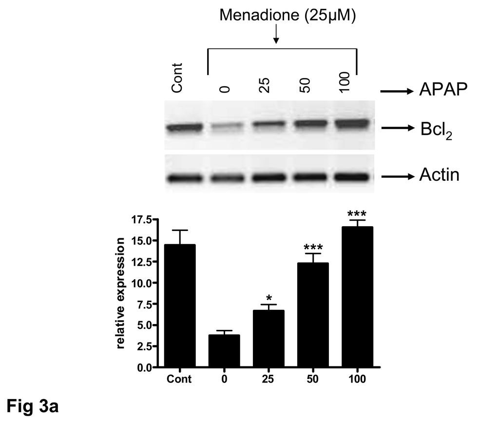
Similar articles
-
Acetaminophen inhibits neuronal inflammation and protects neurons from oxidative stress.J Neuroinflammation. 2009 Mar 16;6:10. doi: 10.1186/1742-2094-6-10. J Neuroinflammation. 2009. PMID: 19291322 Free PMC article.
-
Distinct roles of NF-kappaB p50 in the regulation of acetaminophen-induced inflammatory mediator production and hepatotoxicity.Toxicol Appl Pharmacol. 2006 Mar 1;211(2):157-65. doi: 10.1016/j.taap.2005.06.024. Epub 2005 Aug 2. Toxicol Appl Pharmacol. 2006. PMID: 16081117
-
Grape seed extract ameliorates tumor necrosis factor-α-induced inflammatory status of human umbilical vein endothelial cells.Eur J Nutr. 2011 Sep;50(6):401-9. doi: 10.1007/s00394-010-0151-6. Epub 2010 Nov 28. Eur J Nutr. 2011. PMID: 21113812
-
Age-related decrease in cerebrovascular-derived neuroprotective proteins: effect of acetaminophen.Microvasc Res. 2012 Nov;84(3):278-85. doi: 10.1016/j.mvr.2012.08.004. Epub 2012 Aug 27. Microvasc Res. 2012. PMID: 22944728 Free PMC article.
-
Curcumin, an antioxidant and anti-inflammatory agent, induces heme oxygenase-1 and protects endothelial cells against oxidative stress.Free Radic Biol Med. 2000 Apr 15;28(8):1303-12. doi: 10.1016/s0891-5849(00)00294-x. Free Radic Biol Med. 2000. PMID: 10889462
Cited by
-
Upregulation of Pro-inflammatory Cytokine Expression Following Chronic Paracetamol Treatment in Astrocyte.Neurotox Res. 2018 Jul;34(1):137-146. doi: 10.1007/s12640-018-9875-5. Epub 2018 Feb 14. Neurotox Res. 2018. PMID: 29446054
-
Acetaminophen attenuates lipopolysaccharide-induced cognitive impairment through antioxidant activity.J Neuroinflammation. 2017 Jan 21;14(1):17. doi: 10.1186/s12974-016-0781-6. J Neuroinflammation. 2017. PMID: 28109286 Free PMC article.
-
NQO2 is a reactive oxygen species generating off-target for acetaminophen.Mol Pharm. 2014 Dec 1;11(12):4395-404. doi: 10.1021/mp5004866. Epub 2014 Oct 24. Mol Pharm. 2014. PMID: 25313982 Free PMC article.
-
Acetaminophen inhibits neuronal inflammation and protects neurons from oxidative stress.J Neuroinflammation. 2009 Mar 16;6:10. doi: 10.1186/1742-2094-6-10. J Neuroinflammation. 2009. PMID: 19291322 Free PMC article.
-
P2X7 Cell Death Receptor Activation and Mitochondrial Impairment in Oxaliplatin-Induced Apoptosis and Neuronal Injury: Cellular Mechanisms and In Vivo Approach.PLoS One. 2013 Jun 27;8(6):e66830. doi: 10.1371/journal.pone.0066830. Print 2013. PLoS One. 2013. PMID: 23826152 Free PMC article.
References
-
- Bae MA, Pie JE, Song BJ. Acetaminophen induces apoptosis of C6 glioma cells by activating the c-Jun NH(2)-terminal protein kinase-related cell death pathway. Mol. Pharmacol. 2001;60:847–856. - PubMed
-
- Bisaglia M, Venezia V, Piccioli P, Stanzione S, Porcile C, Russo C, Mancini F, Milanese C, Schettini G. Acetaminophen protects hippocampal neurons and PC12 cultures from amyloid beta-peptides induced oxidative stress and reduces NF-kappaB activation. Neurochem. Int. 2002;41:43–54. - PubMed
-
- Bourdi M, Eiras DP, Holt MP, Webster MR, Reilly TP, Welch KD, Pohl LR. Role of IL-6 in an IL-10 and IL-4 double knockout mouse model uniquely susceptible to acetaminophen-induced liver injury. Chem. Res. Toxicol. 2007;20:208–216. - PubMed
-
- Dambach DM, Durham SK, Laskin JD, Laskin DL. Distinct roles of NF-kappaB p50 in the regulation of acetaminophen-induced inflammatory mediator production and hepatoxicity. Toxicol. Appl. Pharmacol. 2006;211:157–165. - PubMed
Publication types
MeSH terms
Substances
Grants and funding
LinkOut - more resources
Full Text Sources
Medical
