Review
. 2009 Jul;16(7):947-55.
doi: 10.1038/cdd.2009.19. Epub 2009 Mar 6.

Bif-1/endophilin B1: a candidate for crescent driving force in autophagy

Affiliations
Review

Bif-1/endophilin B1: a candidate for crescent driving force in autophagy

Y Takahashi et al. Cell Death Differ. 2009 Jul.

Abstract

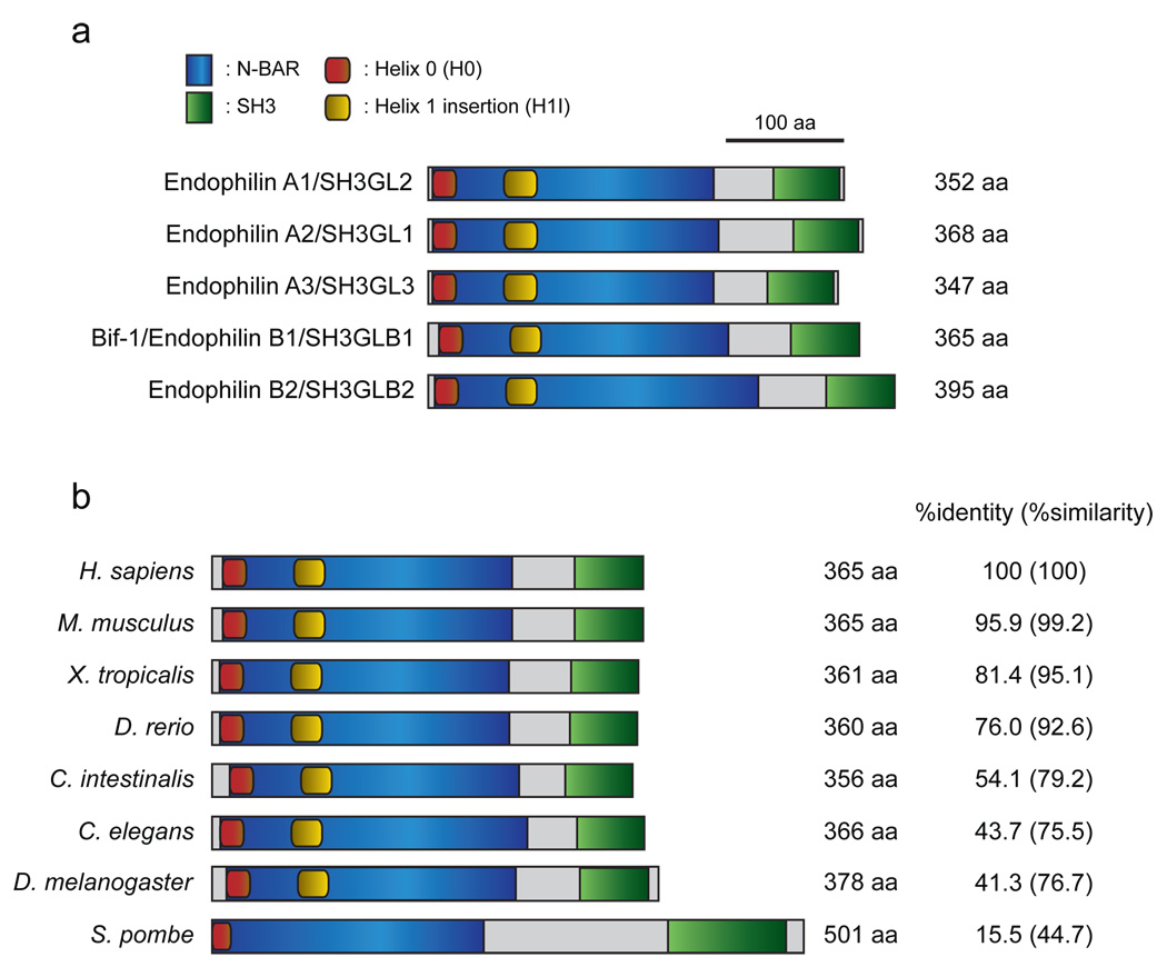
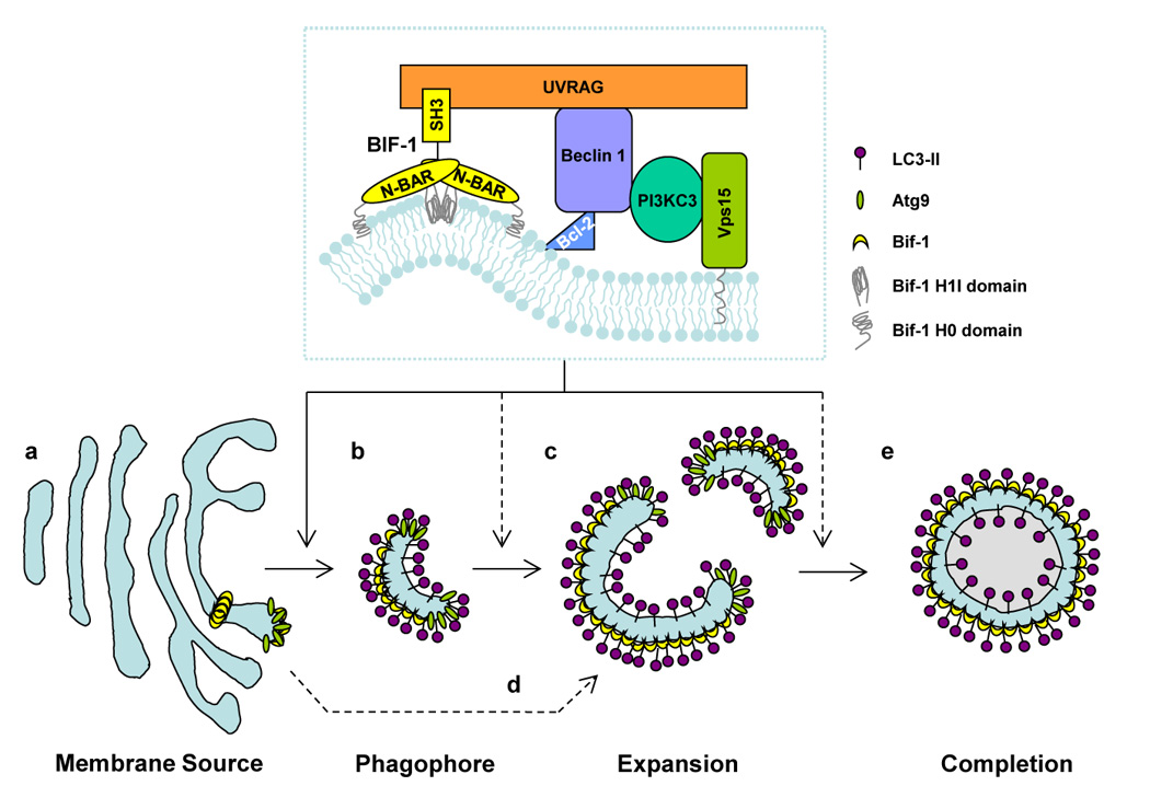
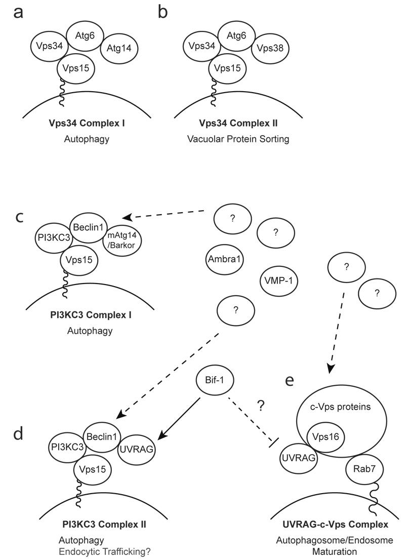
Autophagy is an intracellular bulk degradation system that plays a vital role in maintaining cellular homeostasis. This degradation process involves dynamic membrane rearrangements resulting in the formation of double-membraned autophagosomes. However, the driving force for generating curvature and deformation of isolation membranes remains a mystery. Bax-interacting factor 1 (Bif-1), also known as SH3GLB1 or Endophilin B1, was originally discovered as a Bax-binding protein. Bif-1 contains an amino-terminal N-BAR (Bin-Amphiphysin-Rvs) domain and a carboxy-terminal SH3 (Src-homology 3) domain and shows membrane binding and bending activities. It has been shown that Beclin1 is involved in the nucleation of autophagosomal membranes through an unknown mechanism. It is interesting that, Bif-1 forms a complex with Beclin1 through ultraviolet irradiation resistant-associated gene (UVRAG) and promotes the activation of the class III PI3 kinase, Vps34, in mammalian cells. In response to nutrient starvation, Bif-1 accumulates in punctate foci where it co-localizes with LC3, Atg5, and Atg9. Furthermore, Bif-1-positive, crescent-shaped small vesicles expand by recruiting and fusing with Atg9-positive small membranes to complete autophagosome formation. This review highlights the role of Bif-1 in the regulation of autophagy and discusses the potential involvement of Bif-1 in the biogenesis of membranes for the formation of autophagosomes.


Figures

Figure 1
Figure 1. The domain structure of the endophilin protein family
(a) Schematic representation of human Endophilin A and B proteins. All endophilins are composed of an N-terminal N-BAR domain and a C-terminal SH3 domain. The helix 0 (H0) and helix 1 insertion (H1I) regions located in the N-BAR domain are well-conserved between the Endophilin A and B families. (b) Structure-based alignment of Bif-1/Endophilin B1/SH3GLB1 among species. The N-BAR and SH3 domains, as well as the H0 region are evolutionally conserved. Notably, protein structure prediction analysis revealed that the H1I region is missing in the S. pombe yeast homologue of Bif-1; however, this region is conserved among all multicellular organisms that we investigated. The GenBank accession numbers of endophilins shown in (a) and (b) are: human Endophilin A1/SH3GL2, NP_003017; human Endophilin A2/SH3GL1, NP_003016; human Endophilin A3/SH3GL3, Q99963; human Bif-1/Endophilin B1/SH3GLB1, NP_057093; human Endophilin B2/SH3GLB2, NP_064530; mouse Bif-1, NP_062337; Xenopus Bif-1, NP_001016072; zebrafish Bif-1, NP_001017617; Ciona Bif-1, XP_002125621; Drosophila Bif-1, NP_725873; C. elegans Bif-1, NP_741756; S. pombe Bif-1, NP_595695.
Figure 2
Figure 2. Schematic model of the role of Bif-1 in autophagy
Bif-1 collaborates with PI3KC3 in regulating the biogenesis, expansion and completion of autophagosomes. In response to autophagic signals, Bif-1 tubulates the TGN to generate Atg9-Bif-1-containing vesicles (a). The resultant vesicles serve as the source of phagophores/isolation membranes (b), which are then nucleated and expanded by fusion with other Atg9-Bif-1-containing vesicles (c). Alternatively, Atg9-Bif-1-positive vesicles are delivered to phagophores/isolation membranes, which are de novo synthesized or supplied from unknown donors, as source membranes for the expansion and completion of autophagosomes (d). The membrane is eventually sealed to become a double-membraned autophagosome (e). Atg9 is then released from the vacuole, while Bif-1 remains attached to the autophagosome.
Figure 3
Figure 3. Schematic model of Bif-1 regulation of PI3KC3 and UVRAG complexes
(a, b) The yeast Vps34 complexes. The yeast Vps34 complex I plays an essential role in autophagy at the vesicle nucleation step by recruiting ubiquitin-like Atg proteins to PAS (a), whereas the Vps34 complex II is required for the vacuolar protein sorting system (b). (c–e) The mammalian PI3KC3 complexes and UVRAG complexes. In mammalian systems, the counterparts of yeast Vps34 complex I and II have been identified as the PI3KC3 complex I (c) and II (d), respectively. However, evidence suggests that both PI3KC3 complex I and II are important for autophagy in mammalian systems. (d) Bif-1 has been shown to interact with the complex II and promote the activation of PI3KC3 and the formation of autophagosomes. Moreover, Bif-1 together with the PI3KC3 complex II might be involved in Atg9-containing vesicle formation/trafficking. (e) Bif-1 may regulate UVRAG binding to the class C Vps complex for autophagosome/endosome maturation.

References

    1. Yoshimori T. Autophagy: a regulated bulk degradation process inside cells. Biochem Biophys Res Commun. 2004 Jan 9;313(2):453–458. - PubMed
    1. Levine B, Klionsky DJ. Development by self-digestion: molecular mechanisms and biological functions of autophagy. Dev Cell. 2004 Apr;6(4):463–477. - PubMed
    1. Karantza-Wadsworth V, Patel S, Kravchuk O, Chen G, Mathew R, Jin S, et al. Autophagy mitigates metabolic stress and genome damage in mammary tumorigenesis. Genes Dev. 2007 Jul 1;21(13):1621–1635. - PMC - PubMed
    1. Mathew R, Kongara S, Beaudoin B, Karp CM, Bray K, Degenhardt K, et al. Autophagy suppresses tumor progression by limiting chromosomal instability. Genes Dev. 2007 Jun 1;21(11):1367–1381. - PMC - PubMed
    1. Mizushima N, Levine B, Cuervo AM, Klionsky DJ. Autophagy fights disease through cellular self-digestion. Nature. 2008 Feb 28;451(7182):1069–1075. - PMC - PubMed

Publication types