. 2009 Mar;5(3):e1000327.
doi: 10.1371/journal.ppat.1000327. Epub 2009 Mar 6.

The SET complex acts as a barrier to autointegration of HIV-1

Affiliations

The SET complex acts as a barrier to autointegration of HIV-1

Nan Yan et al. PLoS Pathog. 2009 Mar.

Abstract

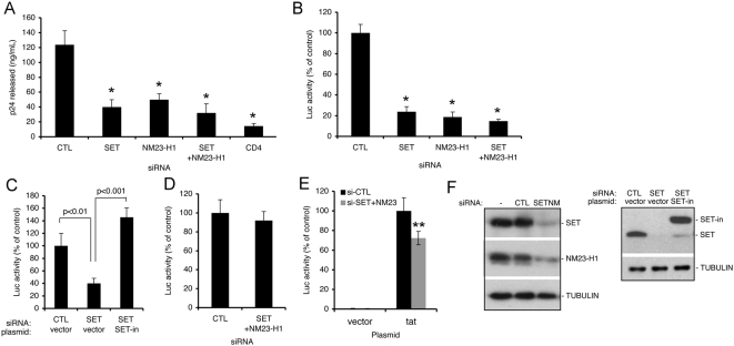
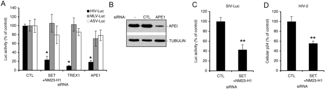
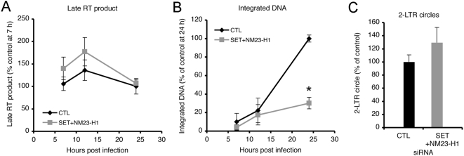
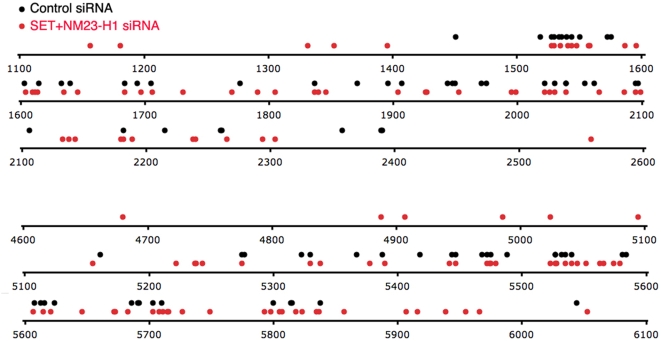
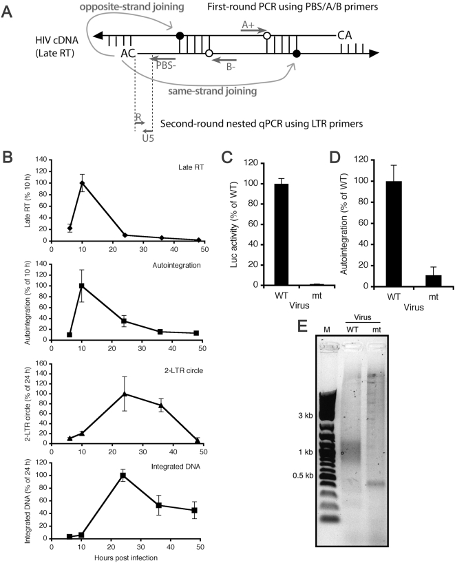
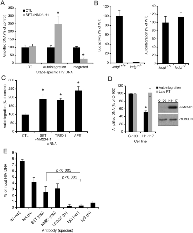
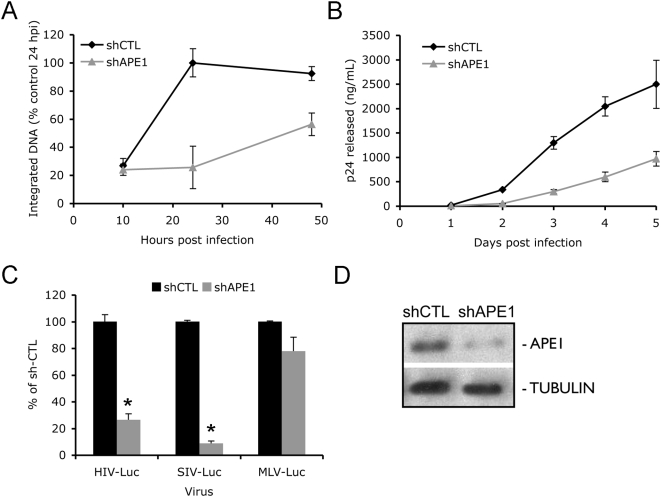
Retroviruses and retrotransposons are vulnerable to a suicidal pathway known as autointegration, which occurs when the 3'-ends of the reverse transcript are activated by integrase and then attack sites within the viral DNA. Retroelements have diverse strategies for suppressing autointegration, but how HIV-1 protects itself from autointegration is not well-understood. Here we show that knocking down any of the components of the SET complex, an endoplasmic reticulum-associated complex that contains 3 DNases (the base excision repair endonuclease APE1, 5'-3' exonuclease TREX1, and endonuclease NM23-H1), inhibits HIV-1 and HIV-2/SIV, but not MLV or ASV, infection. Inhibition occurs at a step in the viral life cycle after reverse transcription but before chromosomal integration. Antibodies to SET complex proteins capture HIV-1 DNA in the cytoplasm, suggesting a direct interaction between the SET complex and the HIV preintegration complex. Cloning of HIV integration sites in cells with knocked down SET complex components revealed an increase in autointegration, which was verified using a novel semi-quantitative nested PCR assay to detect autointegrants. When SET complex proteins are knocked down, autointegration increases 2-3-fold and chromosomal integration correspondingly decreases approximately 3-fold. Therefore, the SET complex facilitates HIV-1 infection by preventing suicidal autointegration.


Conflict of interest statement

The authors have declared that no competing interests exist.

Figures

Figure 1
Figure 1. The SET complex facilitates HIV-1 infection.
(A) SET and/or NM23-H1 knockdown inhibits HIV-1IIIB infection. HeLaCD4 cells transfected with a non-targeting control siRNA (CTL) or siRNAs targeting SET and/or NM23-H1 were infected with HIV-1IIIB, and infection was assayed by p24 release. (B) SET and/or NM23-H1 knockdown blocks single-round HIV-Luc infection. HeLaCD4 cells were transfected with indicated siRNAs and infected with VSV-G pseudotyped HIV-Luc. Luciferase (Luc) activity, measured 48 hpi and normalized to total cellular protein, is compared to Luc activity in cells transfected with control siRNA. (C) Expression of siRNA-insensitive SET (SET-in) rescues the HIV-Luc infection block caused by knocking down endogenous SET. 293T cells, transfected with control or SET siRNAs and then transfected two days later with empty vector or SET-in and pCMV-β-gal plasmids, were infected with HIV-Luc 24 h after transfection, and Luc activity measured 48 h later was normalized to β-galactosidase activity. (D) Expression from transfected HIV-Luc DNA is not affected by SET/NM23-H1 knockdown. HeLaCD4 cells were transfected with HIV-Luc plasmid two days following siRNA transfection. Luc activity was measured 24 h later and normalized as in (D). (E) Expression from a chromatinized HIV-Luc reporter gene is weakly affected by SET/NM23-H1 knockdown. TZM-bl cells that harbor an integrated LTR-Luc reporter gene were first transfected with control or SET/NM23-H1 siRNA, and then transfected 48 h later with either an empty vector or a Tat expression plasmid to activate reporter gene expression. Luc activity was measured 24 h later. (F) Immunoblots showing SET/NM23-H1 knockdown and SET-in over-expression. SET-in is an siRNA-insensitive FLAG-HA-tagged protein. *, p<0.001 relative to control knockdown in (A) and (B). **, p<0.05 relative to control in (E). Mean and standard deviation (S.D.) from at least four independent infections are shown in (A–E).
Figure 2
Figure 2. Knockdown of SET complex proteins inhibits HIV-1 and HIV-2/SIV, but not MLV or ASV infection.
(A) Knockdown of SET/NM23-H1, APE1, or TREX1 inhibits HIV-Luc, but not MLV or ASV single-round reporter viruses. Luc activity was measured 48 hpi. (B) Immunoblot confirms APE1 knockdown. TREX1 knockdown, assayed by qRT-PCR, reduced TREX1 mRNA by 94% (not shown). (C,D) Knockdown of SET/NM23-H1 also inhibits SIV-Luc (C) and HIV-2 (D) infection. *, p<0.01. **, p<0.05.
Figure 3
Figure 3. HIV-1 integration is reduced in SET/NM23-H1 knockdown cells.
(A–C) Late RT products (normalized to mitochondrial DNA) (A) and integrated DNA (normalized to β-globin sequences) (B), measured at indicated times, and 2-LTR circles, measured 24 hpi (C), from control (black) or SET/NM23-H1 (gray) knockdown cells. Data are mean+/−S.D. of triplicate measurements from three independent experiments, normalized to control knockdown cells. *, p<0.001.
Figure 4
Figure 4. Autointegration sites recovered from control and SET/NM23-H1 knockdown libraries.
Each site within selected 1.5-kb regions of the HIV-1 genome (numbering based on the HIV-1NL4-3 strain) is represented as a dot. Autointegrants from control siRNA-treated and SET/NM23-H1 knockdown cells are black and red, respectively.
Figure 5
Figure 5. A PCR-based assay for measuring HIV autointegration (auto-PCR).
(A) Diagram of HIV-1 auto-PCR assay (a more detailed diagram is provided in Figure S4). The common primer binding site (PBS−) (reverse) primer is used to amplify same strand integrants with the A+ (forward) primer or opposite strand integrants with the B− (reverse) primer during first-round PCR. A single-length nested PCR product is then amplified using an LTR primer pair (R-U5) during second-round qPCR. Filled circles, internal viral 5′ phosphates attacked during autointegration. (B) Kinetics of stage-specific HIV-1 DNA product formation. Late RT, autointegrants, and 2-LTR circles were normalized to mitrochondrial DNA; integrated DNA was normalized to β-globin. Values of late RT and autointegration are shown relative to peak values 10 hpi, while 2-LTR and integrated DNA are normalized to peak 24 hpi values. Mean and S.D. from triplicate qPCR measurements are shown. (C–E) Active IN is required for autointegration. HeLaCD4 cells were infected with HIV-Luc carrying wild type (WT) or mutant (mt) IN. Luc activity was measured 48 hpi (C) and auto-PCR was performed 10 hpi (D). First-round PCR products (E) from Hirt supernatant DNA were analyzed by agarose gel electrophoresis.
Figure 6
Figure 6. The SET complex suppresses HIV-1 autointegration.
(A) Stage specific HIV-1 DNAs from control and SET/NM23-H1 knockdown cells infected with HIV-Luc. Late RT (LRT) and autointegration were measured 10 hpi and chromosomal integration was assayed 24 hpi. Mean and S.D. from triplicate qPCR assays of three independent experiments are shown. *, p<0.01. (B) Autointegration is not an obligate by-product of failed integration. Autointegration was measured 10 hpi and Luc activity at 48 hpi. The difference in autointegration is not significant. (C) Knocking down other SET complex proteins, TREX1 or APE1, also increases autointegration. Mean plus S.D. from two independent experiments are shown. *, p<0.01. (D) NM23-H1 over-expression suppresses autointegration. MDA-MB-435, an NM23-H1–deficient metastatic breast cancer cell line, stably transfected with vector (C-100) or an NM23-H1 expression plasmid (H1-117), were infected with VSV-G pseudotyped HIV-Luc, and LRT and autointegrants were measured 10 hpi. *, p<0.01. (E) SET and NM23-H1 associate with HIV-1 cDNA. Cytoplasmic extracts from infected HeLaCD4 cells were immunoprecipitated with the indicated antibodies (mouse (m) or rabbit (rab)), and associated HIV-1 cDNA was quantified by qPCR. Mean plus S.D. from three independent experiments are shown.
Figure 7
Figure 7. APE1 enhances HIV infection in Jurkat T cells.
Jurkat cells expressing sh-CTL or sh-APE1 were infected with HIVIIIB, and integrated DNA (A) and p24 release into the medium (B) were measured at indicated times. Mean plus S.D. from two independent experiments are shown. (C) APE1 knockdown in Jurkat cells inhibits HIV-Luc and SIV-Luc, but not MLV-Luc, infectivity. (D) Immunoblot showing APE1 knockdown in sh-APE1 lentivirus-infected Jurkat cells.

References

    1. Goff SP. Host factors exploited by retroviruses. Nat Rev Microbiol. 2007;5:253–263. - PubMed
    1. Suzuki Y, Craigie R. The road to chromatin–nuclear entry of retroviruses. Nat Rev Microbiol. 2007;5:187–196. - PubMed
    1. Yoder K, Sarasin A, Kraemer K, McIlhatton M, Bushman FD, et al. The DNA repair genes XPB and XPD defend cells from retroviral infection. Proc Natl Acad Sci U S A. 2006;103:4622–4627. - PMC - PubMed
    1. Medzhitov R. Recognition of microorganisms and activation of the immune response. Nature. 2007;449:819–826. - PubMed
    1. Takaoka A, Wang Z, Choi MK, Yanai H, Negishi H, et al. DAI (DLM-1/ZBP1) is a cytosolic DNA sensor and an activator of innate immune response. Nature. 2007;448:501–505. - PubMed

Publication types

MeSH terms