. 2009;26(5):415-24.
doi: 10.1007/s10585-009-9239-x. Epub 2009 Mar 8.

Dietary stearate reduces human breast cancer metastasis burden in athymic nude mice

Affiliations

Dietary stearate reduces human breast cancer metastasis burden in athymic nude mice

Lynda M Evans et al. Clin Exp Metastasis. 2009.

Abstract

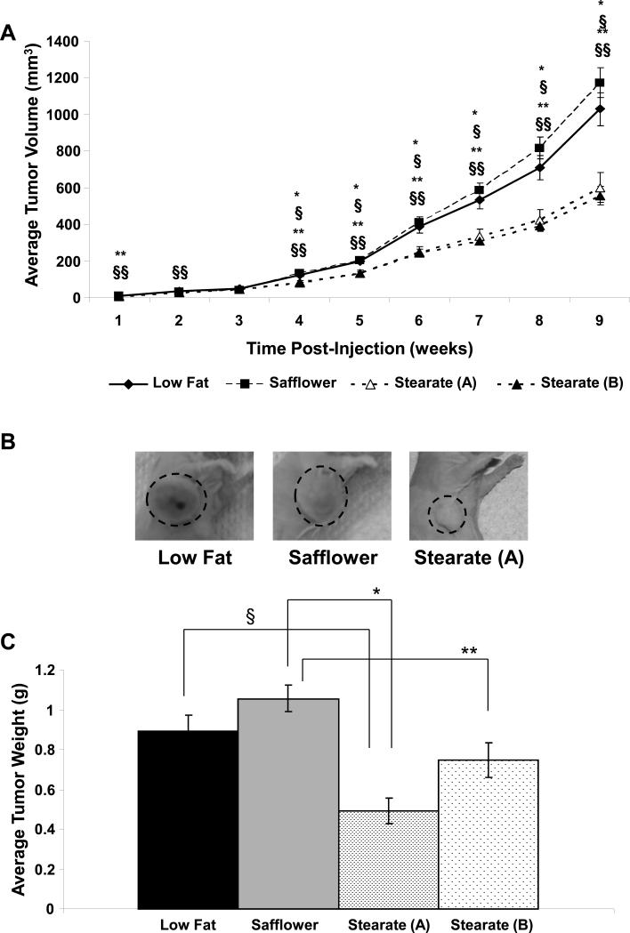
Stearate is an 18-carbon saturated fatty acid found in many foods in the western diet, including beef and chocolate. Stearate has been shown to have anti-cancer properties during early stages of neoplastic progression. However, previous studies have not investigated the effect of dietary stearate on breast cancer metastasis. In this study, we present evidence that exogenously supplied dietary stearate dramatically reduces the size of tumors that formed from injected human breast cancer cells within the mammary fat pads of athymic nude mice by approximately 50% and partially inhibits breast cancer cell metastasis burden in the lungs in this mouse model system. This metastatic inhibition appears to be independent of primary tumor size, as stearate fed animals that had primary tumors comparable in size to littermates fed either a safflower oil enriched diet or a low fat diet had reduced lung metastasis. Also stearate fed mice sub-groups had different primary tumor sizes but no difference in metastasis. This anti-metastasis effect may be due, at least in part, to the ability of stearate to induce apoptosis in these human breast cancer cells. Overall, this study suggests the possibility of dietary manipulation with selected long-chain saturated fatty acids such as stearate as a potential adjuvant therapeutic strategy for breast cancer patients wishing to maximize the suppression of metastatic disease.


Figures

Figure 1
Figure 1. Experimental Timetable
Nude mice were placed on either a low fat diet, a high linoleic diet (Safflower) or a high stearic acid diet (Stearate (A)) for 3 weeks prior to injection with cancer cells. Tumors were allowed to develop for 9 weeks and were removed. Four weeks post-tumor removal, animals were sacrificed and their lungs were analyzed for metastases. A fourth group of animals were placed on the high stearic acid diet (Stearate (B)) and were injected with cancer cells 3 weeks later. The tumors were allowed to grow to the size of the tumors in the low fat and safflower groups before removal. Due to variation within this stearate group, it was broken down into 3 smaller groups – B-i, B-ii, and B-iii – based on when the tumors were removed. After 4 weeks, animals were sacrificed and their lungs were analyzed for metastases.
Figure 2
Figure 2. Food Intake and Weight Gain
A) The average kcal consumed per animal per day was calculated. Of the three diets, the animals on the low fat diet ate the most, followed by the high stearate diet and then the safflower oil diet. Each group's intake was significantly different from the other two diets (§p value of stearate (A) vs. low fat <0.001; ¥p value of low fat vs. safflower <0.001; *p value of stearate (A) vs. safflower <0.001 by ANOVA). B) The weight of the animals was measured once a week for the duration of the experiment. Although differences were observed at individual weeks in the experiment, overall no significant change was seen. (n=14-21 per diet per week; §p value of stearate (A) vs. low fat <0.05; *p value of stearate vs. safflower <0.05 by ANOVA). The second stearate group was not different from the first and was therefore not represented on the graphs. C) The total weight gain of the animals at week 10 was determined. The weight does not include the weight of the tumors removed from the animals. No significant difference was observed between the three groups.
Figure 3
Figure 3. Tumor Weights and Volumes for the Low fat, Safflower, and Stearate (A) Groups
A) Mammary fat pad tumors of the animals were measured weekly after injection. Based on the estimated volumes, stearate began to decrease the average volume of the tumors approximately 4 weeks post-injection. The stearate (A) and stearate (B) primary tumors were not different. (n=20-21 animals per diet; §p value of stearate (A) vs. low fat <0.009; §§p value of stearate (B) vs. low fat<0.021; *p value of stearate (A) vs. safflower <0.003; **p value of stearate (B) vs. safflower<0.015 by ANOVA). When the data were analyzed using a repeated measures model and the curvature of the data points were estimated, all diets were different at week 9 except low fat vs. safflower (p<0.05). B) Immediately prior to removal of the mammary fat pad tumors at week 9, photographs were taken of mice in the low fat, safflower, and stearate (A) groups. All three of these tumors were removed on the same day. Representative images are shown. Animals on the stearate (A) diet tended to have noticeably smaller tumors compared to the other two diets. C) Following tumor removal, tumors were weighed and averages were calculated for each dietary group. Note: safflower oil, low fat and stearate A primary tumors were removed at 9 weeks post injection while the stearate B group tumors were removed and weighed at 11, 12 and 13 weeks post injection of cells. No difference was seen between the low fat and safflower oil treated animals, the low fat and stearate (B) animals, or the stearate (A) and stearate (B) animals. Differences were observed between all other diets (n=20-21; §p value of low fat vs. stearate (A) <0.002; *p value of stearate (A) vs. safflower <0.001; **p value of stearate (B) vs. safflower <0.003 by ANOVA).
Figure 4
Figure 4. Analysis of Lung Metastases from the Low fat, Safflower and Stearate (A) Groups
A) The number of macroscopic lung metastases was counted following necropsy. Stearate significantly decreased the number of macroscopic lung metastases in both the stearate (A) and stearate (B) groups. (n=18-20 animals per diet; §p value of stearate (A) vs. low fat <0.021; *p value of stearate (A) vs. safflower<0.024; §§p value of stearate (B) vs. low fat<0.022; **p value of stearate (B) vs. safflower<0.024 by Cochran-Armitage trend test). B) The animals on the low fat and safflower diets tended to have more metastatic tumors than those on the stearate diet. Interestingly, a large difference was not seen between the two groups of animals fed the stearate diet. C) When the tumor volume to metastases was calculated, no difference was observed.
Figure 5
Figure 5. Tumor Analysis of Animals on the Stearate (B) Diet
A) Tumor volumes were calculated weekly for each of the stearate (B) sub groups. Approximately week 8 post injection, 2c became statistically smaller than B-ii and around week 10 than 2b. At week 11 post injection, all groups were different. (n=7 animals per group; £p value of B-i vs. B-iii <0.001; &p value of B-ii vs. B-iii <0.002; #p value of B-i vs. 2bii <0.02). B) After the tumors were removed, they were weighed. No difference was observed between B-i and B-ii. B-iii tumors were statistically smaller than both B-i and B-ii. C) There was no difference in the metastatic tumor number per animal between the three stearate (B) subgroups.
Figure 6
Figure 6. Stearate Induced Apoptosis of MDA-MB-435 Breast Cancer Cells In Vitro
A) Cells were treated with 50 μM stearate for the times indicated and the presence of cleaved caspase-3 and cleaved PARP was determined by immunblot. As shown, stearate induces cleavage of the two proteins 12-24 hours post-treatment indicating stearate is inducing apoptosis of the breast cancer cells (n=3). B) Cells were treated with 1-100 μM stearate for 12 hours and caspase-3 activity was measured using a fluorescence based assay. Stearate activated caspase-3 in a dose dependent manner (n=4; *p<0.023 ANOVA).

References

    1. Welsch CW. Relationship between dietary fat and experimental mammary tumorigenesis: a review and critique. Cancer Res. 1992;52:2040s–2048s. - PubMed
    1. Lee MM, Lin SS. Dietary fat and breast cancer. Annu Rev Nutr. 2000;20:221–48. - PubMed
    1. Rose DP, Connolly JM, Liu XH. Effects of linoleic acid and gamma-linolenic acid on the growth and metastasis of a human breast cancer cell line in nude mice and on its growth and invasive capacity in vitro. Nutr Cancer. 1995;24:33–45. - PubMed
    1. Rose DP, Connolly JM, Liu XH. Effects of linoleic acid on the growth and metastasis of two human breast cancer cell lines in nude mice and the invasive capacity of these cell lines in vitro. Cancer Res. 1994;54:6557–62. - PubMed
    1. Rose DP, Hatala MA, Connolly JM, Rayburn J. Effect of diets containing different levels of linoleic acid on human breast cancer growth and lung metastasis in nude mice. Cancer Res. 1993;53:4686–90. - PubMed

Publication types