. 2009 Mar;27(3):670-681.
doi: 10.1002/stem.20080742.

Multipotent stromal cells are activated to reduce apoptosis in part by upregulation and secretion of stanniocalcin-1

Affiliations

Multipotent stromal cells are activated to reduce apoptosis in part by upregulation and secretion of stanniocalcin-1

Gregory J Block et al. Stem Cells. 2009 Mar.

Abstract

Multipotent stromal cells (MSCs) have been shown to reduce apoptosis in injured cells by secretion of paracrine factors, but these factors were not fully defined. We observed that coculture of MSCs with previously UV-irradiated fibroblasts reduced apoptosis of the irradiated cells, but fresh MSC conditioned medium was unable reproduce the effect. Comparative microarray analysis of MSCs grown in the presence or absence of UV-irradiated fibroblasts demonstrated that the MSCs were activated by the apoptotic cells to increase synthesis and secretion of stanniocalcin-1 (STC-1), a peptide hormone that modulates mineral metabolism and has pleiotrophic effects that have not been fully characterized. We showed that STC-1 was required but not sufficient for reduction of apoptosis of UV-irradiated fibroblasts. In contrast, we demonstrated that MSC-derived STC-1 was both required and sufficient for reduction of apoptosis of lung cancer epithelial cells made apoptotic by incubation at low pH in hypoxia. Our data demonstrate that STC-1 mediates the antiapoptotic effects of MSCs in two distinct models of apoptosis in vitro.


Figures
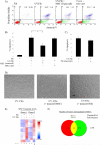
Figure 1
Figure 1
MSCs Reduce Apoptosis of UV Irradiated Fibroblasts. (A) Fibroblasts were incubated on a transwell filter, UV irradiated and then transferred for co-culture with MSCs in a 6-well dish. Forty-eight hrs later, viability and apoptosis were assayed after labeling for annexin-V staining and PI incorporation by flow cytometry. (B) Quantification of annexin-V/PI positive cells. * = p < 0.05. Error bars = SD. (C) Irradiated fibroblasts were incubated alone or in transwell co-cultures with naïve fibroblasts. (D) Representative phase-contrast images of UV irradiated fibroblasts incubated alone and in co-culture with MSCs or naïve fibroblasts. Scale bar = 100 μm. Magnification = 40X. (E) Microarray heat map analysis of shared genes from two MSC donors up-regulated or down-regulated greater than 2.0-fold when incubated either alone (lanes 1,4), in co-cultures with naïve fibroblasts (lanes 2,5), or in co-cultures with irradiated fibroblasts (lanes 3,6) n=1. (F) Venn diagram of genes upregulated 2.0-fold in each MSC donor cell line when co-cultured with UV irradiated fibroblasts versus naïve fibroblasts. Genes upregulated in MSCs from donor 1 (green); donor 2 (red); both donors (yellow) n=1. Abbreviations: Fib, naïve fibroblasts; UV-Fib, irradiated fibroblasts; MSC, MSC in transwell co-culture; MSC CdM = conditioned media from naïve MSCs. Data from experiments performed with MSC donor 1, unless otherwise stated.
Figure 2
Figure 2
Upregulation and Secretion of STC-1 by MSCs Is Required, but not Sufficient for Reduction of Apoptosis of UV Irradiated Fibroblasts. (A) Western blot analyses of cell lysates. Left panel: Fibroblasts incubated alone or with irradiated fibroblasts. Middle panel: MSCs incubated alone, with naïve fibroblasts or with irradiated fibroblasts. A low exposure of 35 kDa band is provided for clarity. Right panel: 70 kDa band was excised, again denatured and reduced before re-electrophoresis. Actin was the loading control. Non-reduced controls taken at the same exposure are shown adjacent to each blot to highlight antibody specificity. (B) Western blot analysis of secreted STC-1 in conditioned media. Albumin on the PVDF membrane was stained with India ink as a loading control. (C) Apoptosis of UV irradiated fibroblasts cultured alone, or co-cultured with MSCs with or without antibodies to STC-1. * = p < 0.05. Error bars = SD. (D) Fibroblasts were treated with 50 ng/mL or 100 ng/mL for 48 hours following irradiation. Apoptosis was measured using flow cytometry. All experiments shown were performed with MSC donor 1.
Figure 3
Figure 3
Up-regulation and Secretion of STC-1 in MSCs Is Required for Reduction of Apoptosis of A549 Cells Incubated Under Hypoxia at Low pH. (A) Apoptosis of A549 cells. Left panel: A549 cells were incubated alone (light bars) or in co-culture with MSC (dark bars) in hypoxia at pHs indicated. Right panel: Representative flow diagram from pH 5.8. * = p<0.05. Error bars = SD. (B) Effects of antibodies to STC-1. A549 cells were incubated alone (light bars) or with MSCs (dark bars) at pH 5.8 under hypoxia. Left panel: Apoptosis of A549 cells. Right panel: Apoptosis of MSCs. Antibodies to STC-1 or non-immune IgG were used at a working dilution of 1: 2,000 * = p<0.05. Error bars = SD. (C) Effects of conditioned medium from A549 cells and co-cultures. Conditioned media was prepared by incubating A549 cells alone (A549 CdM) or in co-culture with MSCs (Co-culture CdM) for 24 hr at pH 5.8 under hypoxia and then transferred to A549 cells incubated under the same conditions for 24 hr with or without addition of antibodies to STC-1 or non-immune IgG (1: 2,000). * = p<0.05. Error bars = SD. (D) Effect of rhSTC-1 on A549 viability after exposure to hypoxia and low pH for 24 hours. rhSTC-1 was used at a final concentration of 50 ng/mL. anti-STC-1 was used at a final dilution of 1:1,000. * = p<0.05. Error bars = SD. (E) Knockdown of STC-1 in A549 cells by siRNA. A549s were co-cultured in transwell with MSCs (MSC transwell), MSCs tranfected with a control siRNA (MSC Control siRNA) or siRNA targeting STC-1 (MSC STC-1 siRNA). Apoptosis was measured using flow cytometry. * = p<0.05. Error bars = SD. All experiments shown were performed with MSC donor 2.
Figure 4
Figure 4
Up-regulation and Secretion of STC-1 in MSCs Is Required for Reduction of Apoptosis of A549 Cells Incubated under Hypoxia at Low pH. (A) Western blot for STC-1 in MSC cell lysates when incubated under hypoxia at the indicated pH. Actin was used as a loading control. (B) Western blots for STC-1 in A549 cell lysates when incubated under normoxia and hypoxia at the indicated pH. Note: Panels in A and B are from the same Western taken at the same exposure. Legend: Norm = pH 7.4 under all conditions. Low = pH 6.3 when cells cultured in normoxic conditions or pH 5.8 in under hypoxia. (C) Analysis of conditioned media for secreted STC-1 from of A549 cells incubated alone (lanes 4-6) or in co-culture with MSCs (lanes 1-3) at the indicated pH in hypoxic conditions. Loading control was albumin. All experiments shown were performed with MSC donor 2.
Figure 5
Figure 5
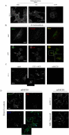
STC-1 Located at Focal Adhesions. (A) Cells were fixed with methanol acetone and labeled with antibodies to STC-1. Magnification = 600X. (B) Cells fixed with paraformaldehyde and co-labeled with antibodies to STC-1 and vinculin, an F-actin binding protein located in focal adhesions. Magnification = 600X for top panels; 200X for bottom panels; 400X for inset. (C) Mouse cells were fixed with methanol/acetone and labeled for STC-1. Magnification = 200X. MEF= Mouse Embryonic Fibroblast; STC-1-MEF=STC-1 overexpressing MEF; mIMCD3=Mouse Inner Medullary Collecting Duct Cell (negative control). Magnification=200X. Inset=400X (D) Left panel: IMCD3 cells co-cultured directly with MSCs and stained with antibodies to STC-1 and human specific PML. Right panel: IMCD3 cells were treated with rhSTC-1 or co-cultured with MSCs in transwell culture and labeled for STC-1. White arrows indicate prominent focal adhesion staining. Magnification=200X. Inset=400X. For all, levels were adjusted linearly for clarity.
Figure 6
Figure 6
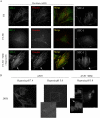
STC-1 Location Was Disrupted in Injured Cells but Preserved When Co-cultured with MSCs. (A) Fibroblasts were fixed with 4% paraformaldehyde, and co-labeled with antibodies to STC-1 (green) and vinculin (red). Irradiation displaced the STC-1 but not the vinculin from focal adhesions. STC-1 was present in focal adhesions in co-cultures. Magnification = 600X. (B) A549 cells were incubated in hypoxia at physiologic or acidic pH, in the absence or presence of MSCs. Cells were stained with antibodies to STC-1. Inset of middle panel is shown with enhanced signal, to display the faint outlines of the cell. Inset of right panel shows distinct focal adhesion staining (arrow). Magnification = 200X upper panels; 400X insets. For all, levels were adjusted linearly for clarity.

References

    1. Owen M, Friedenstein AJ. Stromal stem cells: marrow-derived osteogenic precursors. Ciba Foundation symposium. 1988;136:42–60. - PubMed
    1. Owen M. Marrow stromal stem cells. Journal of cell science. 1988;10:63–76. - PubMed
    1. Spees JL, Olson SD, Ylostalo J, et al. Differentiation, cell fusion, and nuclear fusion during ex vivo repair of epithelium by human adult stem cells from bone marrow stroma. Proceedings of the National Academy of Sciences of the United States of America. 2003;100:2397–2402. - PMC - PubMed
    1. Chopp M, Zhang XH, Li Y, et al. Spinal cord injury in rat: treatment with bone marrow stromal cell transplantation. Neuroreport. 2000;11:3001–3005. - PubMed
    1. Horwitz EM, Gordon PL, Koo WK, et al. Isolated allogeneic bone marrow-derived mesenchymal cells engraft and stimulate growth in children with osteogenesis imperfecta: Implications for cell therapy of bone. Proceedings of the National Academy of Sciences of the United States of America. 2002;99:8932–8937. - PMC - PubMed

Publication types

MeSH terms