

Chemical analysis in vivo and in vitro by Raman spectroscopy--from single cells to humans
- PMID: 19268566
- PMCID: PMC3185305
- DOI: 10.1016/j.copbio.2009.02.006
Chemical analysis in vivo and in vitro by Raman spectroscopy--from single cells to humans
Abstract
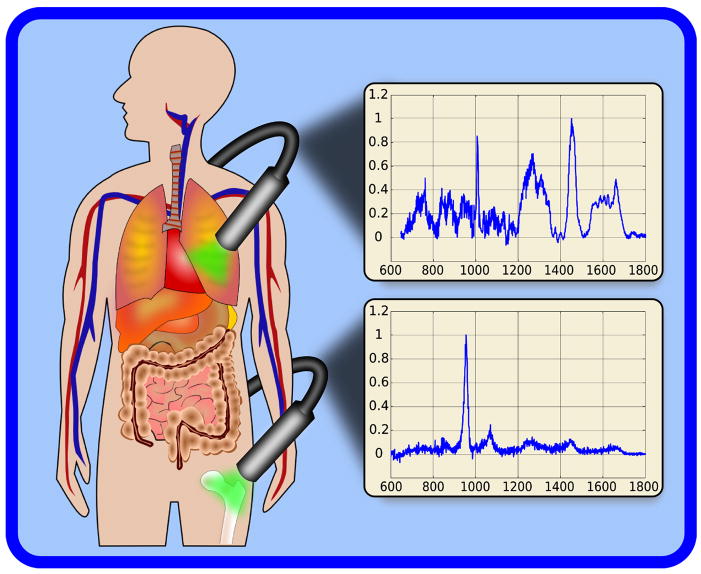
The gold standard for clinical diagnostics of tissues is immunofluorescence staining. Toxicity of many fluorescent dyes precludes their application in vivo. Raman spectroscopy, a chemically specific, label-free diagnostic technique, is rapidly gaining acceptance as a powerful alternative. It has the ability to probe the chemical composition of biological materials in a non-destructive and mostly non-perturbing manner. We review the most recent developments in Raman spectroscopy in the life sciences, detailing advances in technology that have improved the ability to screen for diseases. Its role in the monitoring of biological function and mapping the cellular chemical microenvironment will be discussed. Applications including endoscopy, surface-enhanced Raman scattering (SERS), and coherent Raman scattering (CRS) will be reviewed.
Figures


Similar articles
-
Label-free analysis of cellular biochemistry by Raman spectroscopy and microscopy.Compr Physiol. 2013 Apr;3(2):941-56. doi: 10.1002/cphy.c120025. Compr Physiol. 2013. PMID: 23720335 Review.
-
The many facets of Raman spectroscopy for biomedical analysis.Anal Bioanal Chem. 2015 Jan;407(3):699-717. doi: 10.1007/s00216-014-8311-9. Epub 2014 Nov 27. Anal Bioanal Chem. 2015. PMID: 25428454 Review.
-
Emerging technology: applications of Raman spectroscopy for prostate cancer.Cancer Metastasis Rev. 2014 Sep;33(2-3):673-93. doi: 10.1007/s10555-013-9489-6. Cancer Metastasis Rev. 2014. PMID: 24510129 Review.
-
Coherent Raman Scattering Microscopy in Biology and Medicine.Annu Rev Biomed Eng. 2015;17:415-45. doi: 10.1146/annurev-bioeng-071114-040554. Epub 2015 Oct 22. Annu Rev Biomed Eng. 2015. PMID: 26514285 Free PMC article. Review.
-
Raman and coherent anti-Stokes Raman scattering microspectroscopy for biomedical applications.J Biomed Opt. 2012 Apr;17(4):040801. doi: 10.1117/1.JBO.17.4.040801. J Biomed Opt. 2012. PMID: 22559673
Cited by
-
Raman spectroscopy of microbial pigments.Appl Environ Microbiol. 2014 Jun;80(11):3286-95. doi: 10.1128/AEM.00699-14. Epub 2014 Mar 28. Appl Environ Microbiol. 2014. PMID: 24682303 Free PMC article. Review.
-
Trace cancer biomarker quantification using polystyrene-functionalized gold nanorods.Biomed Opt Express. 2014 Nov 3;5(12):4101-7. doi: 10.1364/BOE.5.004101. eCollection 2014 Dec 1. Biomed Opt Express. 2014. PMID: 25574423 Free PMC article.
-
Label-free visualization of cholesteatoma in the mastoid and tympanic membrane using CARS microscopy.J Otol. 2016 Sep;11(3):127-133. doi: 10.1016/j.joto.2016.09.001. Epub 2016 Sep 10. J Otol. 2016. PMID: 29937821 Free PMC article.
-
Self-reference and random sampling approach for label-free identification of DNA composition using plasmonic nanomaterials.Sci Rep. 2018 May 9;8(1):7398. doi: 10.1038/s41598-018-25444-2. Sci Rep. 2018. PMID: 29743506 Free PMC article.
-
Detection of vancomycin resistances in enterococci within 3 ½ hours.Sci Rep. 2015 Feb 3;5:8217. doi: 10.1038/srep08217. Sci Rep. 2015. PMID: 25645753 Free PMC article.
References
-
- Schwartzberg AM, Oshiro TY, Zhang JZ, Huser T, Talley CE. Improving nanoprobes using surface-enhanced Raman scattering from 30-nm hollow gold particles. Analytical Chemistry. 2006;78:4732–4736. - PubMed
-
- Lu Y, Liu GL, Kim J, Mejia YX, Lee LP. Nanophotonic crescent moon structures with sharp edge for ultrasensitive biomolecular detection by local electromagnetic field enhancement effect. Nano Letters. 2005;5:119–124. - PubMed
-
- Huang XH, El-Sayed IH, Qian W, El-Sayed MA. Cancer cells assemble and align gold nanorods conjugated to antibodies to produce highly enhanced, sharp, and polarized surface Raman spectra: A potential cancer diagnostic marker. Nano Letters. 2007;7:1591–1597. - PubMed
-
- Kim JH, Kim JS, Choi H, Lee SM, Jun BH, Yu KN, Kuk E, Kim YK, Jeong DH, Cho MH, et al. Nanoparticle probes with surface enhanced Raman spectroscopic tags for cellular cancer targeting. Analytical Chemistry. 2006;78:6967–6973. - PubMed
-
- Kim JH, Lee SM, Jun BH, Choi HJ, Kim JS, Cho MH, Kim YK, Jeong DH, Lee YS. Multiplex detection and imaging of cancer markers based on surface-enhanced raman spectroscopic nanoparticle probes (SERS Dots). Nanomedicine-Nanotechnology. Biology and Medicine. 2007;3:341–341.
Publication types
MeSH terms
Grants and funding
LinkOut - more resources
Full Text Sources
Other Literature Sources
Miscellaneous
