. 2009 May;217(1):210-8.
doi: 10.1016/j.expneurol.2009.02.012. Epub 2009 Mar 5.

Sex differences in the response to activation of the poly (ADP-ribose) polymerase pathway after experimental stroke

Affiliations

Sex differences in the response to activation of the poly (ADP-ribose) polymerase pathway after experimental stroke

Mike Yuan et al. Exp Neurol. 2009 May.

Abstract

It is increasingly recognized that histological and functional outcomes after stroke are shaped by biologic sex. Emerging data suggests that ischemic cell death pathways are sexually dimorphic (Hurn, P., Vannucci, S., Hagberg, H. (2005) Adult or perinatal brain injury: does sex matter?. Stroke 36, 193-195 ; Lang, J.T., McCullough, L.D. (2008) Pathways to ischemic neuronal cell death: are sex differences relevant?. J. Transl. Med. 6). Reducing neuronal nitric oxide (NO) or poly-ADP-ribose polymerase (PARP1) activation protects only the male brain (Hagberg, H., et al. PARP-1 gene disruption in mice preferentially protects males from perinatal brain injury. J. Neurochem. 90, 1068-1075 (2004)), and paradoxically enhances ischemic injury in females (McCullough, L.D., et al. Ischemic nitric oxide and poly (ADP-ribose) polymerase-1 in cerebral ischemia: male toxicity, female protection. J. Cereb. Blood Flow Metab. 25, 502-512 (2005)). In this study, we examined downstream mediators of NO/PARP activation to investigate possible mediators of ischemic sexual dimorphism. Nuclear translocation of Apoptosis Inducing Factor (AIF) was equivalent in wild type males and females after stroke and was unaffected by estrogen exposure. Deletion of PARP1 led to a dramatic reduction in stroke-induced poly (ADP-ribose) polymerase (PAR) formation and AIF translocation in both sexes, yet ischemic damage was reduced only in males. Subsequent examination of AIF-deficient Harlequin mice demonstrated that male Harlequin mice had less PAR formation, reduced AIF translocation and less ischemic damage than male wild type mice. In contrast, female Harlequin mice had no neuroprotective effect of gene deletion despite robust reductions in PAR formation and AIF translocation. Although equivalent activation of this cell death pathway occurs in both sexes after ischemia, detrimental effects are only present in males. AIF translocation and PAR formation do not mediate ischemic injury in the female brain, therefore agents designed to reduce PARP1 activation are unlikely to benefit females.


Figures

Figure 1
Figure 1. Nuclear Poly ADP-ribose (PAR) polymer accumulation in WT mice of both sexes after 90 minute MCAO
a. Time course of PAR accumulation in WT mice after stroke. PAR starts to increase from 2 hours after reperfusion, then reaches the highest concentration at 6 hours and gradually decreases by 24 hours. Histone was used as a loading control for the nuclear fraction. b. PAR level at 6 hours after stroke between WT male mice and ovariectomized (OVX) female mice with treatment of Estradiol (E2) or oil implants. PAR levels were higher in male mice regardless of hormone treatment. c. PAR level in male mice were significantly higher than in female mice from 2 hours to 24 hours after stroke. For each group, n = 8 (stroke), 4 (sham), repeated in triplicate. Data represents s.e.m. *p< 0.01 and **p< 0.05, # P< n.s. d. PAR levels in male mice were significantly higher than in OVX female mice after treatment with either Estradiol or oil pellets. There was no difference between the Estradiol and Oil treated OVX female mice in PAR formation *p<0.01, ** p<0.05, # p>0.05.
Figure 2
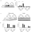
Figure 2. Nuclear AIF Translocation in WT Mice
a. Time course of AIF translocation from the mitochondria to the nuclei in WT mice after stroke. The Mitochondria fraction is shown with MnSOD and the nuclear fraction with Histone showing the purity of the subcellular fractions. Evidence of AIF translocation is seen within 2 hours after MCAO. b. There was no significant difference in AIF translocation between male and female WT mice (n=8 for stroke, 4 for sham, p>.05). c AIF translocation from the mitochondria to the nuclear fragment 6 hours after MCAO in WT male mice and ovariectomized (OVX) female mice treated with Estradiol or oil. d. No significant difference in AIF translocation was seen between the Estradiol and Oil treatment groups in OVX female mice.
Figure 3
Figure 3. Infarct and PAR formation in PARP1 Deficient Mice
a. Infarct size of cerebral cortex, striatum and total hemisphere in wild type (n=8) and PARP1 deficient mice (n=10) in both male and female after a 90 minute MCAO 72 hours after stroke. There is a significant decrease in infarct size in male PARP1 deficient mice as compared with wild type male mice (p<.001). There is a significant increase in infarct size in female PARP deficient mice as compared with wild type female mice (p<.001). b. Both male and female PARP1−/− mice had a lack of stroke induced PAR formation (lane 1 and lane 2) at 6 hours after stroke c. PARP1−/− deficient mice had complete loss of PAR synthesis both in stroke and sham control mice as compared with WT mice. (n=8 per stroke and 4 per sham, samples run in triplicate). Male WT mice had significantly higher PAR formation compared to female WT mice, consistent with Fig 1 (*p<0.01 ).
Figure 4
Figure 4. AIF translocation in PARP1 deficient mice
a. AIF translocation from the mitochondria to the nuclear fraction 6 hours after stroke in WT and PARP1 deficient mice of both sexes. b. Significantly less translocation of AIF from mitochondria to the nuclear fraction occurs in PARP deficient mice as compared with WT mice (Lane 1,2 vs. 3,4), n=8/stroke group, 4 per sham, p<.05. However, no sex differences in AIF translocation are seen either in WT or PARP deficient mice (lane 1 vs. 2 and lane 3 vs. 4, p=n.s).
Figure 5
Figure 5. Infarct in AIF-low expressing (Harlequin) mice
Infarct size of cerebral cortex, striatum and total hemisphere in WT and Hq mice after 90 minute MCAO in both sexes 72 hours after stroke. There is a significant decrease in infarct size in male Harlequin mice as compared with wild type male mice (n=10 gp, *p<.01, #p>.05). There is no significant difference in infarct size in female Hq mice vs. WT female mice.
Figure 6
Figure 6. Infarction (TTC staining) in Hq mice
Representative coronal sections of Hq and WT mice of both sexes 72 hours after MCAO. White color represents infarcted tissue. Cerebral cortical perfusion was continually monitored by Laser-Doppler flowmetry (LDF/Moor Instruments) in wild type and Hq mice. Cortical perfusion was reduced to approximately 90% of baseline after MCAO and recovered to 70%–80% of baseline after reperfusion in all three strains. The temperature of all mice was kept at 37 ± 0.5 ºC throughout the surgery and after care.
Figure 7
Figure 7. PAR Formation and AIF translocation in Harlequin Mice
a. PAR level at 12 hours after stroke in WT mice and Hq mice. b. PAR levels in Hq mice are significantly reduced compared with WT mice. The same sex pattern (males demonstrating enhanced PAR formation) is seen in Hq and WT mice. PAR levels in males are significantly higher than in female mice in both Hq and WT strains 12 hours after stroke. c. AIF translocation from mitochondria to nuclear fraction at 12 hours after stroke (CVA) in WT mice and Hq mice of both sexes. d. Mitochondrial AIF level in Harlequin mice is approximately 20% of WT mice level. Almost no AIF translocation can be detected in the nuclear fraction in Hq mice after stroke.

References

    1. Alkayed N, et al. Gender-linked brain injury in experimental stroke. Stroke. 1998;29:159–165. - PubMed
    1. Andrabi SA, et al. Poly(adp-ribose) (par) polymer is a death signal. Proc Natl Acad Sci U S A. 2006;103:18308–18313. - PMC - PubMed
    1. Brann D, et al. Neurotrophic and neuroprotective actions of estrogen: basic mechanisms and clinical implications. Steroids. 2007;72:381–405. - PMC - PubMed
    1. Bredt DS, et al. Localization of nitric oxide synthase indicating a neural role for nitric oxide. Nature. 1990;347:768–70. - PubMed
    1. Cao G, et al. Translocation of apoptosis-inducing factor in vulnerable neurons after transient cerebral ischemia and in neuronal cultures after oxygen-glucose deprivation. J Cereb Blood Flow Metab. 2003;23:1137–1150. - PubMed

Publication types

MeSH terms