Review
. 2009 May;122(2):125-39.
doi: 10.1016/j.pharmthera.2009.02.003. Epub 2009 Mar 5.

The dynamic effects of nicotine on the developing brain

Affiliations
Review

The dynamic effects of nicotine on the developing brain

Jennifer B Dwyer et al. Pharmacol Ther. 2009 May.

Abstract

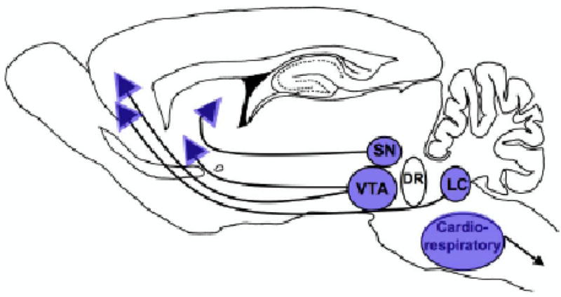
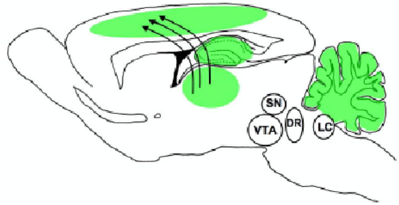
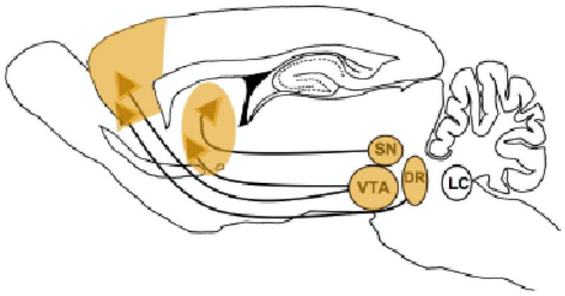
Nicotinic acetylcholine receptors (nAChRs) regulate critical aspects of brain maturation during the prenatal, early postnatal, and adolescent periods. During these developmental windows, nAChRs are often transiently upregulated or change subunit composition in those neural structures that are undergoing major phases of differentiation and synaptogenesis, and are sensitive to environmental stimuli. Nicotine exposure, most often via tobacco smoke, but increasingly via nicotine replacement therapy, has been shown to have unique effects on the developing human brain. Consistent with a dynamic developmental role for acetylcholine, exogenous nicotine produces effects that are unique to the period of exposure and that impact the developing structures regulated by acetylcholine at that time. Here we present a review of the evidence, available from both the clinical literature and preclinical animal models, which suggests that the diverse effects of nicotine exposure are best evaluated in the context of regional and temporal expression patterns of nAChRs during sensitive maturational periods, and disruption of the normal developmental influences of acetylcholine. We present evidence that nicotine interferes with catecholamine and brainstem autonomic nuclei development during the prenatal period of the rodent (equivalent to first and second trimester of the human), alters the neocortex, hippocampus, and cerebellum during the early postnatal period (third trimester of the human), and influences limbic system and late monoamine maturation during adolescence.


Figures

Figure 1
Figure 1
Brain regions impacted by nicotine exposure during the prenatal period. The dopamine-producing cell bodies of the SN/VTA and the noradrenergic cell bodies of the LC are vulnerable to exogenous nicotine-induced deficits during the prenatal period. nAChRs are located on both the cell bodies and terminals of these projections and are able to modulate neurotransmitter release in fetal brain. Additionally, the brainstem nuclei controlling cardiorespiratory and autonomic responses are also sensitive to nicotine exposure during this period. Prenatal nicotine-induced defects in these structures may underlie the increase in dopamine-mediated disorders such as attention deficit hyperactivity disorder and substance abuse, as well as the increased risk for sudden infant death syndrome reported in the clinical literature (see text for detail).
Figure 2
Figure 2
Brain regions impacted by nicotine exposure during the early postnatal period. In the early postnatal period, the cortex, hippocampus, and cerebellum are all in sensitive periods of development and are actively regulated by transient populations of nAChRs. During this time exogenous nicotine may disrupt normal thalmocortical and hippocampal development. Offspring of children whose mothers smoked during pregnancy are at increased risk for cognitive and auditory processing deficits, which are likely related to cortical dysfunction induced by early nicotine exposure (see text for detail).
Figure 3
Figure 3
Brain regions impacted by nicotine exposure during the adolescent period. During adolescence, there is substantial plasticity in limbic circuitry including the amygdala, prefrontal cortex, and nucleus accumbens. The dopamine systems of the SN/VTA also undergo their last major phase of development during this period. These late maturing catecholaminergic projections possess functional nAChRs on the cell bodies and terminals that allow nicotine to alter their excitation and release. nAChRs have also been shown to alter the dopamine and serotonin transporter function in prefrontal cortex and nucleus accumbens. Thus, nicotine exposure during this time produces changes within the limbic and dopaminergic circuitry that underlies motivated behaviors, potentially enhancing the vulnerability to nicotine addiction during this time period. The serotonin system, originating in the dorsal raphe (DR), also appears to be affected by adolescent nicotine exposure. Nicotine-induced alterations in serotonin function may contribute to the increased incidence of mood disorders seen after adolescent smoking (see text for more detail).

References

    1. Adams CE, Broide RS, Chen Y, Winzer-Serhan UH, Henderson TA, Leslie FM, Freedman R. Development of the alpha7 nicotinic cholinergic receptor in rat hippocampal formation. Brain Res Dev Brain Res. 2002;139(2):175–87. - PubMed
    1. Adriani W, Macrì S, Pacifici R, Laviola G. Peculiar vulnerability to nicotine oral self-administration in mice during early adolescence. Neuropsychopharmacology. 2002;27(2):212–24. - PubMed
    1. Adriani W, Spijker S, Deroche-Gamonet V, Laviola G, Le Moal M, Smit AB, Piazza PV. Evidence for enhanced neurobehavioral vulnerability to nicotine during periadolescence in rats. J Neurosci. 2003;23(11):4712–6. - PMC - PubMed
    1. Agulhon C, Charnay Y, Vallet P, Abitbol M, Kobetz A, Bertrand D, Malafosse A. Distribution of mRNA for the alpha4 subunit of the nicotinic acetylcholine receptor in the human fetal brain. Brain Res Mol Brain Res. 1998;58(1–2):123–31. - PubMed
    1. Alkondon M, Pereira EF, Albuquerque EX. alpha-bungarotoxin- and methyllycaconitine-sensitive nicotinic receptors mediate fast synaptic transmission in interneurons of rat hippocampal slices. Brain Res. 1998;810(1–2):257–63. - PubMed

Publication types