

Comparison of ventricular structure and function in Chinese patients with heart failure and ejection fractions >55% versus 40% to 55% versus <40%
- PMID: 19268743
- PMCID: PMC4940025
- DOI: 10.1016/j.amjcard.2008.11.050
Comparison of ventricular structure and function in Chinese patients with heart failure and ejection fractions >55% versus 40% to 55% versus <40%
Abstract
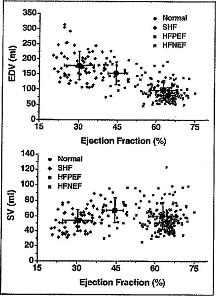
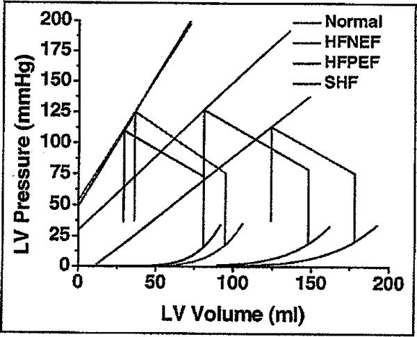
Subjects with heart failure (HF) and a preserved ejection fraction (EF) are heterogenous and the EF used to define this syndrome varies considerably among studies. We sought to determine if physiologic differences exist between subjects with a normal EF (>55%) or mildly decreased EF (40% to 55%). 357 consecutive Chinese patients who were healthy (n = 93) or had HF (n = 264) underwent comprehensive echocardiography, Doppler analysis, and measurement of neurohormones. Subjects with HF were stratified by EF into those with normal EF (>55%, n = 128), mildly decreased EF (40% to 55%, n = 38), or moderate to severely decreased EF (<40%, n = 100). Employing noninvasive pressure-volume analysis, estimated end-systolic and end-diastolic pressure-volume relations were calculated. Subjects with HF and an EF 40% to 55% more often had a previous myocardial infarction and diabetes than those with HF and an EF >55%. Physiologically, the cohort with a mildly decreased EF had eccentrically enlarged ventricles with evidence of remodeling (rightward shifted end-diastolic pressure-volume relation) and decreased chamber contractility (downward shifted end-systolic pressure-volume relation) most comparable to subjects with overt systolic HF. In conclusion, in subjects with HF and a preserved EF, there are distinct physiologic differences between those with a normal (>55%) and a mildly decreased (40% to 55%) EF.
Figures
References
-
- Lloyd-Jones DM, Larson MG, Leip EP, Beiser A, D’Agostino RB, Kannel WB, Murabito JM, Vasan RS, Benjamin EJ, Levy D. Lifetime risk for developing congestive heart failure: the Framingham Heart Study. Circulation. 2002;106:3068–3072. - PubMed
-
- Paulus WJ, Tschope C, Sanderson JE, Rusconi C, Flachskampf FA, Rademakers FE, Marino P, Smiseth OA, De KG, Leite-Moreira AF, et al. How to diagnose diastolic heart failure: a consensus statement on the diagnosis of heart failure with normal left ventricular ejection fraction by the Heart Failure and Echocardiography Associations of the European Society of Cardiology. Eur Heart J. 2007;28:2539–2550. - PubMed
-
- Maron BJ. Hypertrophic cardiomyopathy: a systematic review. JAMA. 2002;287:1308–1320. - PubMed
-
- Maron BJ, Spirito P, Roman MJ, Paranicas M, Okin PM, Best LG, Lee ET, Devereux RB. Prevalence of hypertrophic cardiomyopathy in a population-based sample of American Indians aged 51 to 77 years (the Strong Heart Study) Am J Cardiol. 2004;93:1510–1514. - PubMed
-
- Schiller NB, Shah PM, Crawford M, DeMaria A, Devereux R, Feigenbaum H, Gutgesell H, Reichek N, Sahn D, Schnittger I. Recommendations for quantitation of the left ventricle by two-dimensional echocardiography. American Society of Echocardiography Committee on Standards, Subcommittee on Quantitation of Two-Dimensional Echocardiograms. J Am Soc Echocardiogr. 1989;2:358–367. - PubMed
Publication types
MeSH terms
Grants and funding
LinkOut - more resources
Full Text Sources
Medical
Molecular Biology Databases
Research Materials
Miscellaneous
