

Antagonism of the testis- and ovary-determining pathways during ovotestis development in mice
- PMID: 19269320
- PMCID: PMC2680453
- DOI: 10.1016/j.mod.2009.02.006
Antagonism of the testis- and ovary-determining pathways during ovotestis development in mice
Abstract
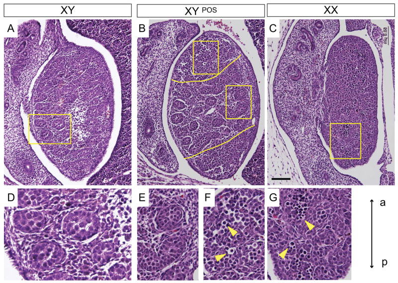
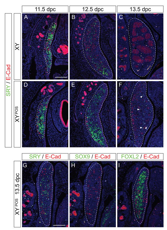
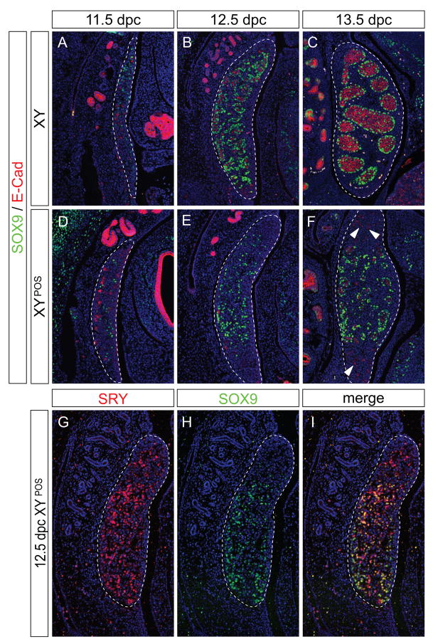
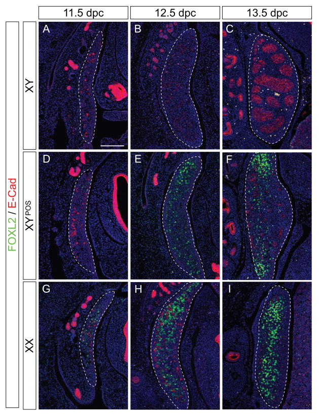
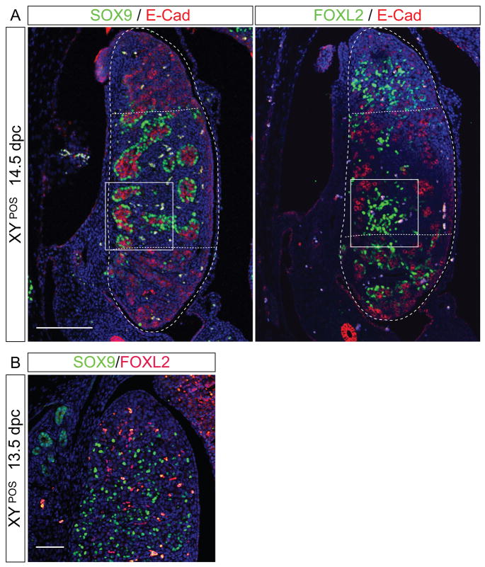
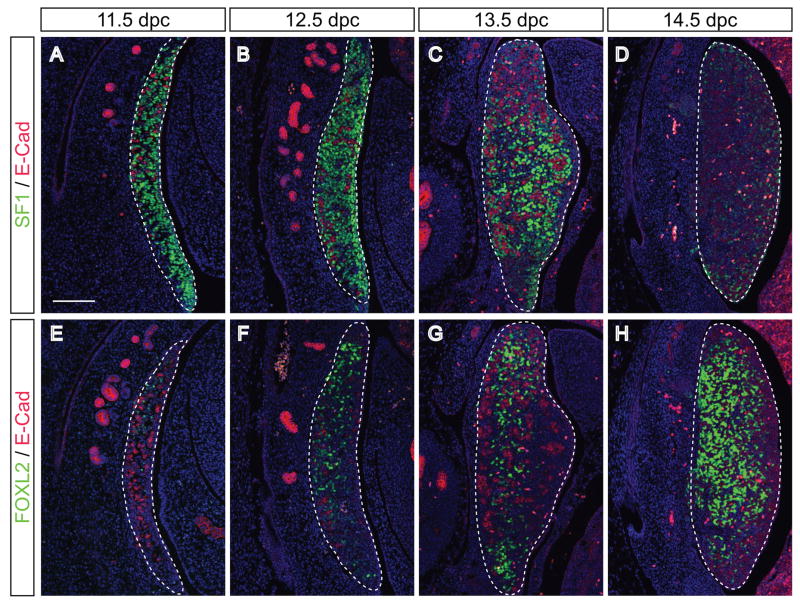
Ovotestis development in B6-XY(POS) mice provides a rare opportunity to study the interaction of the testis- and ovary-determining pathways in the same tissue. We studied expression of several markers of mouse fetal testis (SRY, SOX9) or ovary (FOXL2, Rspo1) development in B6-XY(POS) ovotestes by immunofluorescence, using normal testes and ovaries as controls. In ovotestes, SOX9 was expressed only in the central region where SRY is expressed earliest, resulting in testis cord formation. Surprisingly, FOXL2-expressing cells also were found in this region, but individual cells expressed either FOXL2 or SOX9, not both. At the poles, even though SOX9 was not up-regulated, SRY expression was down-regulated normally as in XY testes, and FOXL2 was expressed from an early stage, demonstrating ovarian differentiation in these areas. Our data (1) show that SRY must act within a specific developmental window to activate Sox9; (2) challenge the established view that SOX9 is responsible for down-regulating Sry expression; (3) disprove the concept that testicular and ovarian cells occupy discrete domains in ovotestes; and (4) suggest that FOXL2 is actively suppressed in Sertoli cell precursors by the action of SOX9. Together these findings provide important new insights into the molecular regulation of testis and ovary development.
Figures
Similar articles
-
Inefficient Sox9 upregulation and absence of Rspo1 repression lead to sex reversal in the B6.XYTIR mouse gonad†.Biol Reprod. 2024 May 9;110(5):985-999. doi: 10.1093/biolre/ioae018. Biol Reprod. 2024. PMID: 38376238 Free PMC article.
-
SRY upregulation of SOX9 is inefficient and delayed, allowing ovarian differentiation, in the B6.Y(TIR) gonad.Differentiation. 2011 Jul;82(1):18-27. doi: 10.1016/j.diff.2011.04.007. Epub 2011 May 17. Differentiation. 2011. PMID: 21592645
-
Testis Determination Requires a Specific FGFR2 Isoform to Repress FOXL2.Endocrinology. 2017 Nov 1;158(11):3832-3843. doi: 10.1210/en.2017-00674. Endocrinology. 2017. PMID: 28938467 Free PMC article.
-
Determination and stability of gonadal sex.J Androl. 2010 Jan-Feb;31(1):16-25. doi: 10.2164/jandrol.109.008201. Epub 2009 Oct 29. J Androl. 2010. PMID: 19875493 Free PMC article. Review.
-
From SRY to SOX9: mammalian testis differentiation.J Biochem. 2005 Jul;138(1):13-9. doi: 10.1093/jb/mvi098. J Biochem. 2005. PMID: 16046443 Review.
Cited by
-
Alterations of sex determination pathways in the genital ridges of males with limited Y chromosome genes†.Biol Reprod. 2019 Mar 1;100(3):810-823. doi: 10.1093/biolre/ioy218. Biol Reprod. 2019. PMID: 30285093 Free PMC article.
-
Regulation of male sex determination: genital ridge formation and Sry activation in mice.Cell Mol Life Sci. 2014 Dec;71(24):4781-802. doi: 10.1007/s00018-014-1703-3. Epub 2014 Aug 20. Cell Mol Life Sci. 2014. PMID: 25139092 Free PMC article. Review.
-
Mouse Gonad Development in the Absence of the Pro-Ovary Factor WNT4 and the Pro-Testis Factor SOX9.Cells. 2020 Apr 29;9(5):1103. doi: 10.3390/cells9051103. Cells. 2020. PMID: 32365547 Free PMC article.
-
A Rare Differences of Sex Development: Male Sex Reversal Syndrome (NonSyndromic 46, XX with Negative Sex-Determining Region of Y Chromosome Gene).J Indian Assoc Pediatr Surg. 2023 Mar-Apr;28(2):154-159. doi: 10.4103/jiaps.jiaps_109_22. Epub 2022 Nov 30. J Indian Assoc Pediatr Surg. 2023. PMID: 37197249 Free PMC article.
-
NEDD4 Promotes Sertoli Cell Proliferation and Adult Leydig Cell Differentiation in the Murine Testis.Endocrinology. 2025 Jul 8;166(9):bqaf115. doi: 10.1210/endocr/bqaf115. Endocrinology. 2025. PMID: 40605617 Free PMC article.
References
-
- Albrecht K, Eicher E. Evidence that Sry is expressed in pre-Sertoli cells and Sertoli and granulosa cells have a common precursor. Dev Biol. 2001;240:92–107. - PubMed
-
- Arango N, Lovell-Badge R, Behringer R. Targeted mutagenesis of the endogenous mouse Mis gene promoter: In vivo definition of genetic pathways of vertebrate sexual development. Cell. 1999;99:409–419. - PubMed
-
- Bardoni B, Zanaria E, Guioli S, Floridia G, Worley KC, Tonini G, Ferrante E, Chiumello G, McCabe ER, Fraccaro M, et al. A dosage sensitive locus at chromosome Xp21 is involved in male to female sex reversal. Nat Genet. 1994;7:497–501. - PubMed
-
- Barrionuevo F, Bagheri-Fam S, Klattig J, Kist R, Taketo MM, Englert C, Scherer G. Homozygous Inactivation of Sox9 Causes Complete XY Sex Reversal in Mice. Biol Reprod. 2006;74:195–201. - PubMed
Publication types
MeSH terms
Substances
Grants and funding
LinkOut - more resources
Full Text Sources
Molecular Biology Databases
Research Materials
