

Atlas-derived perfusion correlates of white matter hyperintensities in patients with reduced cardiac output
- PMID: 19269713
- PMCID: PMC2889176
- DOI: 10.1016/j.neurobiolaging.2009.01.011
Atlas-derived perfusion correlates of white matter hyperintensities in patients with reduced cardiac output
Abstract
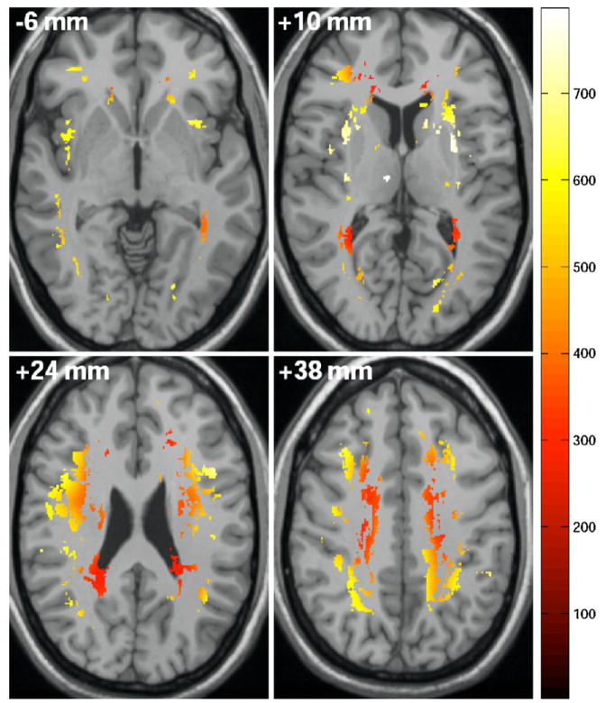
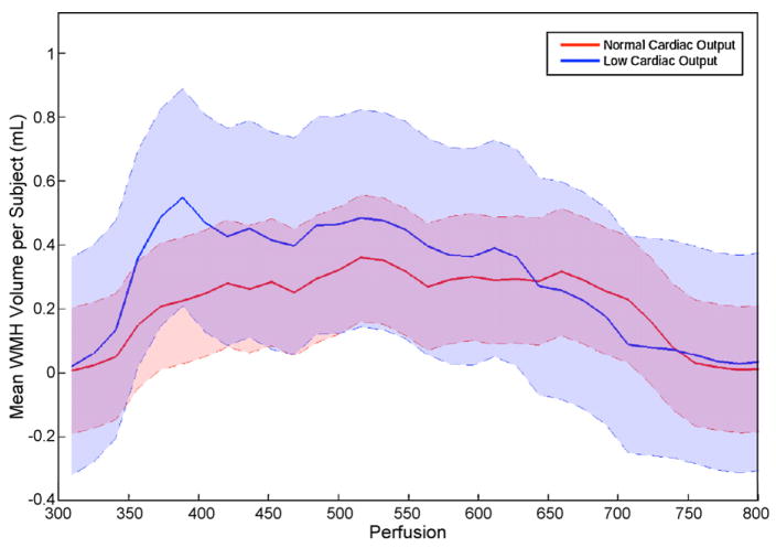
Reduced cardiac output is associated with increased white matter hyperintensities (WMH) and executive dysfunction in older adults, which may be secondary to relations between systemic and cerebral perfusion. This study preliminarily describes the regional distribution of cerebral WMH in the context of a normal cerebral perfusion atlas and aims to determine if these variables are associated with reduced cardiac output. Thirty-two participants (72 ± 8 years old, 38% female) with cardiovascular risk factors or disease underwent structural MRI acquisition at 1.5T using a standard imaging protocol that included FLAIR sequences. WMH distribution was examined in common anatomical space using voxel-based morphometry and as a function of normal cerebral perfusion patterns by overlaying a single photon emission computed tomography (SPECT) atlas. Doppler echocardiogram data was used to dichotomize the participants on the basis of low (n=9) and normal (n=23) cardiac output. Global WMH count and volume did not differ between the low and normal cardiac output groups; however, atlas-derived SPECT perfusion values in regions of hyperintensities were reduced in the low versus normal cardiac output group (p<0.001). Our preliminary data suggest that participants with low cardiac output have WMH in regions of relatively reduced perfusion, while normal cardiac output participants have WMH in regions with relatively higher regional perfusion. This spatial perfusion distribution difference for areas of WMH may occur in the context of reduced systemic perfusion, which subsequently impacts cerebral perfusion and contributes to subclinical or clinical microvascular damage.
Copyright © 2009 Elsevier Inc. All rights reserved.
Conflict of interest statement
Figures
Similar articles
-
Cardiac output as a potential risk factor for abnormal brain aging.J Alzheimers Dis. 2010;20(3):813-21. doi: 10.3233/JAD-2010-100081. J Alzheimers Dis. 2010. PMID: 20413856 Free PMC article. Review.
-
Spatial distribution of white-matter hyperintensities in Alzheimer disease, cerebral amyloid angiopathy, and healthy aging.Stroke. 2008 Apr;39(4):1127-33. doi: 10.1161/STROKEAHA.107.497438. Epub 2008 Feb 21. Stroke. 2008. PMID: 18292383 Free PMC article.
-
Cerebral small vessel disease in aging and Alzheimer's disease: a comparative study using MRI and SPECT.Eur J Neurol. 2013 Feb;20(2):243-50. doi: 10.1111/j.1468-1331.2012.03785.x. Epub 2012 Jun 28. Eur J Neurol. 2013. PMID: 22742818
-
Hyperintensities on T2-weighted images in the basal ganglia of patients with major depression: cerebral perfusion and clinical implications.Psychiatry Res. 2011 May 31;192(2):125-30. doi: 10.1016/j.pscychresns.2010.11.010. Epub 2011 Apr 11. Psychiatry Res. 2011. PMID: 21482458
-
Can white matter hyperintensities based Fazekas visual assessment scales inform about Alzheimer's disease pathology in the population?Alzheimers Res Ther. 2024 Jul 10;16(1):157. doi: 10.1186/s13195-024-01525-5. Alzheimers Res Ther. 2024. PMID: 38987827 Free PMC article.
Cited by
-
Low cardiac index is associated with incident dementia and Alzheimer disease: the Framingham Heart Study.Circulation. 2015 Apr 14;131(15):1333-9. doi: 10.1161/CIRCULATIONAHA.114.012438. Epub 2015 Feb 19. Circulation. 2015. PMID: 25700178 Free PMC article.
-
Inhibition of Extracellular Signal-Regulated Kinase Activity Improves Cognitive Function in Mice Subjected to Myocardial Infarction.Cardiovasc Toxicol. 2024 Aug;24(8):766-775. doi: 10.1007/s12012-024-09877-y. Epub 2024 Jun 8. Cardiovasc Toxicol. 2024. PMID: 38850470
-
Cardiac output as a potential risk factor for abnormal brain aging.J Alzheimers Dis. 2010;20(3):813-21. doi: 10.3233/JAD-2010-100081. J Alzheimers Dis. 2010. PMID: 20413856 Free PMC article. Review.
-
Left ventricular ejection fraction and right atrial diameter are associated with deep regional CBF in arteriosclerotic cerebral small vessel disease.BMC Neurol. 2021 Feb 11;21(1):67. doi: 10.1186/s12883-021-02096-w. BMC Neurol. 2021. PMID: 33573621 Free PMC article.
-
The Relationship Between Pre-existing Coronary Heart Disease and Cognitive Impairment Is Partly Explained by Reduced Left Ventricular Ejection Fraction in the Subjects Without Clinical Heart Failure: A Cross-Sectional Study.Front Hum Neurosci. 2022 May 11;16:835900. doi: 10.3389/fnhum.2022.835900. eCollection 2022. Front Hum Neurosci. 2022. PMID: 35634203 Free PMC article.
References
-
- Beck AT, Ward CH, Mendelson M, Mock J, Erbaugh J. An inventory for measuring depression. Archives of General Psychiatry. 1961;4:561–571. - PubMed
-
- Borenstein AR, Wu Y, Mortimer JA, Schellenberg GD, McCormick WC, Bowen JD, McCurry S, Larson EB. Developmental and vascular risk factors for Alzheimer’s disease. Neurobiology of Aging. 2005;26:325–334. - PubMed
-
- Carmelli D, Swan GE, Reed T, Wolf PA, Miller BL, DeCarli C. Midlife cardiovascular risk factors and brain morphology in identical older male twins. Neurology. 1999;52:1119–1124. - PubMed
-
- De Cristofaro MT, Mascalchi M, Pupi A, Nencini P, Formiconi AR, Inzitari D, Dal Pozzo G, Meldolesi U. Subcortical arteriosclerotic encephalopathy: single photon emission computed tomography-magnetic resonance imaging correlation. American Journal of Physiologic Imaging. 1990;5:68–74. - PubMed
-
- DeCarli C, Miller BL, Swan GE, Reed T, Wolf PA, Garner J, Jack L, Carmelli D. Predictors of brain morphology for the men of the NHLBI twin study. Stroke. 1999;30:529–536. - PubMed
Publication types
MeSH terms
Grants and funding
LinkOut - more resources
Full Text Sources
Medical
