. 2009 May 1;284(18):12318-27.
doi: 10.1074/jbc.M808850200. Epub 2009 Mar 5.

The myosin-binding protein C motif binds to F-actin in a phosphorylation-sensitive manner

Affiliations

The myosin-binding protein C motif binds to F-actin in a phosphorylation-sensitive manner

Justin F Shaffer et al. J Biol Chem. .

Abstract

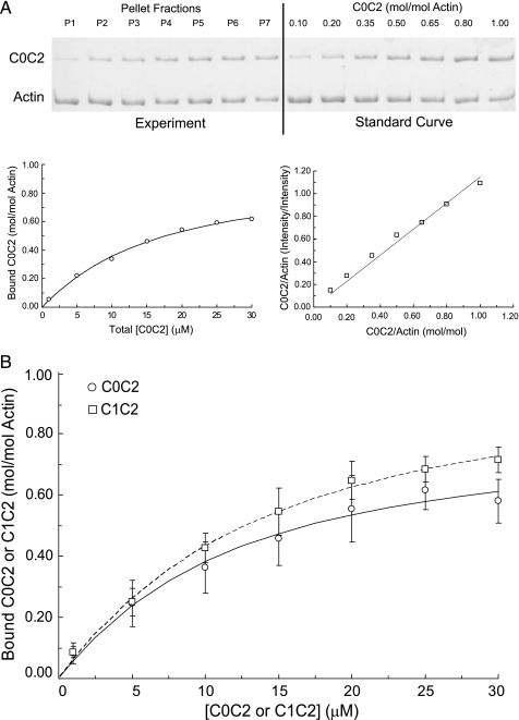
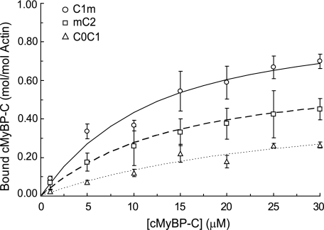
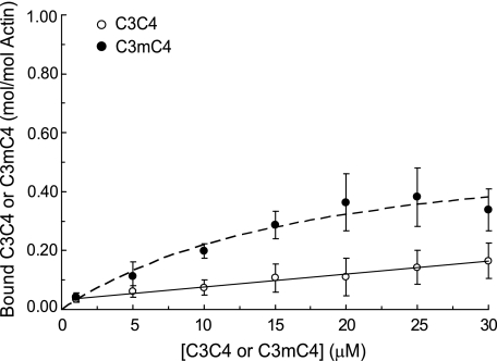
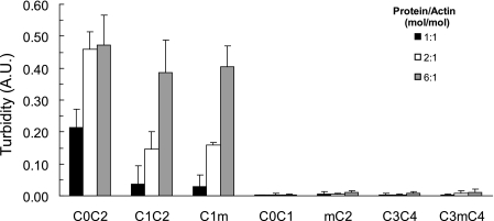
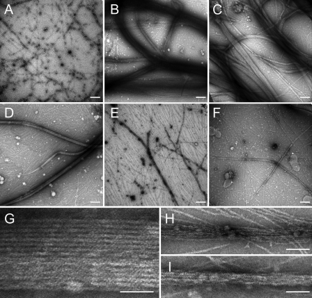
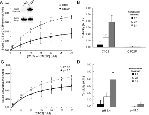
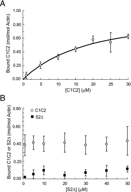
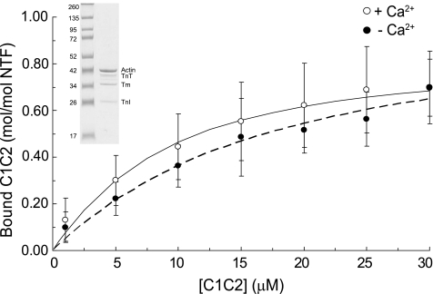
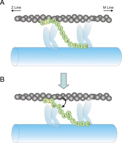
Cardiac myosin-binding protein C (cMyBP-C) is a regulatory protein expressed in cardiac sarcomeres that is known to interact with myosin, titin, and actin. cMyBP-C modulates actomyosin interactions in a phosphorylation-dependent way, but it is unclear whether interactions with myosin, titin, or actin are required for these effects. Here we show using cosedimentation binding assays, that the 4 N-terminal domains of murine cMyBP-C (i.e. C0-C1-m-C2) bind to F-actin with a dissociation constant (K(d)) of approximately 10 microm and a molar binding ratio (B(max)) near 1.0, indicating 1:1 (mol/mol) binding to actin. Electron microscopy and light scattering analyses show that these domains cross-link F-actin filaments, implying multiple sites of interaction with actin. Phosphorylation of the MyBP-C regulatory motif, or m-domain, reduced binding to actin (reduced B(max)) and eliminated actin cross-linking. These results suggest that the N terminus of cMyBP-C interacts with F-actin through multiple distinct binding sites and that binding at one or more sites is reduced by phosphorylation. Reversible interactions with actin could contribute to effects of cMyBP-C to increase cross-bridge cycling.


Figures

FIGURE 1.
FIGURE 1.
Cosedimentation binding data for C0C2 and C1C2. A, representative high speed cosedimentation binding experiment. C0C2 (1-30 μm final concentrations) was mixed with 5 μm F-actin (final concentration) and spun for 30 min at 390,000 × g. The amount of C0C2 and actin in pellet fractions (left) was quantified by SDS-PAGE using a standard curve run on each gel with varying known molar ratios of C0C2 to actin (right). B, summary data (means ± S.D.) for C0C2 and C1C2 binding to actin. The Kd and Bmax values for C0C2 (circles) were 13.7 ± 5.5 μm and 0.92 ± 0.11 mol/mol (n = 7). Kd and Bmax values for C1C2 (squares) were 10.9 ± 2.2 μm and 1.03 ± 0.08 mol/mol (n = 6).
FIGURE 2.
FIGURE 2.
Summary cosedimentation binding data for C1m, mC2, and C0C1. C1m (circles), Kd = 8.7 ± 2.4 μm, Bmax = 0.92 ± 0.14 (n = 4); mC2 (squares), Kd = 10.5 ± 4.7 μm, Bmax = 0.59 ± 0.13 (n = 5); C0C1 (triangles), Kd = 40.4 ± 16.8 μm, Bmax = 0.45 ± 0.10 (n = 7). Data are means ± S.D.
FIGURE 3.
FIGURE 3.
The MyBP-C m-domain binds specifically to actin. Cosedimentation binding data (means ± S.D.) for C3C4 (circles) comprised of domains C3 and C4 of cMyBP-C and for C3mC4 (squares) an engineered protein in which the m-domain sequence was inserted between domains 3 and 4 of cMyBP-C. C3C4 bound nonspecifically to actin as shown by the linear binding relation, whereas C3mC4 bound to actin in a saturable manner with Kd = 11.2 ± 3.3 μm, Bmax = 0.50 ± 0.09 (n = 5).
FIGURE 4.
FIGURE 4.
Bundling of F-actin by C1 and the m-domain. F-actin was combined with recombinant cMyBP-C proteins under conditions identical to those used for cosedimentation binding assays (see “Experimental Procedures”) and solution turbidity was measured as light absorbance at λ = 350 nm. Proteins containing both C1 and the m-domain (i.e. C0C2, C1C2, and C1m) increased solution turbidity in a concentration-dependent manner, whereas C0C1, mC2, C3C4, and C3mC4 did not increase solution turbidity. Data are means ± S.D. A.U., arbitrary units.
FIGURE 5.
FIGURE 5.
Electron micrographs of F-actin in the presence of recombinant cMyBP-C proteins. 5 μm F-actin was combined with 30 μm recombinant cMyBP-C proteins (1:6 ratio) and processed as described under “Experimental Proceduress” for visualization by electron microscopy. Scale bars represent 200 nm in A-F and 50 nm in G-I. A, F-actin alone; B, F-actin plus C0C2; C, F-actin plus C1C2; D, F-actin plus C1m; E, F-actin plus C0C1; F, F-actin plus mC2. Thick bundles of F-actin are apparent in the presence of C0C2 (B), C1C2 (C), and C1m (D), but are either not present in the absence of added proteins (A, F-actin alone) or reduced in the presence of C0C1 (E) or mC2 (F). Panels G-I show higher magnification images of F-actin bundles in the presence of C1m (G), C0C1 (H), and mC2 (I) to illustrate tighter more regular packing of F-actin in the presence of C1m relative to either C0C1 or mC2.
FIGURE 6.
FIGURE 6.
Effects of phosphorylation of the m-domain and alkaline pH on C1C2 binding to actin. A, cosedimentation binding data (means ± S.D.) for phosphorylated C1C2 (i.e. C1C2P, filled circles). Pro Q Diamond staining (inset) confirmed phosphorylation of C1C2P following treatment with PKA. C1C2P bound to F-actin with Kd = 13.3 ± 2.8 μm, Bmax = 0.49 ± 0.16 (n = 7). Binding data for C1C2 (open circles) were redrawn from Fig. 1 for comparison. B, PKA treatment of C1C2 abolished the ability of C1C2P to increase the turbidity of F-actin solutions. C, cosedimentation binding data for C1C2 collected at pH 8.0 (closed circles), Kd = 8.3 ± 3.5 μm, Bmax = 0.65 ± 0.10 (n = 6). Binding data for C1C2 at pH 7.4 (open circles), were redrawn from Fig. 1 for comparison. D, turbidity of solutions containing F-actin and C1C2 was reduced at pH 8.0. A.U., arbitrary units.
FIGURE 7.
FIGURE 7.
Cosedimentation binding of C1C2 to F-actin in the presence of S2Δ. A, cosedimentation binding experiments with 5 μm F-actin performed in the presence of 30 μm S2Δ. The presence of S2Δ did not significantly affect binding of C1C2 to F-actin (Kd = 15.4 ± 3.0 μm and Bmax = 0.97 ± 0.10, n = 4). B, cosedimentation binding assays performed with 5 μm F-actin and 10 μm C1C2 in the presence of 1-50 μm S2Δ. The amount of C1C2 (circles) and S2Δ (squares) found in the pellets did not change in the presence of increasing amounts of S2Δ. Data are means ± S.D.
FIGURE 8.
FIGURE 8.
Cosedimentation binding of C1C2 to native thin filaments. NTF were prepared as described under “Experimental Procedures” from bovine heart and 5 μm purified NTF were used in cosedimentation binding assays. An SDS-PAGE of purified NTF is shown in the inset. Positions of actin, tropomyosin (Tm), troponin I (TnI), and troponin T (TnT) are indicated. Troponin C is not visible on the gel due to weak staining by Coomassie Blue. C1C2 bound to NTF in a saturable manner in the presence (open circles) or absence (closed circle) of Ca2+ (means ± S.D.). Kd and Bmax values were 8.4 ± 4.6 and 0.90 ± 0.13 μm (n = 6) for C1C2 in the presence of Ca2+ and 12.5 ± 4.8 and 0.90 ± 0.13 μm (n = 5) in the absence of Ca2+. Solutions lacking Ca2+ contained (in mmol/liter): 180 KCl, 20 imidazole, pH 7.4, 1 EGTA, 1 MgCl2, and 1 DTT. Solutions with Ca2+ contained additional CaCl2 to achieve 1 mm free Ca2+.
FIGURE 9.
FIGURE 9.
Model of cMyBP-C interactions with F-actin. cMyBP-C (green) is depicted as a series of 12 spherical domains representing domains C0 through C10 and the MyBP-C motif or m-domain (M) between domains C1 and C2. The Pro-Ala-rich region between the C0 and C1 domains is represented as a yellow rectangle. A, the N terminus of cMyBP-C interacts with the thin filament primarily through binding of the C1 domain and the m-domain as demonstrated by data in this study. The cardiac-specific C0 domain is also shown binding to actin based on data from this and other studies (16, 19). B, binding of the M-domain to the thin filament is reduced by phosphorylation of the m-domain or by increasing pH (reduced net positive charge of the m-domain).

References

    1. Richard, P., Charron, P., Carrier, L., Ledeuil, C., Cheav, T., Pichereau, C., Benaiche, A., Isnard, R., Dubourg, O., Burban, M., Gueffet, J. P., Millaire, A., Desnos, M., Schwartz, K., Hainque, B., and Komajda, M. (2003) Circulation 107 2227-2232 - PubMed
    1. Dhandapany, P. S., Sadayappan, S., Xue, Y., Powell, G. T., Rani, D. S., Nallari, P., Rai, T. S., Khullar, M., Soares, P., Bahl, A., Tharkan, J. M., Vaideeswar, P., Rathinavel, A., Narasimhan, C., Ayapati, D. R., Ayub, Q., Mehdi, S. Q., Oppenheimer, S., Richards, M. B., Price, A. L., Patterson, N., Reich, D., Singh, L., Tyler-Smith, C., and Thangaraj, K. (2009) Nat. Genet. 41 187-191 - PMC - PubMed
    1. Harris, S. P., Bartley, C. R., Hacker, T. A., McDonald, K. S., Douglas, P. S., Greaser, M. L., Powers, P. A., and Moss, R. L. (2002) Circ. Res. 90 594-601 - PubMed
    1. Korte, F. S., McDonald, K. S., Harris, S. P., and Moss, R. L. (2003) Circ. Res. 93 752-758 - PubMed
    1. Stelzer, J. E., Fitzsimons, D. P., and Moss, R. L. (2006) Biophys. J. 90 4119-4127 - PMC - PubMed

Publication types

LinkOut - more resources