. 2009 May;30(5):818-23.
doi: 10.1093/carcin/bgp059. Epub 2009 Mar 6.

Penta-O-galloyl-beta-D-glucose induces S- and G(1)-cell cycle arrests in prostate cancer cells targeting DNA replication and cyclin D1

Affiliations

Penta-O-galloyl-beta-D-glucose induces S- and G(1)-cell cycle arrests in prostate cancer cells targeting DNA replication and cyclin D1

Hongbo Hu et al. Carcinogenesis. 2009 May.

Abstract

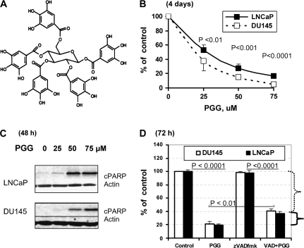
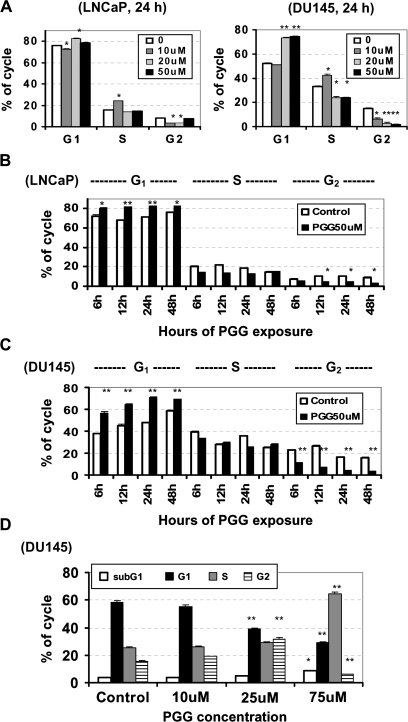
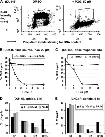
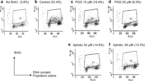
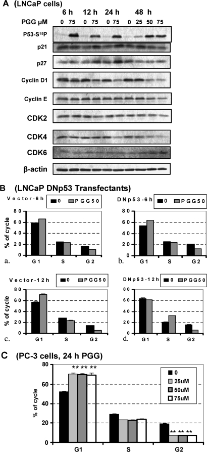
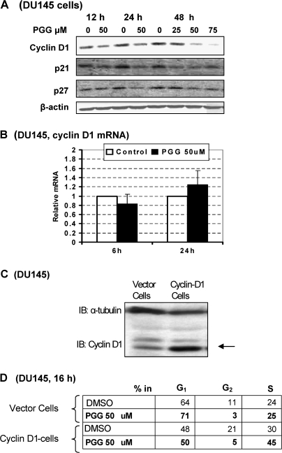
We have recently shown that penta-1,2,3,4,6-O-galloyl-beta-D-glucose (PGG), a naturally occurring hydrolyzable gallotannin, inhibited the in vivo growth of human androgen-independent p53-mutant DU145 prostate cancer (PCa) xenograft in athymic nude mice without adverse effect on their body weight. We have also shown that PGG induced caspase-mediated apoptosis in the DU145 cells and the androgen-dependent human p53-wild-type LNCaP cells. Here, we investigated the cell cycle effects of PGG in these and other PCa cells. Our data show that treatment with subapoptotic doses of PGG induced S-arrest, whereas higher doses of PGG induced not only S-arrest but also G(1) arrest. We show, for the first time, that irrespective of the p53 functional status of the PCa cell lines, PGG exerted a rapid (within 2 h) and potent inhibition (inhibitory concentration by 50% approximately 6 microM) of 5-bromo-2'-deoxyuridine incorporation into S phase cells. In isolated nuclei, PGG inhibited DNA replicative synthesis with superior efficacy than a known DNA polymerase alpha inhibitor, aphidocolin. In addition to the S-arrest action, we have found a close association of downregulation of cyclin D1 with G(1) arrest induced by PGG. Overexpressing this G(1) cyclin abolished G(1) arrest, but hastened the S-arrest induction by PGG. Together, our data indicate that PGG induced PCa S-arrest probably through DNA replicative blockage and induced G(1) arrest via cyclin D1 downregulation to contribute to anticancer activity. Our data raise the hypothesis that PGG may be a novel inhibitor of DNA polymerases.


Figures

Fig. 1.
Fig. 1.
Effect of a general caspase inhibitor zVADfmk on the growth inhibitory action of PGG in DU145 and LNCaP cells. (A) Chemical structure of PGG. (B) Overall inhibitory effects on LNCaP and DU145 cell number after 4 days of daily treatment. Expressed as % of control cell set as 100 (mean ± SEM, n = 3 wells of 6-well plates). P values indicate statistical difference from controls. (C) Detection of cleaved poly (ADP-ribose) polymerase in LNCaP and DU145 cells as an indicator of caspase-mediated apoptosis after 48 h of exposure to increasing doses of PGG. (D) Effect of zVADfmk on the growth inhibitory action of PGG (75 μM) in LNCaP and DU145 cells treated in the presence or absence of 40 μM zVADfmk for 3 days without medium change. Expressed as % of control cell set as 100 (mean ± SEM, n = 3 wells of 12-well plates). Difference between/among key groups are indicated by P values.
Fig. 2.
Fig. 2.
The effect of PGG on cell cycle distribution in LNCaP and DU145 PCa cells. (A) Propidium iodide flowcytometric analysis of the cell cycle distribution of LNCaP and DU145 cells exposed to increasing concentration of PGG for 24 h. Values are mean ± SEM, n = 3 T25 flasks. Difference from control *<0.05, **<0.01. (B and C) Propidium iodide flowcytometric analysis of the cell cycle distribution of LNCaP cells (B) and DU145 cells (C) exposed to 50 μM PGG for 6, 12, 24 and 48 h. Values are mean ± SEM, n = 3 T25 flasks. Difference from respective control *<0.05, **<0.01 (D) Propidium iodide flowcytometric analysis of subG1 (apoptotic) and the cell cycle distribution of DU145 cells exposed to increasing concentration of PGG for 24 h and then returned to drug-free medium for another 24 h. Values are mean ± SEM, n = 3 T25 flasks. Difference from respective control *<0.05, **<0.01.
Fig. 3.
Fig. 3.
BrdU incorporation detection by immunofluorescence in (A) control DU145 cells and cells treated with PGG for 24 h. R3 region encompasses S phase cells that were active in DNA replicative synthesis. (B) Acute time course of PGG (20 μM)-induced inhibition of BrdU incorporation (R3 region) in DU145 cells. (C) Dose-dependent inhibition of BrdU incorporation (R3 region) by PGG exposure for 6 h in DU145 cells. The % of S-phase cells based on DNA content determined by propidium iodide remained unaffected. (D and E) Effects of aphidocolin, a known inhibitor of DNA polymerase alpha, on cell cycle distribution and BrdU incorporation in DU145 cells (D) and LNCaP cells (E). Similar patterns were observed for 12 and 24 h. Each bar represents the average of two T25 flasks, the deviation within 2%.
Fig. 4.
Fig. 4.
The direct effect of PGG or aphidocolin on BrdUTP incorporation into S phase nuclei (in vitro DNA synthesis) of isolated nuclei preparations from DU145 cells. Nuclei were treated with PGG (c and d) or aphidocolin (e and f) for 2 h in the test tubes in the presence of BrdUTP to label those active in replicative synthesis. BrdU detection was carried out by immunofluoresence flowcytometry as for whole cells (Figure 3). LNCaP nuclei showed the same responses.
Fig. 5.
Fig. 5.
Role of P53-p21 axis for G1 and S-arrests by PGG. (A) Effect of PGG on expression of phospho-Ser15-p53, P21Cip1 and cyclin D1 and selected cell cycle proteins in LNCaP cells detected by western blot. (B) Comparison of PGG-induced cell cycle arresting effects in dominant-negative (DN) p53 transfectant (P151S) (panels b and d) versus vector-transfectant LNCaP cells (panels a and c). Each value represents the average of two T25 flasks, with deviation range indicated. (C) Propidium iodide flowcytometric analysis of the cell cycle distribution of p53-null PC-3 cells exposed to increasing concentration of PGG for 24 h. Values are mean ± SEM, n = 3 T25 flasks. Difference from control, **<0.01.
Fig. 6.
Fig. 6.
Effect of PGG on cyclin D1 and P21Cip1 and P27Kip1 expression in DU145 cells and impact of ectopic expression of cyclin D1 on PGG-induced G1 and S-arrest in DU145 cells. (A) Western blot analyses of cyclin D1 and P21Cip1 and P27Kip1 expression. (B) Lack of effect of PGG on cyclin D1 steady state messenger RNA (mRNA) level detected by real-time reverse transcription–polymerase chain reaction. (C) Western blot verification of stable forced overexpression of cyclin D1 in DU145 cells. (D) Cell cycle analyses after 16 h treatment with/without PGG (50 μM) of the vector-transfected cells and cyclin D1-transfected cells. Each value represents the average of two T25 flasks.

Similar articles

Cited by

References

    1. Gupta S, et al. Inhibition of prostate carcinogenesis in TRAMP mice by oral infusion of green tea polyphenols. Proc. Natl Acad. Sci. USA. 2001;98:10350–10355. - PMC - PubMed
    1. Lambert JD, et al. Inhibition of carcinogenesis by polyphenols: evidence from laboratory investigations. Am. J. Clin. Nutr. 2005;81:284S–291S. - PubMed
    1. Singh RP, et al. Mechanisms of action of novel agents for prostate cancer chemoprevention. Endocr. Relat. Cancer. 2006;13:751–778. - PubMed
    1. Hu H, et al. Penta-1,2,3,4,6-O-galloyl-beta-D-glucose induces p53 and inhibits STAT3 in prostate cancer cells in vitro and suppresses prostate xenograft tumor growth in vivo. Mol. Cancer Ther. 2008;7:2681–2691. - PubMed
    1. Pan MH, et al. Induction of apoptosis by penta-O-galloyl-beta-D-glucose through activation of caspase-3 in human leukemia HL-60 cells. Eur. J. Pharmacol. 1999;381:171–183. - PubMed

Publication types

Substances