. 2009 Jun;94(6):739-48.
doi: 10.1113/expphysiol.2008.046136. Epub 2009 Mar 6.

Fibroblast growth factor 2-stimulated proliferation is lower in muscle precursor cells from old rats

Affiliations

Fibroblast growth factor 2-stimulated proliferation is lower in muscle precursor cells from old rats

Seth S Jump et al. Exp Physiol. 2009 Jun.

Abstract

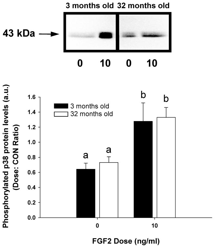
In aged skeletal muscle, impairments in regrowth and regeneration may be explained by a decreased responsiveness of muscle precursor cells (MPCs) to environmental cues such as growth factors. We hypothesized that impaired responsiveness to fibroblast growth factor 2 (FGF2) in MPCs from old animals would be explained by impaired FGF2 signalling. We determined that 5-bromo-2'-deoxyuridine (BrdU) incorporation and cell number increase less in MPCs from 32- compared with 3-month-old rats. In the presence of FGF2, we demonstrated that there were age-associated differential expression patterns for FGF receptor 1 and 2 mRNAs. Measurement of downstream signalling revealed that that mitogen-activated protein kinase/ERK kinase 1/2 (MEK1/2)-extracellular signal-regulated kinase 1/2, protein kinase C and p38 were FGF2-driven pathways in MPCs. Uniquely, protein kinase C signalling was shown to play the largest role in FGF2-stimulated proliferation in MPCs. c-Jun N-terminal kinase (JNK) signalling was ruled out as an FGF2-stimulated proliferation pathway in MPCs. Inhibition of JNK had no effect on FGF2 signalling to BrdU incorporation, and FGF2 treatment was associated with increased phosphorylation of p38, which inhibits, rather than stimulates, BrdU incorporation in MPCs. Surprisingly, the commonly used vehicle, dimethyl sulphoxide, rescued proliferation in MPCs from old animals. These findings provide insight for the development of effective treatment strategies that target the age-related impairments of MPC proliferation in old skeletal muscle.


Figures

Figure 1
Figure 1. Fibroblast growth factor 2-stimulated MPC proliferation
A, CyQUANT assay for cell number in MPCs from 3- and 32-month-old animals. The assay was performed after 48 h of FGF2 treatment (n = 4). The cell number at each dose of FGF2 stimulation was normalized to control cell number (CON; 0 ng ml−1 FGF2). B, BrdU incorporation data of MPCs from 3- and 32-month-old animals treated with different doses of FGF2 for 24 h (n = 4 for 1.0, 2.5 and 10 ng ml−1, n = 6 for 5 ng ml−1 and n = 2 for 100 ng ml−1). Values are means ± S.E.M. * Significant difference for cell number compared with 32-month-old animals (P < 0.05).
Figure 2
Figure 2. Quantification of mRNA for FGFR isoforms
A, mRNA for FGFRs determined in MPCs from 3- and 32-month-old animals after 24 h in GM (n = 4 for young and old). Relative quantities for each isoform were determined based on the relative quantity value for FGFR1 from 3-month-old animals set to 1. B, quantification of mRNA in MPCs from 3- and 32-month-old animals after 24 h of FGF2 treatment (n = 3 for young and old). Relative quantities for each isoform were determined based on the relative quantity of FGFR1 from 3-month-old animals in GM set to 1. All relative quantities of FGFR1–4 mRNAs in low-serum conditions were significantly less than their respective high-serum values. The relative expression levels of the FGFR isoform mRNAs were significantly different in the rank order of FGFR4 ≫ FGFR1 = FGFR2 = FGFR3. Different letters denotes a significant difference between groups.
Figure 3
Figure 3. Cell signalling measured by Western blot
A, Western blot analysis of the increase in phosphorylated MEK1/2 (Ser217/Ser221) in MPC cell lysates with FGF2 treatment (0, 0.5 or 10 ng ml−1) with a representative Western blot; n = 8 for young and n = 7 for old. Muscle precursor cells were grown for 48 h in GM. Then, the medium was changed to low-serum medium for 3 h. After 3 h, the MPCs were treated with FGF2 for 10 min. B, Western blot analysis of the increase in phosphorylated ERK1/2 (Thr202/Tyr204) in MPC cell lysates with FGF2 treatment (0, 0.5 or 10 ng ml−1) with a representative Western blot; n = 9 for young and n = 8 for old. Cell culture conditions were same as in A. Different letters denote a significant difference between FGF2 doses (P < 0.05).
Figure 4
Figure 4. Representative Western blot showing levels of phosphorylated p38 protein (Thr180/Tyr182) in MPC cell lysates
The figure shows quantitative Western blot data (n = 6 for young and old) from MPCs treated with FGF2 (0 or 10 ng ml−1). Muscle precursor cells were grown in GM for 24 h. After 24 h, GM was changed to low-serum medium for a 3 h equilibration period. After 3 h of treatment, FGF2 was added to the medium for a 10 min treatment. Data were analysed by two-way ANOVA. Different letters denote statistical significance (P < 0.05).
Figure 5
Figure 5. Bromodeoxyuridine-positive nuclei with MEK1/2 and PKC inhibition in the presence of FGF2 (10 ng ml−1) and without or with DMSO (0.1%)
Muscle precursor cells were grown for 24 h in GM. After 24 h, the medium was changed to low-serum medium with or without DMSO (n = 4 without DMSO and n = 6 with DMSO). After 1 h of exposure to DMSO, FGF2 (10 ng ml−1) was added to the medium. At 23 h of treatment with FGF2, MPCs were pulse-labelled with BrdU for 1 h. Muscle precursor cells were harvested and fixed after 24 h of treatment. Dissimilar letters denote significant differences between young and old without DMSO (P < 0.05).
Figure 6
Figure 6. MEK1/2 inhibitor Western blot and proliferation data
A, representative Western blot for phosphorylated ERK1/2 (Thr202/Tyr204) in MPC cell lysates treated with FGF2 (10 ng ml−1) in the presence or absence of U0126. Muscle precursor cells were grown for 24 h in GM. After 24 h, the medium was changed to 2% HS, and chemical inhibitors were added to the medium (0.4% DMSO) 1 h prior to a 10 min FGF2 treatment. B, representative Western blot for phosphorylated ERK1/2 (Thr202/Tyr204) in MPC cell lysates treated with FGF2 (10 ng ml−1) the presence of MEK1/2 inhibitor SL327 with the same cell culture conditions as in A. C, BrdU-positive nuclei with MEK1/2 and PKC inhibition in the presence of FGF2; n = 6 for young and old. CON = no FGF2 + DMSO (0.1%). Passage 2 MPCs were grown for 24 h in GM. After 24 h, the medium was changed to low-serum medium, and MEK1/2 inhibitors (10 μM U0126 and 40 μM MEK1/2 inhibitor) and PKC inhibitior (10 μM bisindolylmaleimide I) were added for 1 h, prior to treatment with FGF2 for 23 h. Different letters denote significant differences between inhibitor types or conditions (P < 0.05).
Figure 7
Figure 7. Bromodeoxyuridine-positive nuclei with MEK1/2 and p38 pathway inhibition in the presence of FGF2 (10 ng ml−1)
CON = No FGF2 + DMSO (0.4%). Passage 2 MPCs were grown for 24 h in GM. After 24 h, the medium was changed to low-serum medium containing 0.4% DMSO and with two different chemical inhibitors alone and in combination (10 μM U0126 and 10 μM SB203580). After 1 h of treatment with inhibitors, FGF2 was added to the media. After 23 h of treatment with FGF2, MPCs were pulse-labelled with BrdU (10 μM) for 1 h. Muscle precursor cells were harvested and fixed after 24 h of treatment. Different letters denote significant differences between inhibitor types or conditions (P < 0.05). n = 3 for no inhibitor, n = 6 for U0126, n = 2 for SB203580 + U0126 and CON and n = 4 for SB20580.

References

    1. Allen RE, Boxhorn LK. Regulation of skeletal muscle satellite cell proliferation and differentiation by transforming growth factor-β, insulin-like growth factor I, and fibroblast growth factor. J Cell Physiol. 1989;138:311–315. - PubMed
    1. Allen RE, Temm-Grove CJ, Sheehan SM, Rice G. Skeletal muscle satellite cell cultures. Methods Cell Biol. 1997;52:155–176. - PubMed
    1. Anderson JE, Mitchell CM, McGeachie JK, Grounds MD. The time course of basic fibroblast growth factor expression in crush-injured skeletal muscles of SJL/J and BALB/c mice. Exp Cell Res. 1995;216:325–334. - PubMed
    1. Brien S, Prescott P, Bashir N, Lewith H, Lewith G. Systematic review of the nutritional supplements dimethyl sulfoxide (DMSO) and methylsulfonylmethane (MSM) in the treatment of osteoarthritis. Osteoarthritis Cartilage. 2008;16:1277–1288. - PubMed
    1. Brooks SV, Faulkner JA. Contraction-induced injury: recovery of skeletal muscles in young and old mice. Am J Physiol Cell Physiol. 1990;258:C436–C442. - PubMed

Publication types

MeSH terms