. 2009 May;37(8):2596-606.
doi: 10.1093/nar/gkp115. Epub 2009 Mar 6.

Plasmodium falciparum heterochromatin protein 1 binds to tri-methylated histone 3 lysine 9 and is linked to mutually exclusive expression of var genes

Affiliations

Plasmodium falciparum heterochromatin protein 1 binds to tri-methylated histone 3 lysine 9 and is linked to mutually exclusive expression of var genes

Karla Pérez-Toledo et al. Nucleic Acids Res. 2009 May.

Abstract

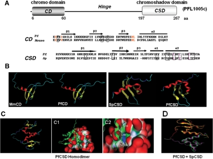
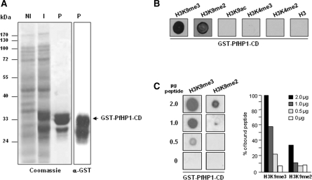
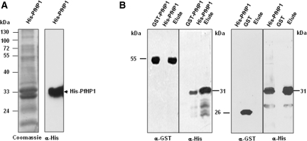
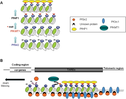
Increasing experimental evidence shows a prominent role of histone modifications in the coordinated control of gene expression in the human malaria parasite Plasmodium falciparum. The search for the histone-mark-reading machinery that translates histone modifications into biological processes, such as formation of heterochromatin and antigenic variation is of foremost importance. In this work, we identified the first member of a histone modification specific recognition protein, an orthologue of heterochromatin protein 1 (PfHP1). Analysis of the PfHP1 amino-acid sequence revealed the presence of the two characteristic HP1 domains: a chromodomain (CD) and a chromo shadow domain (CSD). Recombinant CD binds to di- and tri-methylated lysine 9 from histone H3, but not to unmodified or methylated histone H3 in lysine 4. PfHP1 is able to interact with itself to form dimers, underlying its potential role in aggregating nucleosomes to form heterochromatin. Antibodies raised against PfHP1 detect this molecule in foci at the perinuclear region. ChIP analysis using anti-PfHP1 shows that this protein is linked to heterochromatin of subtelomeric non-coding repeat regions and monoallelic expression of the major virulence var gene family. This is the first report implicating an HP1 protein in the control of antigenic variation of a protozoan parasite.


Figures

Figure 1.
Figure 1.
Structural modelling of the chromo domain (CD) and chromoshadow domain (CSD) of PfHP1. (A) Schematic diagram showing the complete ORF and location of CD and CSD of PfHP1 separated by the hinge. Numbers correspond to residue positions for each domain. The primary sequence alignment of CD from mouse HP1ß (19–73 aa) and PfHP1 (6–60 aa) was used for the modelling of the CD domain. The boxes indicate the aromatic cage residues potentially involved in the recognition of the H3K9me peptide. Conserved residues that form a complementary surface responsible for H3 peptide recognition in the CD from Drosophila are indicated in red (24). The primary sequence alignment of CSD from S. pombe (261–320 aa) and from PfHP1 (197–267 aa) was used to perform the model of the CSD domains. Residues implicated in the formation of the dimer interface are enclosed in boxes. Residues that have shown to be important for dimer formation in S. pombe (25) are indicated with pink letters. Both alignments were performed with ClustalW and used for modelling with the program MOE (www.chemcomp.com). The secondary structure elements are shown above the alignment: bars represent α-helices (α-1 and α-2) and arrows represent β-strands (β1–β3). (B) Tertiary structure of CD and CSD domains of PfHP1. The structure is conserved between mouse and P. falciparum CD domains, and between S. pombe and P. falciparum CSD domains. (C) Cartoon representation of the CSD dimer. The two monomers are indicated in red. The molecular surface of the putative PfHP1-CSD dimer is shown. (C1) A putative monomer is shown by cartoon and the other one by molecular surface. Surface convex zones are shown in red (protuberances); concave zones that correspond to binding or recognition sites are shown in green (hydrophobic regions) and the polar surface is denoted in blue. (C2) The hydrophobic region and the crucial L residues that contact the other subunit in the formation of the dimer are indicated. (D) Superposition of the 3D models of the CSD domains homodimers from Swi6 (magenta–yellow) and PfHP1 homodimers (green–red). The CSD architecture is strongly conserved between S. pombe and P. falciparum.
Figure 2.
Figure 2.
PfHP1 binds to histone H3 peptide methylated at lysine 9. (A) Total proteins from DH5α strain expressing the CD of PfHP1 before (NI) and after IPTG induction (I), as well as the purified GST-PfHP1-CD fusion protein (P), were separated by SDS–PAGE and stained with Coomassie blue (left). An anti-GST antibody was used to detect the presence of the fusion protein (right). (B) Purified protein GST-PfHP1-CD was assayed for binding to histone H3 peptides. 200 ng of GST-CD protein (GST-PfHP1-CD) were dotted to a nitrocellulose membrane and incubated with 2 µg of the following biotinylated peptides: H3K9me3, H3K9me2, H3K9ac, H3K4me3, H3K4me2 and unmodified H3. (C) Two hundred nanograms of GST-PfHP1-CD fusion protein were dotted to nitrocellulose and incubated with decreasing amounts of H3K9me3 and H3K9me2 biotinylated peptides (left panels). A representative result of two independent experiments is shown. The right panel shows a quantitative presentation of the GST-PfHP1-CD affinity to the peptides.
Figure 3.
Figure 3.
In vitro dimerization of the PfHP1 protein. (A) His-PfHP1 protein purified using nickel-nitriloacetic acid resin (left). Western blot analysis with anti-histidine antibodies identified the monomer of PfHP1 (right). (B) Interaction between His-PfHP1 (∼30 kDa) and GST-PfHP1 fusion proteins (∼55 kDa). Purified His-PfHP1 was mixed with total bacterial lysate overexpressing GST-PfHP1 fusion protein (left panels) or only GST (right panels). Protein complexes were recovered, separated by SDS–PAGE and detected by Western blot with anti-histidine and anti-GST antibodies.
Figure 4.
Figure 4.
Nuclear expression and cellular localization of PfHP1. (A) Western blot analysis of nuclear extracts from P. falciparum using the following antibodies: anti-GST antibody (left), preimmune serum, anti-GST-PfHP1 antibody (middle) and anti-acetyl histone 4 antibodies (right). (B) IFA analysis of PfHP1 (green) throughout the parasite asexual blood stage cycle (ring, trophozoite and schizont stages). (C) In ring stage, partial co-localization between PfHP1 (red) and PfSir2 (green) antibodies (yellow spots) is observed. (D) Co-localization with a nucleolar marker PfNop1 shows minimal overlap of PfHP1 with this compartment. Scale bars represent 1 µm.
Figure 5.
Figure 5.
PfHP1 is associated to constitutive and facultative heterochromatin. (A) Schematic representation of telomeric and subtelomeric regions of P. falciparum. In this parasite, chromosome ends are composed of telomeres and telomere associated repetitive elements (TAREs 1–6). The subtelomeric coding region contains members of several multigene families important for virulence, such as the var genes. The var gene repertoire is classified in different types according to the conserved upstream sequences. Ups A, B and E types are located on subtelomeric regions, whereas UpsC types are found in central chromosomal position. The positions of the probes used in ChIP assays are shown. (B) Dot blot based ChIP analysis of PfHP1 abundance at telomeric and non-coding subtelomeric regions. DNA probes corresponding to TARE 1, 2, 3, 6 and a sequence between TARE 2–3 were fixed to nylon membranes and hybridized to DNA immunoprecipitated with anti-PfHP1 antibodies and preimmune serum (PI) (left panels). The 5′-UTR region of the histidine rich protein 1 (HRP1) gene, expressed during blood stage, was used as control (left panels). (C) ChIP analysis of FCR3-CSA parasites, in which the var2CSA gene is active (UpsE ON). (D) ChIP analysis of FCR3-CD36 parasites, in which the var2CSA gene is repressed (UpsE OFF). In (C) and (D) DNA corresponding to different types of var 5′-UTR regions (UpsE, UpsC and UpsB), 5′-UTR region of HRP1 and TARE6 were fixed on nylon membranes and hybridized to DNA immunoprecipitated with anti-PfHP1 antibodies and PI serum (left panels). The right panels show a quantitative representation of PfHP1 levels at the different DNA regions. In all cases, ChIP analysis was performed in ring stage parasites and a representative result is shown. Error bars represent the standard deviation from three independent experiments each performed in duplicate. Input corresponds to DNA prepared from fragmented chromatin prior to immunoprecipitation.
Figure 6.
Figure 6.
Hypothetical model for heterochromatin formation at P. falciparum chromosome ends. (A) PfHP1-mediated heterochromatin model depends on the action of the histone deacetylase PfSir2 and histone methyltransferase PfKMT1 (10,15,37). (B) General view of the known chromatin components at P. falciparum subtelomeres. Spreading of heterochromatin along the different TAREs into adjacent coding regions probably involves the cooperation of PfHP1, PfSir2 and PfKMT1. The role of PfOrc1 in this process remains unknown.

Similar articles

Cited by

References

    1. Bozdech Z, Llinas M, Pulliam BL, Wong ED, Zhu J, DeRisi JL. The transcriptome of the intraerythrocytic developmental cycle of Plasmodium falciparum. PLoS Biol. 2003;1:E5. - PMC - PubMed
    1. Le Roch KG, Zhou Y, Blair PL, Grainger M, Moch JK, Haynes JD, De la Vega P, Holder AA, Batalov S, Carucci DJ, et al. Discovery of gene function by expression profiling of the malaria parasite life sycle. Science. 2003;301:1503–1508. - PubMed
    1. Aravind L, Iyer LM, Wellems TE, Miller LH. Plasmodium biology: genomic gleanings. Cell. 2003;115:771–785. - PubMed
    1. Templeton TJ, Iyer LM, Anantharaman V, Enomoto S, Abrahante JE, Subramanian GM, Hoffman SL, Abrahamsen MS, Aravind L. Comparative analysis of apicomplexa and genomic diversity in eukaryotes. Genome Res. 2004;14:1686–1695. - PMC - PubMed
    1. Callebaut I, Prat K, Meurice E, Mornon JP, Tomavo S. Prediction of the general transcription factors associated with RNA polymerase II in Plasmodium falciparum: conserved features and differences relative to other eukaryotes. BMC Genomics. 2005;6:100. - PMC - PubMed

Publication types

MeSH terms