Comparative Study
. 2009 Jun;19(6):665-73.
doi: 10.1093/glycob/cwp036. Epub 2009 Mar 6.

Secondary cell wall polysaccharides of Bacillus anthracis are antigens that contain specific epitopes which cross-react with three pathogenic Bacillus cereus strains that caused severe disease, and other epitopes common to all the Bacillus cereus strains tested

Affiliations
Comparative Study

Secondary cell wall polysaccharides of Bacillus anthracis are antigens that contain specific epitopes which cross-react with three pathogenic Bacillus cereus strains that caused severe disease, and other epitopes common to all the Bacillus cereus strains tested

Christine Leoff et al. Glycobiology. 2009 Jun.

Abstract

The immunoreactivities of hydrogen fluoride (HF)-released cell wall polysaccharides (HF-PSs) from selected Bacillus anthracis and Bacillus cereus strains were compared using antisera against live and killed B. anthracis spores. These antisera bound to the HF-PSs from B. anthracis and from three clinical B. cereus isolates (G9241, 03BB87, and 03BB102) obtained from cases of severe or fatal human pneumonia but did not bind to the HF-PSs from the closely related B. cereus ATCC 10987 or from B. cereus type strain ATCC 14579. Antiserum against a keyhole limpet hemocyanin conjugate of the B. anthracis HF-PS (HF-PS-KLH) also bound to HF-PSs and cell walls from B. anthracis and the three clinical B. cereus isolates, and B. anthracis spores. These results indicate that the B. anthracis HF-PS is an antigen in both B. anthracis cell walls and spores, and that it shares cross-reactive, and possibly pathogenicity-related, epitopes with three clinical B. cereus isolates that caused severe disease. The anti-HF-PS-KLH antiserum cross-reacted with the bovine serum albumin (BSA)-conjugates of all B. anthracis and all B. cereus HF-PSs tested, including those from nonclinical B. cereus ATCC 10987 and ATCC 14579 strains. Finally, the serum of vaccinated (anthrax vaccine adsorbed (AVA)) Rhesus macaques that survived inhalation anthrax contained IgG antibodies that bound the B. anthracis HF-PS-KLH conjugate. These data indicate that HF-PSs from the cell walls of the bacilli tested here are (i) antigens that contain (ii) a potentially virulence-associated carbohydrate antigen motif, and (iii) another antigenic determinant that is common to B. cereus strains.


Figures

Fig. 1
Fig. 1
The repeating unit structure of the HF-PS from B. anthracis Sterne, Pasteur, and Ames as previously reported (top) and the consensus structure that is indicated for the HF-PS from members of the B. cereus group (bottom). This consensus repeating unit structure can be substituted by Gal, Glc as well as by acetyl groups (indicated by “X”).
Fig. 2
Fig. 2
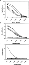
Immunoreactivity of the HF-PSs to antisera raised in rabbits against B. anthracis Sterne (A) live spores and (B) killed spores, and to (C) B. cereus ATCC 14579 spores. The ELISA microtiter plate wells were coated with B. anthracis Pasteur HF-PS-BSA (▴), B. cereus G9241 HF-PS-BSA (□), B. cereus 03BB87 HF-PS-BSA (•), B. cereus 03BB102 HF-PS-BSA (⧫), B. cereus ATCC 14579 HF-PS (x); the chemically synthesized AntRha2-BSA conjugate (Mehta et al. 2006) was used as a positive control (◊), and maltoheptaose-BSA (○) and BSA (|) alone as negative controls. Even though the antisera were raised against spores from B. anthracis Sterne and the HF-PS-BSA conjugate was prepared from B. anthracis Pasteur HF-PS, it should be noted that the HF-PSs from B. anthracis Ames, Pasteur, and Sterne all have identical structures (Leoff, Saile, et al. 2008).
Fig. 3
Fig. 3
An ELISA inhibition assay showing the ability of various HF-PSs to inhibit the binding of antiserum to live B. anthracis spores to the HF-PS-BSA conjugate of B. anthracis Pasteur. The microtiter plates were coated with the HF-PS-BSA and the ability to bind antiserum that had been incubated with various concentrations of the different HF-PS preparation was determined as described in Material and methods. B. anthracis Pasteur HF-PS (◊); B. anthracis Ames HF-PS (▪); B. cereus G9241 HF-PS (Δ); B. cereus ATCC 14579 HF-PS (○); B. cereus ATCC 10987 HF-PS (▴); AntRha2 (•); maltoheptaose (x); BSA (□).
Fig. 4
Fig. 4
Reactivity of rabbit anti-B. anthracis HF-PS-KLH antiserum with HF-PS from other species. ELISA microtiter plates were coated with the various HF-PS-BSA conjugates and the ability of the HF-PS-KLH antiserum to bind these conjugates was determined as described in Materials and methods. B. anthracis HF-PS-BSA (•); B. cereus ATCC 14579 HF-PS-BSA (▴); chemically synthesized AntRha2 trisaccharide-BSA (⧫); maltoheptaose-BSA (x); and BSA only (▪).
Fig. 5
Fig. 5
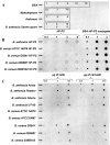
An immuno-dot blot assay shows the binding of the antiserum to B. anthracis HF-PS-KLH conjugate to the indicated amount (μg) of (A) BSA, maltoheptaose, chemically synthesized AntRha2 trisaccharide (labeled as Anthrose), and B. anthracis Sterne spores; (B) unconjugated and BSA-conjugated HF-PS from the indicated B. anthracis and B. cereus strains; and (C) cells and cell walls from the indicated B. anthracis and B. cereus strains.
Fig. 6
Fig. 6
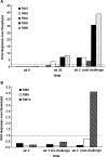
Reactivity of Rhesus macaque sera with B. anthracis HF-PS. Pre- and postexposure sera from five AVA-vaccinated (A) and three naïve (B) animals were tested with ELISA for the presence of anti-HF-PS IgG on plates coated with the B. anthracis Pasteur HF-PS conjugated to KLH. The dashed line indicates the reactivity threshold (RT), corresponding to the value 1, which was determined by testing the sera from 88 true negative Rhesus macaques (RM) as described in Materials and methods. Based on the RT, OD values were transformed to represent the fold rise over the RT. For all animals baseline, sera drawn in week 0 were tested. For the vaccinated animals (A) sera from week 30, after a course of three AVA shots, and 14 days after exposure to B. anthracis Ames spores were tested. For the naïve Rhesus macaques (B) preexposure sera drawn 3 weeks before exposure and postexposure sera from day 14 were tested.

References

    1. Avashia SB, Riggins WS, Lindley C, Hoffmaster AR, Drumgoole R, Nekomoto T, Jackson PJ, Hill KK, Williams K, Lehman L, et al. Fatal pneumonia among metalworkers due to inhalation exposure to Bacillus cereus containing Bacillus anthracis toxin genes. Clin Infect Dis. 2007;44:414. - PubMed
    1. Baillie LW. Bacillus anthracis, a story of nature subverted by man. Lett Appl Microbiol. 2005;41:227–229. - PubMed
    1. Bhavsar AP, Erdman LK, Schertzer JW, Brown ED. Teichoic acid is an essential polymer in Bacillus subtilis that is functionally distinct from teichuronic acid. J Bacteriol. 2004;186:7865–7873. - PMC - PubMed
    1. Bystricky S, Machova E, Bartek P, Kolarova N, Kogan G. Conjugation of yeast mannans with protein employing cyanopyridinium agent (CDAP) – An effective route of antifungal vaccine preparation. Glycoconj J. 2000;17:677–680. - PubMed
    1. Choudhury B, Leoff C, Saile E, Wilkins P, Quinn CP, Kannenberg EL, Carlson RW. The structure of the major cell wall polysaccharide of Bacillus anthracis is species specific. J Biol Chem. 2006;281:27932–27941. - PubMed

Publication types

MeSH terms