. 2009 Mar 31;106(13):5252-7.
doi: 10.1073/pnas.0900691106. Epub 2009 Mar 6.

A mouse model for nonsyndromic deafness (DFNB12) links hearing loss to defects in tip links of mechanosensory hair cells

Affiliations

A mouse model for nonsyndromic deafness (DFNB12) links hearing loss to defects in tip links of mechanosensory hair cells

Martin Schwander et al. Proc Natl Acad Sci U S A. .

Abstract

Deafness is the most common form of sensory impairment in humans and is frequently caused by single gene mutations. Interestingly, different mutations in a gene can cause syndromic and nonsyndromic forms of deafness, as well as progressive and age-related hearing loss. We provide here an explanation for the phenotypic variability associated with mutations in the cadherin 23 gene (CDH23). CDH23 null alleles cause deaf-blindness (Usher syndrome type 1D; USH1D), whereas missense mutations cause nonsyndromic deafness (DFNB12). In a forward genetic screen, we have identified salsa mice, which suffer from hearing loss due to a Cdh23 missense mutation modeling DFNB12. In contrast to waltzer mice, which carry a CDH23 null allele mimicking USH1D, hair cell development is unaffected in salsa mice. Instead, tip links, which are thought to gate mechanotransduction channels in hair cells, are progressively lost. Our findings suggest that DFNB12 belongs to a new class of disorder that is caused by defects in tip links. We propose that mutations in other genes that cause USH1 and nonsyndromic deafness may also have distinct effects on hair cell development and function.


Conflict of interest statement

The authors declare no conflict of interest.

Figures

Fig. 1.
Fig. 1.
Analysis of auditory function. (A) Diagram of a developing hair bundle. Stereocilia are connected to each other and to the kinocilium by various linkages. Linkages that contain PCDH15 and CDH23 are indicated. (B) The diagram shows the localization of CDH23 and PCDH15 at tip links. (C) Click-evoked ABR for a 2-month-old wild-type (black lines) and a salsa mouse (red line) at different sound intensities (dB). ABR waves I–IV are indicated. (D) Average auditory thresholds for 3-week-old and 2-month-old mice [wild-type n = 4 for 3 weeks (white) and 2 months (gray); salsa n = 5 for 3 weeks (orange) and 2 months (red)]. The mean ± SD is indicated; a Student's t test was performed. (E) Auditory thresholds in 3-week-old (triangles) and 2-month-old (circles) mice as determined by pure tone ABR recordings. In contrast to wild-type (gray and black lines) salsa mutants (orange and red lines) showed progressive hearing loss. (F) Analysis of movement in the open field. salsa mice (red) showed normal numbers of small-diameter rotations (>2.75-cm radius) and were not hyperactive. As a positive control, the sirtaki mouse line (gray) that has vestibular defects (35) is shown. Values are mean ± SD. A Student t test was performed. **, P < 0.01; ***, P > 0.001.
Fig. 2.
Fig. 2.
The mutation in salsa mice maps to a Ca2+-binding motif in CDH23. (A) Sequence chromatograph from wild-type and salsa mice reveals an A-to-T mutation in exon 22 of Cdh23. (B) The C-terminal part of EC7 of CDH23 from different species is shown. The Glu737Val substitution in salsa affects a conserved Ca2+-binding motif (yellow boxes). CDH1 and CDH2 are shown for comparison. (C) CDH23 EC7/8 wild-type (blue) and salsa mutant (red) sequences threaded onto the E-cadherin EC1/2 by using the automodel class and a sequence alignment produced by T-Coffee. Energy-minimized model shows that the Glu737Val mutation affects Ca2+ coordination. (D) Domain structure of CDH23 indicating the 27 extracellular cadherin repeats (blue). Missense mutations in Ca2+-binding motifs in the CDH23 extracellular domain cause DFNB12 in humans (purple shaded box). Nonsense and splice site mutations have been identified in waltzer mice and USH1D (gray shaded box). SS indicates signal sequence; TM, transmembrane domain.
Fig. 3.
Fig. 3.
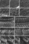
Preserved hair bundle morphology in salsa mice. (A–F) Scanning electron microscopy micrographs of cochlear whole mounts from P5 wild-type mice and salsa and waltzer mutants. (A and B) The organ of Corti in salsa mice was patterned normally in 3 rows of outer and 1 row of inner hair cells. (C–F) Hair bundles in salsa displayed the characteristic polarized morphology with a single kinocilium. Hair bundles in waltzer mice were fragmented. (G–L) At P28, hair bundle morphology was indistinguishable in wild-type and salsa mice. [Scale bars: A and B (5 μm), C–F (2 μm), G and H (10 μm), and I–L (2 μm).]
Fig. 4.
Fig. 4.
Progressive loss of CDH23 expression in salsa mice. (A–J) Cochlear whole mounts of wild-type and salsa mice were stained with an antibody against the CDH23 cytoplasmic domain (green) and with phalloidin (red). (A and B) Levels of CDH23 expression in homozygous salsa and wild-type mice at P5 were similar. (C and D) Higher-magnification view of CDH23 expression in hair bundles at P5. (E and F) At P10, CDH23 staining was maintained at stereociliary tips. (G–J) At P30, CDH23 staining was barely detectable in salsa. Arrowheads point to CDH23 in wild types and residual staining in salsa. (K) Quantification of CDH23 staining in hair cells at P10 and P30. salsa mice (black) showed reduced numbers of CDH23 puncta in hair bundles at P30. Values are mean ± SD. A Student's t test was performed. ***, P < 0.001. [Scale bar: A and B (8 μm), C–J (2 μm).]
Fig. 5.
Fig. 5.
Progressive loss of tenting at stereociliary tips. (A and B) Scanning electron microscopy analysis in P5 wild-type and salsa mice revealed tip tenting in stereocilia of cochlear hair cells (arrowheads). (C and D) Defects in tenting in salsa mice at P60 (arrowheads in C indicate tenting in wild-type). (E–G) Tips in vestibular hair cells display normal tenting. Tip links (arrowhead in G) were preserved. (H) Quantification of tip tenting at P5 and P21. Tenting was reduced in hair cells from the medial and basal cochlear turns at P21. *, P < 0.05. [Scale bars: A–D (300 nm), E and F (500 nm), and G (150 nm).]
Fig. 6.
Fig. 6.
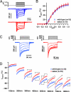
Mechanotransduction currents. Data from wild type are represented in blue and from salsa in red. (A) Examples of transduction currents in cochlear outer hair cells at P7 in response to 5-ms mechanical stimulation. (B) Current-displacement [I(X)] relationships revealed no obvious difference between wild type and salsa. (C) Examples of transduction currents in cochlear outer hair cells at P7 in response to 100-ms mechanical stimulation. (D) Averaged transduction currents for deflections between 100 and 800 nm expressed as a percentage of peak currents at 800 nm. Current amplitude was lower in homozygous salsa mice between 300-nm and 800-nm deflection. Data are shown as mean ± SEM. Student's 2-tailed unpaired t test was performed (*, P < 0.05; **, P < 0.01).
Fig. 7.
Fig. 7.
salsa and DFNB12 mutations affect interactions between CDH23 and PCDH15. (A) Diagram of CDH23 and PCDH15 constructs. The extracellular domains were fused to a His or Fc tag. (B) CDH23Fc and the mutant derivatives were incubated with PCDH15-His in the presence of 1 mM EDTA or in the presence of 10 μM and 100 μM Ca2+. Protein complexes were purified and analyzed by Western blotting. Complex formation was observed in the presence but not absence of Ca2+. The salsa and DFNB12 mutations weakened interactions between CDH23 and PCDH15. (Right) Controls for input amounts of CDH23Fc and mutant derivatives.

Comment in

References

    1. Astuto LM, et al. CDH23 mutation and phenotype heterogeneity: A profile of 107 diverse families with Usher syndrome and nonsyndromic deafness. Am J Hum Genet. 2002;71:262–275. - PMC - PubMed
    1. Baux D, et al. UMD-USHbases: A comprehensive set of databases to record and analyse pathogenic mutations and unclassified variants in seven Usher syndrome causing genes. Hum Mutat. 2008;29:E76–E87. - PubMed
    1. Becirovic E, et al. Usher syndrome type 1 due to missense mutations on both CDH23 alleles: Investigation of mRNA splicing. Hum Mutat. 2008;29:452. - PubMed
    1. Bolz H, et al. Mutation of CDH23, encoding a new member of the cadherin gene family, causes Usher syndrome type 1D. Nat Genet. 2001;27:108–112. - PubMed
    1. Bork JM, et al. Usher syndrome 1D and nonsyndromic autosomal recessive deafness DFNB12 are caused by allelic mutations of the novel cadherin-like gene CDH23. Am J Hum Genet. 2001;68:26–37. - PMC - PubMed

Publication types