. 2009 May;191(9):3024-40.
doi: 10.1128/JB.01505-08. Epub 2009 Mar 6.

Influences of capsule on cell shape and chain formation of wild-type and pcsB mutants of serotype 2 Streptococcus pneumoniae

Affiliations

Influences of capsule on cell shape and chain formation of wild-type and pcsB mutants of serotype 2 Streptococcus pneumoniae

Skye M Barendt et al. J Bacteriol. 2009 May.

Abstract

PcsB is a protein of unknown function that plays a critical role in cell division in Streptococcus pneumoniae and other ovococcus species of Streptococcus. We constructed isogenic sets of mutants expressing different amounts of PcsB in laboratory strain R6 and virulent serotype 2 strain D39 to evaluate its cellular roles. Insertion mutagenesis in parent and pcsB(+) merodiploid strains indicated that pcsB is essential in serotype 2 S. pneumoniae. Quantitative Western blotting of wild-type and epitope-tagged PcsB showed that all PcsB was processed into cell-associated and secreted forms of the same molecular mass and that cell-associated PcsB was moderately abundant and present at approximately 4,900 monomers per cell. Controlled expression and complementation experiments indicated that there was a causative relationship between the severity of defects in cell division and decreasing PcsB amount. These experiments also showed that perturbations of expression of the upstream mreCD genes did not contribute to the cell division defects of pcsB mutants and that mreCD could be deleted. Unexpectedly, capsule influenced the cell shape and chain formation phenotypes of the wild-type D39 strain and mutants underexpressing PcsB or deleted for other genes involved in peptidoglycan biosynthesis, such as dacA. Underexpression of PcsB did not result in changes in the amounts or composition of lactoyl-peptides, which were markedly different in the R6 and D39 strains, and there was no correlation between decreased PcsB amount and sensitivity to penicillin. Finally, microarray analyses indicated that underexpression of PcsB may generate a signal that increases expression of the VicRK regulon, which includes pcsB.


Figures

FIG. 1.
FIG. 1.
Genetic constructs used in this study (drawn to scale). See Table 1 for strains. (A) The native pcsB locus showing the upstream mreCD genes and the downstream rpsB gene that encodes a ribosomal protein. The ΔpcsB<>ermAM and ΔpcsB<>Pc-ermAM reading frame replacements of pcsB are indicated along with the mapped internal PpcsB promoter (L.-T. Sham, unpublished result) and putative terminators (lollipops). The location of the intercistronic Pc-ermAM insertion used as a marker in some constructs is indicated 1,180 nucleotides downstream from the pcsB start codon. (B) Structures of the ΔbgaA′:: kant1t2-Pc-pcsB+ and ΔbgaA′:: kant1t2-PfcsK-pcsB+ constructs used for ectopic expression of PcsB in strains IU1979, IU2801, and IU2537. The kant1t2-Pc-pcsB+ or kant1t2-PfcsK-pcsB+ constructs contain the constitutive synthetic Pc promoter and ermAM ribosome binding site (37) or the fucose-inducible PfcsK promoter and fcsK ribosome binding site (10), respectively, and the t1t2 ribosome operon terminators from E. coli to block transcription from upstream of Pc and PfcsK (46). The constructs were inserted between the end of pts-eII and a deletion of the promoter-proximal region of bgaA that extended 6297 nucleotides from the translation start codon of bgaA. (C) Insertion of the kant1t2-Pc-pcsB+ and kant1t2-PfcsK-pcsB+ constructs into the native pcsB locus. The constructs replaced the intercistronic region between mreCD and pcsB, which included the putative terminator and PpcsB internal promoter shown in panel A. No known transcription terminator follows the divergently transcribed kan gene in these constructs.
FIG. 2.
FIG. 2.
Western immunoblot analyses of cell-associated (pellet) and secreted (supernatant) PcsB. (A) Demonstration that all cell-associated and secreted PcsB is processed to the same Mr. IU1690 (D39 parent) and IU2105 expressed wild-type (41.9 kDa) and C-terminal FLAG-tagged PcsB (42.8 kDa), respectively. The left four lanes or right four lanes were probed with affinity-purified anti-PcsB or commercial anti-FLAG antibody, respectively, as described in Materials and Methods. The autoradiogram is representative of several independent experiments but was not used for quantitation, because the signal was not strictly linear with concentration. Instead, blots were quantitated directly by using an IVIS imaging system (see Materials and Methods). (B) Production of cell-associated unprocessed (SP-PcsB; Mr, 45.0) and processed (PcsB; Mr, 41.9) PcsB by R6 pcsB+ ΔbgaA′::PfcsK-pcsB (A27D) merodiploid strain IU2718 grown in CDM containing 0.8% (wt/vol) fucose. A 28-μg aliquot of pellet extract from IU2718 was loaded and Western blots were probed with purified anti-PcsB antibody (see above). A pellet extract from R6 grown in CDM was included as a marker on the same gel. Intervening lanes were removed from the figure. (C) Quantitation of the absolute amount of cell-associated PcsB was performed as described in Materials and Methods using affinity-purified anti-PcsB antibody. A representative autoradiogram is shown of the PcsB produced from 7.5 and 15 μg (used in quantitation) of extract prepared from cell pellets (p) of D39 Δcps strain IU1945 and the corresponding amount of secreted (s) PcsB recovered from the medium (see Materials and Methods). Purified PcsB was spiked into extracts of strain IU2537, which greatly underexpressed PcsB (Table 2), at the following amounts in lanes 1 to 6: 0.001 μg, 0.005 μg. 0.008 μg, 0.010 μg, 0.050 μg, and 0.10 μg, respectively. Quantitation was performed directly from blots by using an IVIS imaging system and not from autoradiograms, such as the one shown. See text for additional details.
FIG. 3.
FIG. 3.
Representative micrographs of R6-derived strains expressing various amounts of PcsB. See Table 2 for relative expression levels. Phase-contrast microscopy and staining with FL-Van and DAPI are described in Materials and Methods. Bar, 2 μm. (A, B, C, and E) Cells from cultures growing exponentially in BHI broth at 37°C (with 1% [wt/vol] maltose for panel E); (D) a culture depleted of PcsB for 5 h by removal of fucose. See the text for additional details.
FIG. 4.
FIG. 4.
Representative micrographs of D39-derived strains growing exponentially in BHI broth at 37°C and expressing various amounts of PcsB. See Table 2 for relative expression levels. Phase-contrast microscopy and staining with FL-Van and DAPI are described in Materials and Methods. Bar, 2 μm. The arrow in panel B indicates a typical large, more spherical cell formed in strain IU1807. See the text for additional details.
FIG. 5.
FIG. 5.
Length-to-width ARs and chain formation distributions of R6- and D39-derived strains expressing various amounts of PcsB growing exponentially in BHI broth at 37°C. Phase-contrast microscopy and determinations of ARs from micrographs, such as those in Fig. 3 and 4, were performed as described in Materials and Methods. Average ARs (listed at tops of bars) were based on ≈25 individual cells from multiple independent cultures (usually ≥3). Standard errors of the mean ARs are indicated. ***, P < 0.0001; **, P < 0.005, determined by two-tailed t tests relative to the following parent strains: bars 2 and 3 versus 1; bars 5 to 9 versus 4, and bar 10 versus 9. The chain formation distributions of strains are indicated above the bars and are based on ≈100 separate chains of cells from multiple independent cultures (usually ≥3). The upper and lower numbers correspond to the percentage of chains that contain two (diplococcus) or four cells and chains that contain greater than five cells, respectively. The average chain lengths for R6 (bar 1) and D39 (bar 4) were 2.5 ± 0.1 and 11 ± 1 cells per chain, respectively. 1807 Large refers to the larger cells in chains of strain IU1807. See text for additional details.
FIG. 6.
FIG. 6.
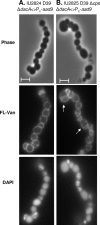
Representative micrographs of encapsulated D39 ΔdacA and isogenic unencapsulated D39 Δcps ΔdacA growing exponentially in BHI broth at 37°C. Phase-contrast microscopy and staining with FL-Van and DAPI are described in Materials and Methods. Bar, 2 μm. The arrows in panel B indicate typical cells containing multiple or misplaced equatorial rings in strain IU2825. See text for additional details.
FIG. 7.
FIG. 7.
Lactoyl-peptide compositions of strains R6 (top) and D39 (bottom). PG was isolated from cells growing exponentially in BHI broth at 37°C, lactoyl-peptides were released by base hydrolysis, and RP-HPLC was performed as described in Materials and Methods. The total yields of all lactoyl-peptides were similar for the unencapsulated R6 and encapsulated D39 strains (2.9 × 10⁷ and 3.3 × 10⁷ arbitrary mvolt area units, respectively). The structures of the lactoyl-peptides in each peak were determined by MS, and representative structures of species containing (monomer M7 and dimer D9) or lacking (monomer M1 and dimer D1) Ala-Ser additions and cross-bridges are shown. Complete peak assignments and other lactoyl-peptide structures are provided in the supplemental material. Quantitation of the relative amounts of each lactoyl-peptide from the R6 and D39 chromatograms can be found in Table S2 of the supplemental material. Underexpression or severe depletion of PcsB or the absence of MreCD did not affect the relative amounts or composition of the lactoyl-peptides, and the same chromatogram was obtained for unencapsulated strain D39 Δcps as shown for its encapsulated parent D39 (see text).

References

    1. Anantharaman, V., and L. Aravind. 2003. Evolutionary history, structural features and biochemical diversity of the NlpC/P60 superfamily of enzymes. Genome Biol. 4R11. - PMC - PubMed
    1. Arbeloa, A., J. E. Hugonnet, A. C. Sentilhes, N. Josseaume, L. Dubost, C. Monsempes, D. Blanot, J. P. Brouard, and M. Arthur. 2004. Synthesis of mosaic peptidoglycan cross-bridges by hybrid peptidoglycan assembly pathways in gram-positive bacteria. J. Biol. Chem. 27941546-41556. - PubMed
    1. Bateman, A., and N. D. Rawlings. 2003. The CHAP domain: a large family of amidases including GSP amidase and peptidoglycan hydrolases. Trends Biochem. Sci. 28234-237. - PubMed
    1. Battig, P., and K. Muhlemann. 2007. Capsule genes of Streptococcus pneumoniae influence growth in vitro. FEMS Immunol. Med. Microbiol. 50324-329. - PubMed
    1. Bendezu, F. O., and P. A. de Boer. 2008. Conditional lethality, division defects, membrane involution, and endocytosis in mre and mrd shape mutants of Escherichia coli. J. Bacteriol. 1901792-1811. - PMC - PubMed

Publication types