

doi: 10.1101/gad.1761309.
Regulated degradation of FANCM in the Fanconi anemia pathway during mitosis
Affiliations
- PMID: 19270156
- PMCID: PMC2658523
- DOI: 10.1101/gad.1761309
Item in Clipboard
Regulated degradation of FANCM in the Fanconi anemia pathway during mitosis
Genes Dev.
.
Erratum in
- Genes Dev. 2009 Apr 15;23(8):1025.. D'Andrea, Alan [corrected to D'Andrea, Alan D]
Abstract
The 13 Fanconi anemia (FA) proteins cooperate in a common DNA repair pathway. Eight of these proteins are assembled into a multisubunit E3 ligase called the FA core complex. During S phase, the FA core complex is loaded by the FANCM protein into chromatin where it monoubiquitinates its substrates. In mitosis, the FA core complex is released from FANCM by an unknown mechanism. Here we show that FANCM is hyperphosphorylated and degraded during mitosis. beta-TRCP and Plk1 are the key regulators of FANCM degradation. Nondegradable mutant forms of FANCM retain the FA core complex in the chromatin and disrupt the FA pathway. Our data provide a novel mechanism for the cell cycle-dependent regulation of the FA pathway.
Figures
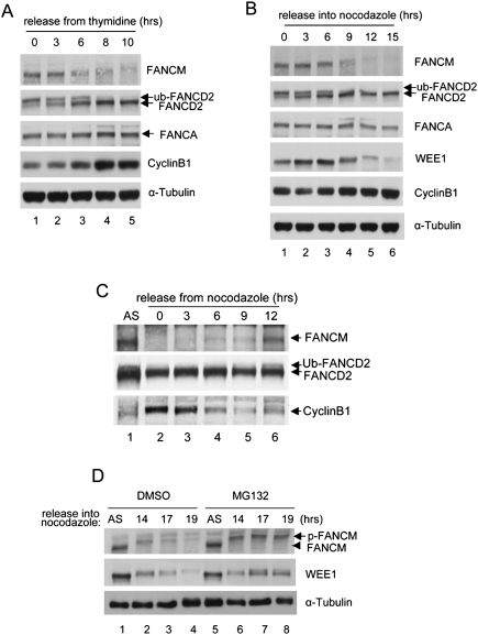
FANCM is degraded at G2/M phase. (A) HeLa cells were synchronized by double thymidine block and released into regular media and samples were harvested at indicated time points. (B) HeLa cells were synchronized by the same procedure as A, but released into nocodazole-containing media. Note the disappearance of phosphorylated FANCM. (C) Nocodazole-arrested HeLa cells were released into fresh medium and the status of FANCM was monitored at indicated time points. (D) HeLa cells were arrested using nocodazole for indicated times and 10 μM MG132 was added during the last 4 h of the nocodazole treatment before the cells were harvested.
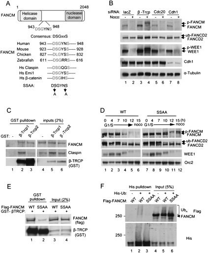
β-TRCP mediates degradation of FANCM. (A) A schematic for FANCM protein and alignment of amino acids corresponding to the DSGxxS sequence with FANCM orthologs and other β-TRCP substrates. (B) HeLa cells were treated with indicated siRNAs before being arrested using nocodazole. The higher migrated bands of FANCM and WEE1 indicate phosphorylated forms. (C) GST-β-TRCP proteins were overexpressed in 293T cells and pull-down assay was performed using glutathione sepharose. (D) Flag-FANCM wild type or SSAA mutant were expressed in HeLa cells that were synchronized by double thymidine block followed by release into nocodazole-containing media for indicated times. (E) GST-β-TRCP1 was coexpressed with either Flag-FANCM wild type or SSAA mutant in 293T cells and pull-down assay was performed using glutathione sepharose. (F) 293T cells were cotransfected with 6XHis-Ub plasmid and Flag-FANCM wild type or SSAA plasmids, followed by nocodazole and MG132 treatment. Cells lysates were subjected to pull-down using Ni-NTA beads and the eluates were analyzed by Western blots.
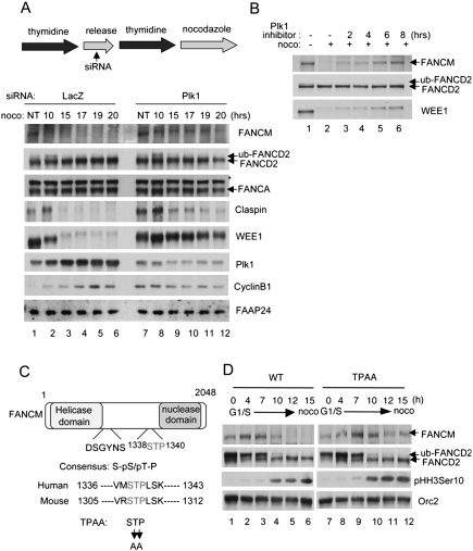
Plk1 mediates degradation of FANCM (A, top panel). A schematic for the synchronization assay. siRNAs were treated during a release period after the first thymidine block. (A, bottom panel) HeLa cells were synchronized at G1/S, and released into nocodazole-containing media. Samples were harvested at indicated time points and analyzed by Western blots. Asterisks indicate cross-reacting bands. (B) Nocodazole-arrested HeLa cells were treated with Plk1-specific inhibitor BI2536 for indicated times and the samples were analyzed by Western blots. (C) A schematic for FANCM protein and alignment of amino acids corresponding to the STP sequence with mouse Fancm ortholog. (D) Flag-FANCM wild type or TPAA mutant were expressed in HeLa cells that were synchronized by double thymidine block followed by release into nocodazole-containing media for indicated times.
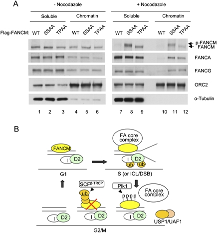
Degradation of FANCM regulates localization of the FA core complex (A) Flag-FANCM wild type, SSAA, or TPAA mutant were expressed in HeLa cells that were synchronized by double thymidine block followed by release into nocodazole-containing media. At 15 h from nocodazole treatment, mitotic cells were collected and fractionated to nucleo/cytoplasmic soluble fractions (S) and chromatin-enriched fractions (P). (B) Model. During G1/S phase, the FA core complex is recruited to chromatin by FANCM, where FANCD2 is monoubiquitinated. During G2/M phase, FANCM is degraded and subsequently the FA core complex is released, leading to inactivation of the FA pathway.
References
-
- Branzei D., Foiani M. Regulation of DNA repair throughout the cell cycle. Nat. Rev. Mol. Cell Biol. 2008;9:297–308. - PubMed
-
- Brummelkamp T.R., Bernards R., Agami R. A system for stable expression of short interfering RNAs in mammalian cells. Science. 2002;296:550–553. - PubMed
-
- Busino L., Donzelli M., Chiesa M., Guardavaccaro D., Ganoth D., Dorrello N.V., Hershko A., Pagano M., Draetta G.F. Degradation of Cdc25A by β-TrCP during S phase and in response to DNA damage. Nature. 2003;426:87–91. - PubMed
-
- Ciccia A., Ling C., Coulthard R., Yan Z., Xue Y., Meetei A.R., Laghmani el H., Joenje H., McDonald N., de Winter J.P., Wang W., West S.C. Identification of FAAP24, a Fanconi anemia core complex protein that interacts with FANCM. Mol. Cell. 2007;25:331–343. - PubMed
Publication types
MeSH terms
Substances
Grants and funding
LinkOut - more resources
Full Text Sources
Other Literature Sources
Miscellaneous