. 2009 May;8(9):808-19.
doi: 10.4161/cbt.8.9.8131. Epub 2009 May 8.

Bcl-2 antagonists interact synergistically with bortezomib in DLBCL cells in association with JNK activation and induction of ER stress

Affiliations

Bcl-2 antagonists interact synergistically with bortezomib in DLBCL cells in association with JNK activation and induction of ER stress

Girija Dasmahapatra et al. Cancer Biol Ther. 2009 May.

Erratum in

  • Correction.
    [No authors listed] [No authors listed] Cancer Biol Ther. 2019;20(9):1300. doi: 10.1080/15384047.2019.1618578. Epub 2019 May 22. Cancer Biol Ther. 2019. PMID: 31116086 Free PMC article. No abstract available.

Abstract

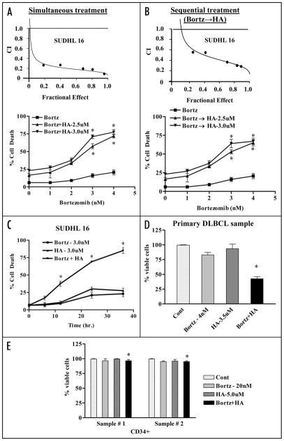
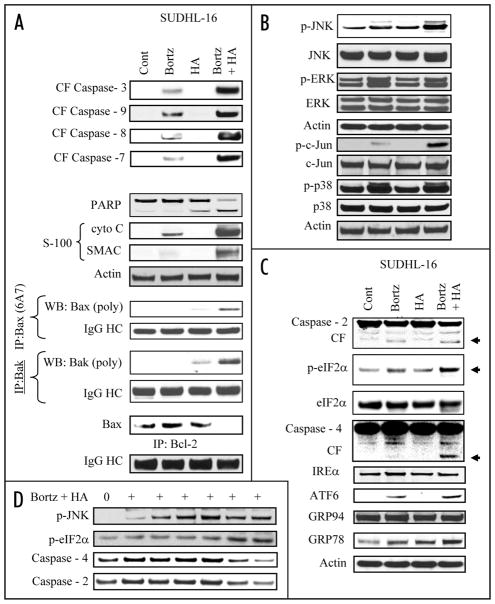
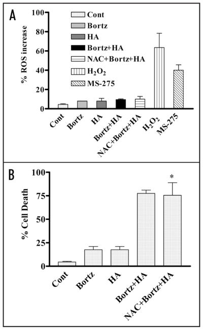
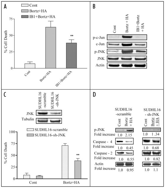
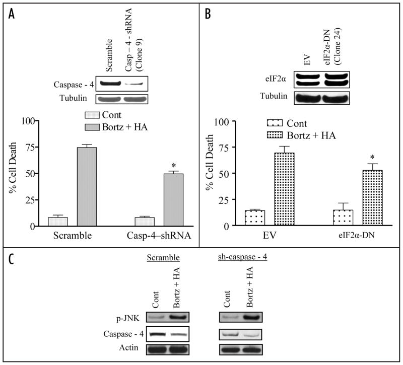
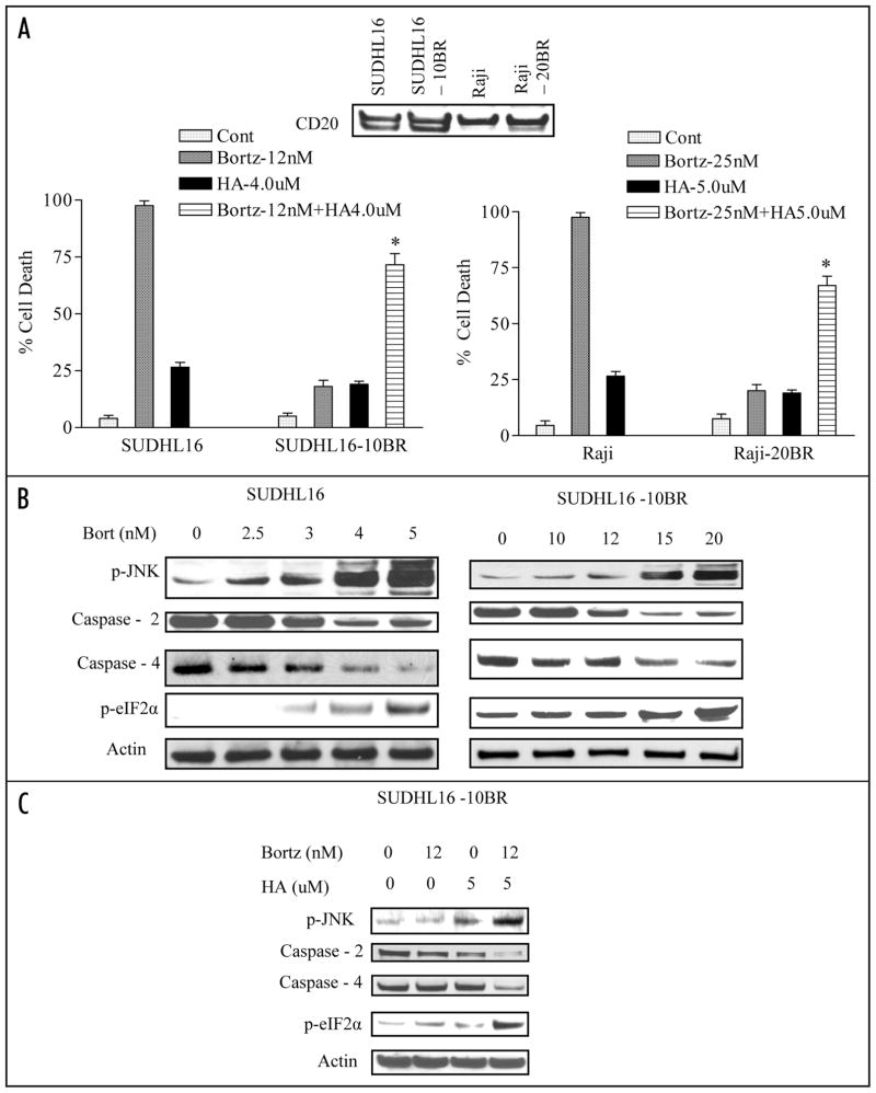
Mechanisms underlying interactions between the proteasome inhibitor bortezomib and small molecule Bcl-2 antagonists were examined in GC- and ABC-type human DLBCL (diffuse lymphocytic B-cell lymphoma) cells. Concomitant or sequential exposure to non- or minimally toxic concentrations of bortezomib or other proteasome inhibitors and either HA14-1 or gossypol resulted in a striking increase in Bax/Bak conformational change/translocation, cytochrome c release, caspase activation and synergistic induction of apoptosis in both GC- and ABC-type cells. These events were associated with a sharp increase in activation of the stress kinase JNK and evidence of ER stress induction (e.g., eIF2alpha phosphorylation, activation of caspases-2 and -4, and Grp78 upregulation). Pharmacologic or genetic (e.g., shRNA knockdown) interruption of JNK signaling attenuated HA14-1/bortezomib lethality and ER stress induction. Genetic disruption of the ER stress pathway (e.g., in cells expressing caspase-4 shRNA or DN-eIF2alpha) significantly attenuated lethality. The toxicity of this regimen was independent of ROS generation. Finally, HA14-1 significantly increased bortezomib-mediated JNK activation, ER stress induction, and lethality in bortezomib-resistant cells. Collectively these findings indicate that small molecule Bcl-2 antagonists promote bortezomib-mediated mitochondrial injury and lethality in DLBCL cells in association with enhanced JNK activation and ER stress induction. They also raise the possibility that such a strategy may be effective in different DLBCL sub-types (e.g., GC- or ABC), and in bortezomib-resistant disease.


Figures

Figure 1
Figure 1
Co-treatment of bortezomib and Bcl-2 antagonist leads to synergistic induction of cell death in DLBCL cells in a time dependent manner but not in normal hematopoietic cells. SUDHL16 cells were treated with indicated concentration of bortezomib ± HA14-1. (A) simultaneously or (B) sequentially (8 h bortezomib pretreatment followed by HA14-1) for a total of 36 h, after which cell death was monitored by flow cytometry with 7AAD staining. Insets (A and B): Fractional Effect (FA) values were determined by comparing results to those of untreated controls, and Median Dose Effect analysis was employed to characterize the nature of the interaction. Combination Index (C.I.) values less than 1.0 denote a synergistic interaction. Two additional studies yielded equivalent results. (C) SUDHL6 cells were treated with bortezomib ± HA14-1 at the indicated concentrations for various intervals, and cell death was determined by flow-cytometry with 7AAD staining (D) Primary human bone marrow DLBCL cells were isolated as described in Methods and suspended in medium containing 10% FCS at a cell density of 0.75 × 10⁶/ml cells in the presence of 4 nM bortezomib ± 3.5 uM of HA14-1 for 14 h. At the end of drug exposure, apoptotic cells were monitored by Annexin/PI staining. Apoptotic cell death for controls was <25–20%. The percentage of non apoptotic cells were considered to represent the viable fraction, and values were normalized to controls. (E) CD34+ cells obtained from the bone marrow of two patients undergoing routine diagnostic procedures for non-myeloid hematologic disorders were isolated by an immunomagnetic bead separation technique as described in Methods and exposed to bortezomib (20 nM) ± HA14-1 (5.0 μM) for 48 h. At the end of this period, the percentage of apoptotic cells was determined by Annexin V/PI staining and flow cytometry. The percentage of viable cells in each sample was normalized to controls. Values represent the means ± S.D. for triplicate determination. For (A–C), * = significantly greater than bortezomib alone; p < 0.01. For (D), * = not significantly different from values for untreated controls; p > 0.05.
Figure 2
Figure 2
Combined exposure to bortezomib and HA14-1 leads to a dramatic increase in caspase activation, mitochondrial damage, Bax and Bak translocation and conformational change, in association with JNK activation and ER stress induction in SUDHL16 cells. SUDHL16 cells were treated with 3 nM bortezomib ± 3.0 μM of HA14-1 for 14 h. (A) cytosolic (S-100) fractions were obtained as described in Materials and Methods, and expression of cytochrome c, AIF and Smac/DIABLO were monitored by western blot. Proteins from whole cell lysates were prepared and expression of the indicated proteins were determined by western blotting. Bax and Bak translocation and conformational change, as well as the association between Bax and Bcl-2 were monitored by immunoprecipitation followed by western blotting as described in Methods (B and C). At the end of the drug exposure (14 h) as (A) above, cells were lysed, sonicated, the proteins denatured, and subjected to western blot analysis using the indicated primary antibodies. (D) SUDHL 16 cells were treated with 3 nM bortezomib ± 3 μM HA14-1 for various intervals and changes in the expression of the indicated protein expression were monitored by western blotting. For these and all other studies, each lane was loaded with 30 μg of protein; blots were stripped and reprobed with antibodies directed against actin to ensure equivalent loading and transfer. Results are representative of three separate experiments.
Figure 3
Figure 3
Co-administration of bortezomib and HA14-1 fails to induce ROS generation and pretreatment with the antioxidant (NAC) fails to circumvent lethality in SUDHL4 cells. (A) SUDHL4 cells (preteated with or without 5 mM NAC for 3 h) were exposed to 5 nM bortezomib ± 4 μM HA14-1 for 4 h. Cells were also exposed to MS-275 (2 μM) or H2O2 (0.5 mM) for 30 min to serve as positive control for ROS generation. At the end of the exposure interval, ROS generation was monitored as described in Methods. (B) SUDHL4 cells (preteated with or without 5 mM NAC for 3 h) were exposed to 5 nM bortezomib ± 4 μM HA14-1 for 36 h. At the end of drug treatment, cell death was monitored by 7 AAD staining as described in methods. For (B), * = not significantly different from values for cells treated with bortezomib + HA14-1 in the absence of NAC; p > 0.05.
Figure 4
Figure 4
Pharmacologic and genetic interruption of the JNK pathways significantly diminishes bortezomib/HA14-1 lethality in SUHDL16 cells. (A) SUDHL16 cells pretreated with the JNK inhibitor IB1 (ALX 159–600; 10 μM) for 2 hr were exposed to 3 nM bortezomib ± 3.0 μM HA14-1 for 36 hrs. At the end of drug exposure, apoptosis was monitored by 7 AAD staining and flow cytometry. ** = significantly less than values for cells treated in the absence of IB1; p < 0.01. (B) Cells were treated as above (A) for 14 hrs and western blot analysis was employed to monitor the effect of drugs on expression of the indicated proteins. Each lane was loaded with 30 μg of protein; blots were stripped and reprobed with antibodies directed against actin to ensure equivalent loading and transfer. The results of a representative study are shown; two additional studies yielded equivalent results. (C) SUDHL16 cells stably transfected with JNK shRNA or vectors encoding a scrambled sequence were exposed to 4.0 nM bortezomib + 4.0 μM HA14-1. After 36 hr of drug exposure, apoptotic cells were monitored by 7 AAD staining and flow cytometry. inset: relative expression of JNK protein in SUDHL16-scrambled sequence and shJNK clones (D) Following 14 hr of drug exposure as (C) above, western blot analysis was employed to monitor protein expression of phospho-JNK, caspase-4 and caspase 2. Blots were stripped and reprobed with anti-actin antibodies to ensure equal loading and transfer of protein For (A),** = significantly less than values for scrambled sequence clone; p < 0.01. For (C), ** = significantly less than values for empty-vector controls; p < 0.05.
Figure 5
Figure 5
Knockdown of caspase-4 expression or ecotopic expression of eIF2α-DN significantly diminishes bortezomib/HA14-induced lethality in SUDHL16 cells. (A) SUDHL16 cells stably transfected with caspase-4 shRNA or a scrambled sequence vector were exposed to 4.0 nM bortezomib + 4.0 μM HA14-1. Following 36 h of drug exposure, apoptotic cells were monitored by annexin V/PI staining and flow cytometry. Inset: relative expression of caspase 4 protein in scrambled sequence and shJNK clones. (B) SUDHL16 cells stably transfected with an eIF2α-DN or empty vector (pcDNA3.1) construct were incubated with 4 nM bortezomib + 4.0 μM HA14-1. After 36 h of drug exposure, apoptotic cells were monitored by annexin V/PI staining and flow cytometry. (C) Following 14 h of drug exposure to SUDHL16-casp4 shRNA cells as described in (A) above, western blot analysis was employed to monitor protein expression of caspase-4 and phospho-JNK. Blots were stripped and reprobed with anti-actin antibodies to ensure equal loading and transfer of protein (30 μg each lane). For (A and B),* = significantly less than values for control cells; p < 0.05. Two additional studies yielded equivalent results.
Figure 6
Figure 6
HA14-1 increases the ability of bortezomib to induce JNK activation, evidence of ER stress, and lethality in bortezomib-resistant cells. (A) SUDHL16, SUDHL16-10BR, Raji and Raji-20BR cells were treated with the indicated concentrations of bortezomib ± HA14-1 for 36 and 48 h respectively after which cell death was assessed by flow cytometry using 7AAD staining. Inset: immuno-blotting depicting the expression of CD20 in parental and bortezomib-resistant cells. (B) SUDHL16, SUDHL16-10BR cells were treated with indicated concentration of bortezomib for 14 h. At the end of drug exposure, cells were lysed and equivalent amounts (30 μg) of protein subjected to immunoblotting with the indicated antibodies (C) SUDHL16-10BR cells were treated with the indicated concentration of bortezomib ± HA14-1 for 14 h. At the end of drug exposure, cells were lysed and equivalent amounts (20 μg) of protein were subjected to immunoblotting with antibodies as indicated. In each case, blots were stripped and probed with antibodies directed against actin to ensure equivalent loading and transfer of proteins. For (A), * = significantly greater than values for bortezomib alone; p < 0.01.

Comment in

  • Piling up the JNK: drug synergy through ER stress.
    Hertan LM, Koumenis C. Hertan LM, et al. Cancer Biol Ther. 2009 May;8(9):820-2. doi: 10.4161/cbt.8.9.8403. Cancer Biol Ther. 2009. PMID: 19458484 No abstract available.
  • Findings of Research Misconduct.
    [No authors listed] [No authors listed] NIH Guide Grants Contracts (Bethesda). 2015 Dec 18:NOT-OD-16-040. NIH Guide Grants Contracts (Bethesda). 2015. PMID: 26693581 Free PMC article. No abstract available.
  • Findings of Research Misconduct.
    [No authors listed] [No authors listed] Fed Regist. 2015 Dec 10;80(237):76703-76704. Fed Regist. 2015. PMID: 27737268 Free PMC article. No abstract available.

References

    1. Coiffier B. Non-Hodgkin’s lymphomas. In: Cavalli F, Hansen HH, Kaye SB, editors. Textbook of medical oncology. London: Martin Dunitz; 1997. pp. 34–5.
    1. Coiffier B, Lepage E, Briere J, Herbrecht R, Tilly H, Bouabdallah R, et al. CHOP chemotherapy plus rituximab compared with CHOP alone in elderly patients with diffuse large-B-cell lymphoma. N Engl J Med. 2002;346:235–42. - PubMed
    1. Alizadeh AA, Eisen MB, Davis RE, Ma C, Lossos IS, Rosenwald A, et al. Distinct types of diffuse large B-cell lymphoma identified by gene expression profiling. Nature. 2000;403:503–11. - PubMed
    1. Rosenwald A, Wright G, Chan WC, Connors JM, Campo E, Fisher RI, et al. The use of molecular profiling to predict survival after chemotherapy for diffuse large-B-cell lymphoma. N Engl J Med. 2002;346:1937–47. - PubMed
    1. Lossos IS, Jones CD, Warnke R, Natkunam Y, Kaizer H, Zehnder JL, et al. Expression of a single gene, BCL-6, strongly predicts survival in patients with diffuse large B-cell lymphoma. Blood. 2001;98:945–51. - PubMed

Publication types

MeSH terms