

Epithelial-to-mesenchymal transition in the development and progression of adenocarcinoma and squamous cell carcinoma of the lung
- PMID: 19270647
- PMCID: PMC2675657
- DOI: 10.1038/modpathol.2009.19
Epithelial-to-mesenchymal transition in the development and progression of adenocarcinoma and squamous cell carcinoma of the lung
Abstract
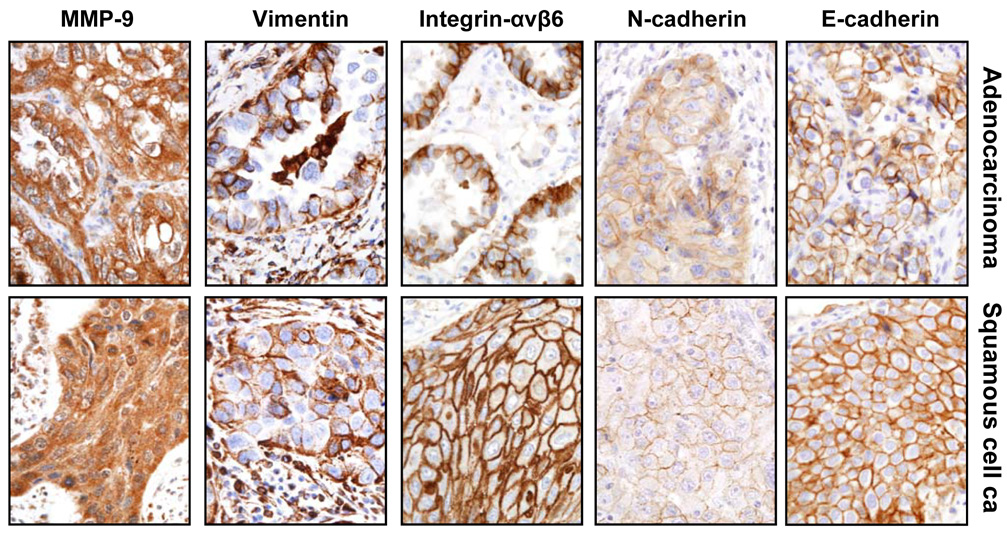
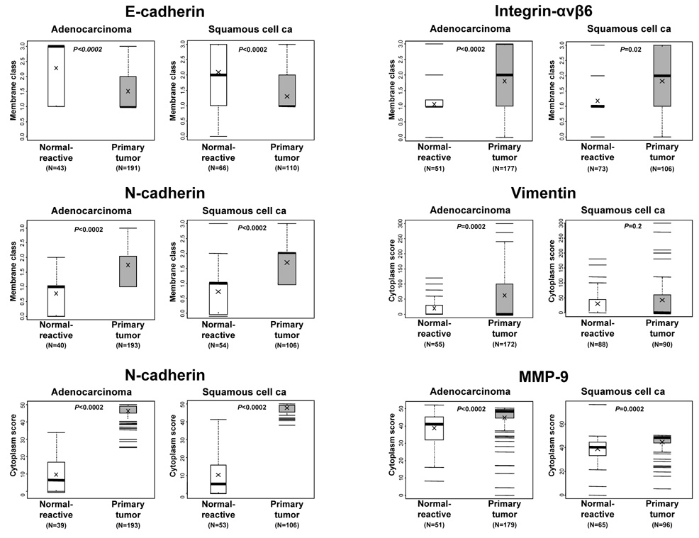
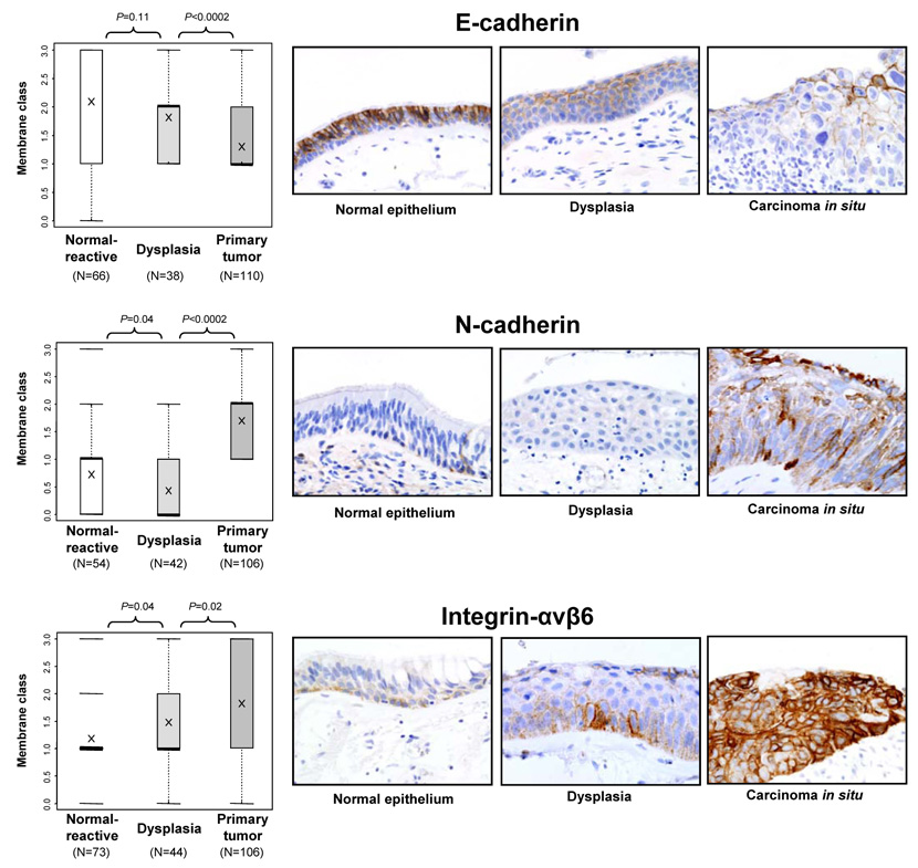
Epithelial-to-mesenchymal transition is a process in which cells undergo a developmental switch from an epithelial to a mesenchymal phenotype. We investigated the role of this phenomenon in the pathogenesis and progression of adenocarcinoma and squamous cell carcinoma of the lung. Archived tissue from primary tumors (n=325), brain metastases (n=48) and adjacent bronchial epithelial specimens (n=192) were analyzed for immunohistochemical expression by image analysis of E-cadherin, N-cadherin, integrin-alpha v beta 6, vimentin, and matrix metalloproteinase-9. The findings were compared with the patients' clinicopathologic features. High expression of the epithelial-to-mesenchymal transition phenotype (low E-cadherin and high N-cadherin, integrin-alpha v beta 6, vimentin, and matrix metalloproteinase-9) was found in most lung tumors examined, and the expression pattern varied according to the tumor histologic type. Low E-cadherin membrane and high N-cadherin cytoplasmic expression were significantly more common in squamous cell carcinoma than in adenocarcinoma (P=0.002 and 0.005, respectively). Dysplastic lesions had significantly lower expression of the epithelial-to-mesenchymal transition phenotype than the squamous cell carcinomas, and integrin-alpha v beta 6 membrane expression increased stepwise according to the histopathologic severity. Brain metastases had decreased epithelial-to-mesenchymal transition expression compared with primary tumors. Brain metastases had significantly lower integrin-alpha v beta 6 membrane (P=0.04), N-cadherin membrane, and cytoplasm (P<0.0002) expression than the primary tumors. The epithelial-to-mesenchymal transition phenotype is commonly expressed in primary squamous cell carcinoma and adenocarcinoma of the lung; this expression occurs early in the pathogenesis of squamous cell carcinoma. Brain metastases showed characteristics of reversed mesenchymal-to-epithelial transition. Our findings suggest that epithelial-to-mesenchymal transition is a potential target for lung cancer chemoprevention and therapy.
Figures
References
-
- Jemal A, Siegel R, Ward E. Cancer statistics 2008. CA Cancer J Clin. 2008;58:71–96. - PubMed
-
- Minna JD, Gazdar A. Focus on lung cancer. Cancer Cell. 2002;1:49–52. - PubMed
-
- Travis WD, Brambilla E, Muller-Hermelink HK, Harris CC. Tumours of the lung. In: Travis WD, Brambilla E, Muller-Hermelink HK, Harris CC, editors. Pathology and Genetics: Tumours of the Lung, Pleura, Thymus and Heartedn, Vol. Lyon: International Agency for Research on Cancer (IARC); 2004. pp. 9–124.
-
- van Zandwijk N. Neoadjuvant strategies for non-small cell lung cancer. Lung Cancer. 2001;34(Suppl 2):S145–S150. - PubMed
-
- Wistuba II. Genetics of preneoplasia: lessons from lung cancer. Curr Mol Med. 2007;7:3–14. - PubMed
Publication types
MeSH terms
Substances
Grants and funding
LinkOut - more resources
Full Text Sources
Other Literature Sources
Medical
Research Materials
