. 2009;4(3):e4751.
doi: 10.1371/journal.pone.0004751. Epub 2009 Mar 9.

MMPs regulate both development and immunity in the tribolium model insect

Affiliations

MMPs regulate both development and immunity in the tribolium model insect

Eileen Knorr et al. PLoS One. 2009.

Abstract

Background: Matrix metalloproteinases (MMPs) are evolutionarily conserved and multifunctional effector molecules in development and homeostasis. In spite of previous, intensive investigation in vitro and in cell culture, their pleiotrophic functions in vivo are still not well understood.

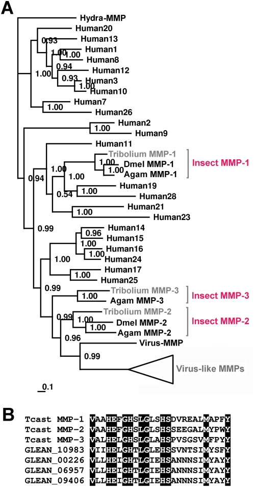
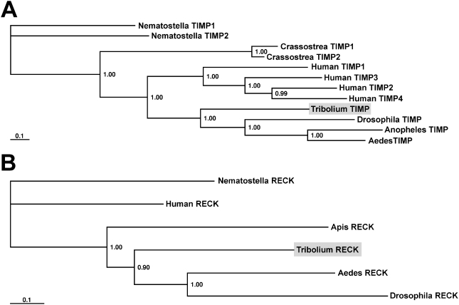
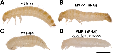
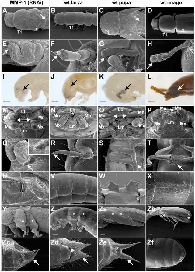
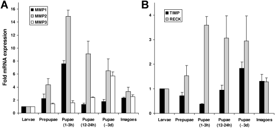
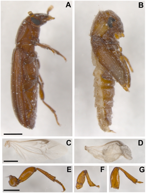
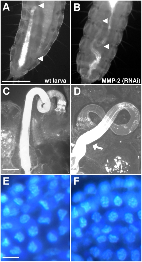
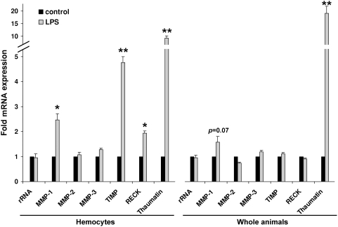
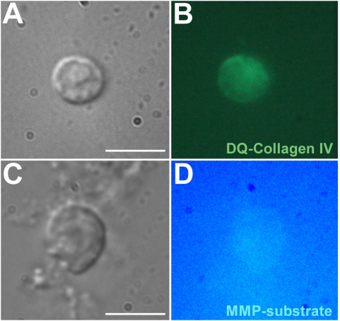
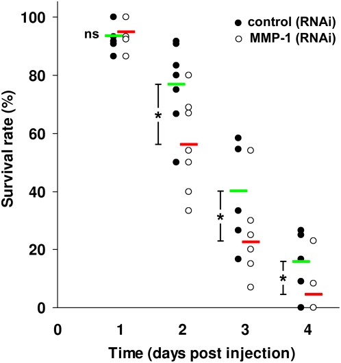
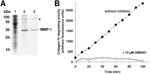
Methodology/principal findings: We show that the genetically amenable beetle Tribolium castaneum represents a feasible model organism to explore MMP functions in vivo. We silenced expression of three insect-type Tribolium MMP paralogs and their physiological inhibitors, TIMP and RECK, by dsRNA-mediated genetic interference (RNAi). Knock-down of MMP-1 arrested development during pupal morphogenesis giving phenotypes with altered antennae, compound eyes, wings, legs, and head. Parental RNAi-mediated knock-down of MMP-1 or MMP-2 resulted in larvae with non-lethal tracheal defects and with abnormal intestines, respectively, implicating additional roles of MMPs during beetle embryogenesis. This is different to findings from the fruit fly Drosophila melanogaster, in which MMPs have a negligible role in embryogenesis. Confirming pleiotrophic roles of MMPs our results also revealed that MMPs are required for proper insect innate immunity because systemic knock-down of Tribolium MMP-1 resulted in significantly higher susceptibility to the entomopathogenic fungus Beauveria bassiana. Moreover, mRNA levels of MMP-1, TIMP, and RECK, and also MMP enzymatic activity were significantly elevated in immune-competent hemocytes upon stimulation. To confirm collagenolytic activity of Tribolium MMP-1 we produced and purified recombinant enzyme and determined a similar collagen IV degrading activity as observed for the most related human MMP, MMP-19.

Conclusions/significance: This is the first study, to our knowledge, investigating the in vivo role of virtually all insect MMP paralogs along with their inhibitors TIMP and RECK in both insect development and immunity. Our results from the Tribolium model insect indicate that MMPs regulate tracheal and gut development during beetle embryogenesis, pupal morphogenesis, and innate immune defense reactions thereby revealing the evolutionarily conserved roles of MMPs.


Conflict of interest statement

Competing Interests: The authors have declared that no competing interests exist.

Figures

Figure 1
Figure 1. Phylogenetic analysis of Tribolium MMPs along with MMPs from other organisms.
A Bayesian protein tree, with posterior probabilities, was generated using aligned MMP sequences from T. castaneum, Anopheles gambiae, Drosophila melanogaster, Homo sapiens, and Heliothis zea virus 1. A Hydra vulgaris MMP was used as outgroup. This analysis revealed that Tribolium MMP-1 is orthologous to other insect MMP-1s and Tribolium MMP-2 to MMP-2 from Anopheles and Drosophila. Interestingly, Tribolium MMP-3 clades with MMP-3 from Anopheles, but a MMP-3 orthologue is obviously absent in Drosophila. Insect MMP-1 proteins show highest relation to human MMP-19 and MMP-28 whereas the other insect MMPs group together with human membrane-anchored MMPs (MMP-14 to MMP-17, MMP-24 and MMP-25). In contrast to Anopheles and Drosophila, Tribolium has numerous genes that are most closely related to a MMP from Heliothis zea virus 1. The scale bar represents the substitutions per site according to the model of amino acid evolution applied. (B) Three Tribolium MMPs and virus-derived Tribolium MMP proteins have catalytic domains with conserved MMP specific active site sequences (MMP active site consensus sequence: HEXGHXXGXXHSX6M). For better overview sequences of only four virus-derived MMPs are shown.
Figure 2
Figure 2. Phylogenetic analysis of Tribolium TIMP and RECK along with homologs from other organisms.
(A) Aligned TIMP and (B) RECK protein sequences from T. castaneum along with homologs from other animals, were used to generate bayesian protein trees. Tribolium TIMP and RECK (indicated by gray shading) group well with other insect orthologs. Humans and some other animals have two or more TIMP homologs. Posterior probabilities are plotted at the nodes.
Figure 3
Figure 3. RNAi (MMP-1) knock-down Tribolium larvae arrest during larval to pupal transformation.
Lateral views of whole bodies of wild-type larva (A) and arrested MMP-1 knock-down animals at the larval-pupal transformation (B) are shown. For comparison wild-type pupa (C) and MMP-1 knock-down animals that were removed from the puparium (D) are also shown. Scale bar, 1 mm.
Figure 4
Figure 4. MMP-1 knock-down phenotype.
MMP-1 knock-down animals were analyzed in detail in comparison to wild-type larva, pupa and imago. (A–D) MMP-1 knock-down animals had early pupa-like dorsal features including thorax segment 1 (T1), wings (asterisks) and a pupa-specific abdominal line of setae (arrows). (E–H) Their antennae were thick and un-segmented (tips of antennae are indicated by arrows). (I–L) MMP-1 (RNAi) larvae displayed premature compound eyes (pupal ommatidia) and exhibited reduced larval stemmata (arrows). (M–P) The mouth of MMP-1 (RNAi) phenotype animals exhibited a pupa-like morphology; mandible (Mn), maxilla (Mx), labrum (Lb), labium (Lm), gap between mandibles (indicated by arrow lines in pupa and MMP-1 knock-down phenotype), interlocking of mandibles (indicated by asterisks in larva and imago). (Q–T) MMP-1 (RNAi) larvae had shortened legs with a double inchoate claw similar to that observed in pupa (tip of legs are indicated by arrows). (U–X) Gin-traps were present in MMP-1 knock-down animals and pupae but were absent in wild-type larvae and imagoes. (Y–Zb) In MMP-1 (RNAi) animals wings were rudimentally developed (asterisks) (Zc–Zf), but genital papillae (asterisks, covered in adults) developed into those of pupae, replacing larval pygopods (arrow-head). Furthermore, urogomphi were elongated and pupa-like (arrows, not present in imago). Scale bars: A–D, 500 µm; E–H and M–T, 100 µm; I–L and Y–Zf, 200 µm; U–X, 50 µm.
Figure 5
Figure 5. Muscle development in MMP-1 (RNAi) animals.
(A) Pig-19 larva showed muscle specific EGFP-fluorescence throughout the whole body, with a notably strong expression at its lateral sides. (B) In control (RNAi) Pig-19 pupa the muscle EGFP-fluorescence was predominantly localized in the abdomen and visible as parallel strands. (C) MMP-1 (RNAi) larva showed a pupa-like localization of EGFP-fluorescence of muscles in the abdomen with additionally significant fluorescence in the thorax. Scale bar, 1 mm.
Figure 6
Figure 6. Quantitative real time RT-PCR analysis of transcriptional levels of Tribolium MMPs during metamorphosis.
(A) The mRNA levels of MMP-1 (black bars), MMP-2 (gray bars), and MMP-3 (white bars) of different metamorphosis stages is shown relative to their expression levels in last instar larvae. Expression rates of MMP-1 increased 4-fold during prepupal stage and 8-fold during early pupal stage (1–3 h) and declined soon after this to basal levels. In contrast, MMP-2 mRNA levels were determined to be elevated at all analyzed prepupal and pupal stages with the highest elevation (15-fold) at the early pupal stage when compared to larval stage. Interestingly, MMP-3 expression levels were not induced during early stages but in a later stage of metamorphosis. In 3 day old pupae, we determined a 6-fold elevated transcript level. (B) TIMP gene expression was repressed 2-fold in early metamorphosis and induced in later stages, whereas RECK gene expression was induced during all tested stages of metamorphosis. Expression levels of MMP-4 (Glean 10983) and 18S rRNA genes were not significantly different at all analyzed stages (data not shown). Results represent mean values of three independent determinations±S.D. from ten animals per stage that were pooled for analysis.
Figure 7
Figure 7. Phenotype arising upon Dpp knock-down in last instar larvae.
(A) Lateral views of wild-type imago (A) and arrested dpp (RNAi) animal at pupal-adult transformation (B) are shown. This phenotype is dominant, however, some animals exhibited a milder phenotype, in which they ecdysed to imagoes but had affected wings (D) and leg deformations (F,G) of mainly the tarsus when compared to wild-type wing (C) and leg (E). Scale bar, A–B, 500 µm; C–G, 250 µm.
Figure 8
Figure 8. MMP-2 knock-down in adults result in offspring with gut defects.
Tribolium larvae that are exposed to UV-light, resulting in auto-fluorescence of their cuticle and intestine. (A) Auto-fluorescence of the posterior end of a wild-type first instar larva showed an intestine that is normally s-shaped in a typical way in up to three segments (arrow-heads). (B) In contrast, examination of the posterior end of a first instar MMP-2 (RNAi) larva revealed an abnormal intestine with a twisted hind gut and a half truncated ileum loop. Closer examination of dissected hind guts from wild-type (C) and MMP-2 (RNAi) (D) larvae revealed no obvious differences except a kink at the end of the colon of MMP-2 (RNAi) larvae (indicated by an arrow). Gut epithelium seems not to be affected since cell appearance by DAPI-mediated nucleus staining indicated no differences. Scale bars, A–B, 100 µm; C–D, 50 µm; E–F, 5 µm.
Figure 9
Figure 9. Quantitative real time RT-PCR analysis of transcriptional levels of Tribolium MMPs, TIMP, and RECK, along with immune-inducible thaumatin during innate immune responses.
The mRNA levels of MMP-1, TIMP, and RECK were significantly induced 2 to 5-fold in hemocytes in response to immune-challenge along with the immune-inducible thaumatin gene. In contrast, in whole animal preparations containing mainly fat body tissue no significantly induced expression of tested genes could be observed except immune-inducible thaumatin, although a tendency towards MMP-1 induction was detected. 18S rRNA gene expression was not influenced by treatment, and expression levels of MMP-2 or MMP-3 were not significantly different upon immune-challenge. Results represent mean values of three independent determinations±S.D. from ten animals that were pooled for analysis. Statistically significant differences were determined using Student's t-test and are indicated (*, p<0.05; **, p<0.01).
Figure 10
Figure 10. Detection of MMP-like collagenolytic activity on the surface of stimulated Tribolium hemocytes.
Typical granular hemocytes from Tribolium (A,C) exhibited MMP-like enzymatic activity localized at their surface by hydrolyzing DQ™ collagen type IV resulting in green fluorescence (B) and of quenched, fluorogenic MMP-1/MMP-9 peptidic substrate resulting in bright blue fluorescence (D), respectively. Scale bar, 10 µm.
Figure 11
Figure 11. RNAi (MMP-1) knock-down Tribolium imagoes are more susceptible to infection by the fungal entomopathogen B. bassiana.
Injection of Beauveria bassiana spores into the hemocoel of Tribolium imagoes resulted in killing within 4 to 5 days. MMP-1 knock-down animals died significantly faster than wild type animals. Values of seven independent trials including 10–15 animals per trial are shown for control (RNAi) animals and MMP-1 (RNAi) animals. Mean values of surviving control (RNAi) animals are shown in green and of surviving MMP-1 (RNAi) animals in red. Statistically significant differences were determined using Student's t-test and are indicated (*, p<0.05). Ns, not significant.
Figure 12
Figure 12. Purification and refolding of recombinant Tribolium MMP-1 and determination of its collagenolytic activity.
(A) MMP-1 (lane 2) was purified to apparent homogeneity from the urea-soluble E. coli inclusion bodies fraction (lane 1) as observed by SDS–PAGE analysis. After refolding in appropriate buffer, we observed the MMP-1 protein band (according to the calculated molecular mass of about 30.3 kDa) and an additional band with an estimated two-fold molecular weight that may correspond to a dimer of MMP-1 (indicated by an asterisk). Molecular mass standards are indicated in kDa. (B) Collagen-IV degrading activity of recombinant Tribolium MMP-1 was monitored using DQ™ collagen (type IV from human placenta). The reaction was found to be linear for at least 100 min under examined conditions. This activity was abolished in the presence of 10 µM GM6001.

References

    1. Nagase H, Woessner JF. Matrix metalloproteinases. J Biol Chem. 1999;274:21491–21494. - PubMed
    1. Page-McCaw A, Ewald AJ, Werb Z. Matrix metalloproteinases and the regulation of tissue remodelling. Nat Rev Mol Cell Biol. 2007;8:221–233. - PMC - PubMed
    1. Sternlicht MD, Werb Z. How matrix metalloproteinases regulate cell behavior. Annu Rev Cell Dev Biol. 2001;17:463–516. - PMC - PubMed
    1. Vu TH, Werb Z. Matrix metalloproteinases: effectors of development and normal physiology. Genes Dev. 2000;14:2123–2133. - PubMed
    1. Bode W, Maskos K. Structural basis of the matrix metalloproteinases and their physiological inhibitors, the tissue inhibitors of metalloproteinases. Biol Chem. 2003;384:863–872. - PubMed

Publication types

MeSH terms

Substances