. 2009;4(3):e4756.
doi: 10.1371/journal.pone.0004756. Epub 2009 Mar 9.

Silencing of PINK1 expression affects mitochondrial DNA and oxidative phosphorylation in dopaminergic cells

Affiliations

Silencing of PINK1 expression affects mitochondrial DNA and oxidative phosphorylation in dopaminergic cells

Matthew E Gegg et al. PLoS One. 2009.

Abstract

Background: Mitochondrial dysfunction has been implicated in the pathogenesis of Parkinson's disease (PD). Impairment of the mitochondrial electron transport chain (ETC) and an increased frequency in deletions of mitochondrial DNA (mtDNA), which encodes some of the subunits of the ETC, have been reported in the substantia nigra of PD brains. The identification of mutations in the PINK1 gene, which cause an autosomal recessive form of PD, has supported mitochondrial involvement in PD. The PINK1 protein is a serine/threonine kinase localized in mitochondria and the cytosol. Its precise function is unknown, but it is involved in neuroprotection against a variety of stress signalling pathways.

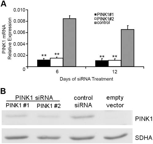
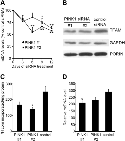
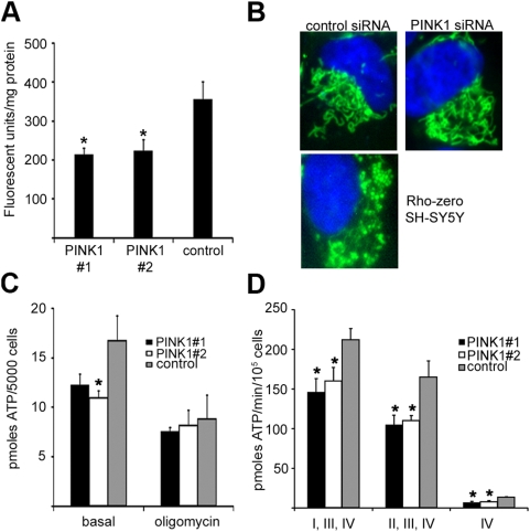
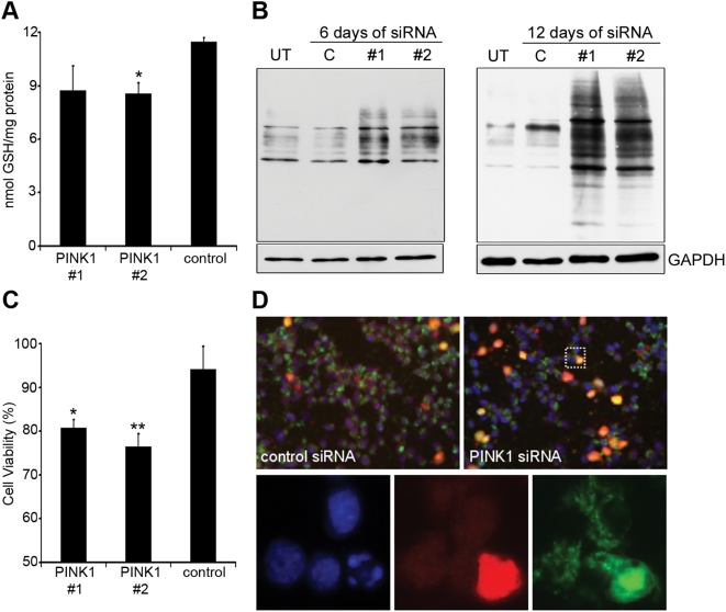
Methodology/principal findings: In this report we have investigated the effect of silencing PINK1 expression in human dopaminergic SH-SY5Y cells by siRNA on mtDNA synthesis and ETC function. Loss of PINK1 expression resulted in a decrease in mtDNA levels and mtDNA synthesis. We also report a concomitant loss of mitochondrial membrane potential and decreased mitochondrial ATP synthesis, with the activity of complex IV of the ETC most affected. This mitochondrial dysfunction resulted in increased markers of oxidative stress under basal conditions and increased cell death following treatment with the free radical generator paraquat.

Conclusions: This report highlights a novel function of PINK1 in mitochondrial biogenesis and a role in maintaining mitochondrial ETC activity. Dysfunction of both has been implicated in sporadic forms of PD suggesting that these may be key pathways in the development of the disease.


Conflict of interest statement

Competing Interests: The authors have declared that no competing interests exist.

Figures

Figure 1
Figure 1. Silencing of PINK1 mRNA and protein expression in SH-SY5Y cells.
A, SH-SY5Y cells were transfected with PINK1 siRNA (PINK1 #1 or PINK1 #2) or scrambled control siRNA for 6 or 12 days and PINK1 mRNA levels measured by reverse transciption and quantitative real-time PCR. Relative expression was determined against GAPDH mRNA levels and data were expressed as mean±s.e.m. (n = 4). **P<0.01 vs. scrambled control siRNA. B, SH-SY5Y cells over-expressing recombinant PINK1 protein were transfected with either PINK1 siRNA (PINK1 #1 or PINK1 #2) or scrambled control siRNA for 40 hours. The expression of full-length recombinant PINK1 protein was determined in cell lysates by western blotting. No band was detected in SH-SY5Y transfected with empty vector. Protein loading was verified with antibody against succinate dehydrogenase subunit SDHA.
Figure 2
Figure 2. Measurement of mtDNA levels and synthesis in PINK1-silenced SH-SY5Y cells.
A, SH-SY5Y cells were treated with PINK1 or scrambled control siRNA for 3, 6, 9 or 12 days and mtDNA levels measured by quantitative real-time PCR. MtDNA levels were expressed relative to the single copy nuclear gene TK2. Data are expressed as percentage of control siRNA mtDNA levels (n = 4). B, cells were treated with siRNA for 12 days and cell lysates probed for TFAM, GAPDH and porin by western blot. C, SH-SY5Y cells were treated with siRNA for 6 days and de novo mtDNA synthesis measured by quantifying the incorporation of [methyl-3H]thymidine into mtDNA over an 18-hour period in the presence of aphidicolin. Data are expressed as 103 counts per minute incorporated per mg of protein and are the mean±s.e.m (n = 4). D, SH-SY5Y were treated with PINK1 siRNA for 6 days in the presence of 10 µM ddC, and then the ddC washed away, and the cells incubated for a further 3 days in the presence of siRNA before mtDNA levels were measured (n = 4). * P<0.05 vs. scrambled control siRNA; **P<0.01 vs. scrambled control siRNA.
Figure 3
Figure 3. Mitochondrial membrane potential, morphology and ATP synthesis in PINK1-silenced SH-SY5Y cells.
Cells were treated with PINK1 or scrambled control siRNA for 12 days. A, mitochondrial membrane potential was assessed by measuring the fluorescence of mitochondrial JC-1 aggregates. Data are expressed against protein and are the mean±s.e.m. (n = 5). B, live cells were treated with 250 nM MitoTracker Green and 5 µg/ml DAPI, and color images were captured by fluorescent microscopy. Typical mitochondrial networks found in single cells following control or PINK1 siRNA treatment are shown in green. The far right panel shows the disruption of the mitochondrial network in cells pharmacologically depleted of mtDNA (Rho zero cells). The nucleus of each cell is shown in blue. C, steady state cellular ATP levels were measured in siRNA treated cells under basal conditions or following pre-treatment with 1 µg/ml of oligomycin. Data are the mean±s.e.m. (n = 4). D, ATP synthesis was measured in permeabilized siRNA-treated cells at 37°C using substrates that feed electrons into the electron transport chain at particular points (glutamate+malate (complex I); succinate+rotenone (complex II) or ascorbate+N,N,N′,N′-tetramethyl-p-phenylenediamine (complex IV)). Data are the mean±s.e.m. (n≥5). * P<0.05 vs. scrambled control siRNA.
Figure 4
Figure 4. Oxidative stress and cell death in PINK1 silenced cells.
SH-SY5Y cells were treated with PINK1 or scrambled control siRNA for 12 days. A, reduced glutathione was measured by reverse-phase high performance liquid chromatography. Data are expressed against protein and are the mean±s.e.m. (n = 4). B, the carbonylation of protein residues following PINK1 silencing (#1, #2), or in untreated cells (UT), or cells treated with control siRNA (C), was determined by Oxyblot. Cells were treated with siRNA for either 6 or 12 days. Loading of Triton X-100 cell lysates was assessed by re-probing the western blot with GAPDH antibody. C, cell death was measured in siRNA-treated cells by the Cell Titer-Blue assay following treatment with 0.5 mM paraquat for 24 hours. Data are expressed as percentage of fluorescence of vehicle-treated cells grown in sister wells (n = 6). D, immunofluorescence of cells following treatment with 0.5 mM paraquat for 24 hours. Cells probed for active caspase-3 (red) and cytochrome-c (green). Cell nuclei were counterstained with DAPI (blue). The inset is shown below. * P<0.05; ** P<0.01 vs. scrambled control siRNA.

References

    1. Schapira AHV. Mitochondria in the aetiology and pathogenesis of Parkinson's disease. Lancet Neurol. 2008;7:97–109. - PubMed
    1. Schapira AH, Cooper JM, Dexter D, Jenner P, Clark JB, et al. Mitochondrial complex I deficiency in Parkinson's disease. Lancet. 1989;1 (8649):1269. - PubMed
    1. Betarbet R, Sherer TB, MacKenzie G, Garcia-Osuna M, Panov AV, et al. Chronic systemic pesticide exposure reproduces features of Parkinson's disease. Nat Neurosci. 2000;3:1301–1306. - PubMed
    1. Bender A, Krishnan KJ, Morris CM, Taylor GA, Reeve AK, et al. High levels of mitochondrial DNA deletions in substantia nigra neurons in aging and Parkinson disease. Nat Genetics. 2006;38:515–517. - PubMed
    1. Ekstrand MI, Terzioglu M, Galter D, Zhu SW, Hofstetter C, et al. Progressive parkinsonism in mice with respiratory-chain-deficient dopamine neurons. Proc Natl Acad Sci U S A. 2007;104:1325–1330. - PMC - PubMed

Publication types

MeSH terms