Review
. 2009 Mar-Apr;10(2):176-84.
doi: 10.3348/kjr.2009.10.2.176. Epub 2009 Mar 3.

Congenital anomalies of the aortic arch: evaluation with the use of multidetector computed tomography

Affiliations
Review

Congenital anomalies of the aortic arch: evaluation with the use of multidetector computed tomography

Aysel Türkvatan et al. Korean J Radiol. 2009 Mar-Apr.

Abstract

Congenital anomalies of the aortic arch have clinical importance, as the anomalies may be associated with vascular rings or other congenital cardiovascular diseases. Multidetector computed tomography (MDCT) angiography enables one to display the detailed anatomy of vascular structures and the spatial relationships with adjacent organs; this ability is the greatest advantage of the use of MDCT angiography in comparison to other imaging modalities in the evaluation of the congenital anomalies of the aortic arch. In this review article, we illustrate 16-slice MDCT angiography appearances of congenital anomalies of the aortic arch.

Keywords: Angiography; Aortic arch anomalies; Multidetector computed tomography.


Figures

Fig. 1
Fig. 1
Diagram of Edward's developmental model of aortic arch. A. According to hypothetical double aortic arch system in which there is aortic arch and ductus arteriosus on each side, carotid and subclavian arteries arise from their respective arches. Descending aorta is in midline. B. Normal branching arch results from interruption of dorsal segment of right arch between right subclavian artery and descending aorta, with regression of right ductus arteriosus. AAo = ascending aorta, DAo = descending aorta, PA = pulmonary artery, T = trachea, E = esophagus, RSA = right subclavian artery, RCA = right carotid artery, LCA = left carotid artery, LSA = left subclavian artery
Fig. 2
Fig. 2
Diagram of development of left aortic arch with aberrant right subclavian artery is presented. This anomaly results from interruption of dorsal segment of right arch between right carotid artery and right subclavian arteries with regression of right ductus arteriosus in hypothetical double aortic arch. AAo = ascending aorta, DAo = descending aorta, PA = pulmonary artery, T = trachea, E = esophagus, RSA = right subclavian artery, RCA = right carotid artery, LCA = left carotid artery, LSA = left subclavian artery, ARSA = aberrant right subclavian artery
Fig. 3
Fig. 3
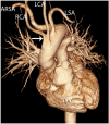
Left aortic arch (LAA) with aberrant right subclavian artery (ARSA) in 66-year-old woman with dysphagia is shown. Anterior (A) and posterior (B) volume rendering images show ARSA with Kommerell's diverticulum at its origin (white arrows). Right carotid artery (RCA) arises as first branch from aortic arch, which is followed by left carotid artery (LCA), left subclavian artery (LSA) and ARSA. Axial (C) and coronal multiplanar reformatted (D) images show presence of aneurysm (An) of Kommerell's diverticulum with mural thrombus (T) and calcifications. There is compression of esophagus (black arrow).
Fig. 4
Fig. 4
Left aortic arch (LAA) with aberrant right subclavian artery in 48-year-old woman with anomalous origin of right vertebral artery from right common carotid artery is shown. Axial image shows retroesophageal course of aberrant right subclavian artery (arrow).
Fig. 5
Fig. 5
Aberrant right subclavian artery in 53-year-old woman with severe aortic coarctation presenting with hypertension and dysphagia. Axial (A) and sagittal multiplanar reformatted (B) images show left aortic arch (LAA) with aberrant right subclavian artery (long arrows) causing esophageal compression (short arrows). Paravertebral collaterals and prominent internal mammarian arteries are also seen. AAo = ascending aorta
Fig. 6
Fig. 6
Aberrant right subclavian artery in 48-year-old man with aortic coarctation presenting with hypertension. Axial oblique (A) and posterior volume rendering (B) images show aberrant right subclavian artery (ARSA) arising just distal to aortic coarctation (arrows). LAA = left aortic arch, LSA = left subclavian artery, LCA = left carotid artery, RCA = right carotid artery
Fig. 7
Fig. 7
Aberrant right subclavian artery in 51-year-old woman with dysphagia is shown. Anterior volume rendering image shows aberrant right subclavian artery (ARSA) and common origin of carotid arteries (arrow). RCA = right carotid artery, LCA = left carotid artery, LSA = left subclavian artery
Fig. 8
Fig. 8
Diagram of development of right aortic arch with aberrant left subclavian artery is shown. Anomaly results from interruption of dorsal segment of left arch between left common carotid artery and left subclavian arteries with regression of right ductus arteriosus in hypothetical double aortic arch. AAo = ascending aorta, DAo = descending aorta, PA = pulmonary artery, T = trachea, E = esophagus, RSA = right subclavian artery, RCA = right carotid artery, LCA = left carotid artery, LSA = left subclavian artery, ALSA = aberrant left subclavian artery
Fig. 9
Fig. 9
Right aortic arch with aberrant left subclavian artery in 45-year-old asymptomatic woman is shown. Anterior (A) and posterior (B) volume rendering images show right aortic arch (RAA) with aberrant left subclavian artery (ALSA) associated with Kommerell's diverticulum (arrows) at its origin. First branch arising from aortic arch is left carotid artery (LCA), which is followed by right carotid artery (RCA), right subclavian artery (RSA) and ALSA.
Fig. 10
Fig. 10
Right aortic arch (RAA) with aberrant left subclavian artery in 63-year-old woman without dysphagia. Axial (A) and sagittal multiplanar reformatted (B) images show aneurysmal Kommerell's diverticulum (KD) with 3.2 cm diameter causing esophageal compression (arrows). AAo = ascending aorta
Fig. 11
Fig. 11
Right aortic arch (RAA) with aberrant left subclavian artery in 52-year-old-woman with dysphagia is presented. Axial image shows aneurysmal Kommerell's diverticulum (KD) with 4 cm diameter.
Fig. 12
Fig. 12
Diagram of development of right aortic arch with mirror image branching. Anomaly results from interruption of dorsal segment of left arch between left subclavian artery and descending aorta with regression of right ductus arteriosus in hypothetical double aortic arch. AAo = ascending aorta, DAo = descending aorta, PA = pulmonary artery, T = trachea, E = esophagus, RSA = right subclavian artery, RCA = right carotid artery, LCA = left carotid artery, LSA = left subclavian artery, LIA = left innominate artery
Fig. 13
Fig. 13
Right aortic arch with mirror image branching in 20-year-old woman with tetralogy of Fallot, aortopulmonary collateral arteries, partial anomalous pulmonary venous connection, double superior vena cava and double inferior vena cava with azygos continuation. Coronal volume rendering (A) and axial (B, C) images show left innominate artery (LIA) is first branch arising from arch, which is followed by right carotid artery (RCA) and right subclavian artery (RSA). Prominent left internal mammarian artery, mediastinal collateral vessels and dilatation of ascending and arcus aorta are also seen. AAo = ascending aorta, LCA = left carotid artery, LSA = left subclavian artery, RAA = right aortic arch
Fig. 14
Fig. 14
Diagram of development of right aortic arch with isolated left subclavian artery is shown. Anomaly results from interruption of left arch at two levels; one level is between left common carotid and left subclavian arteries and other level is distal to attachment of left ductus. Left subclavian artery does not have connection with aorta, but is connected to pulmonary artery by left ductus arteriosus. AAo = ascending aorta, DAo = descending aorta, PA = pulmonary artery, T = trachea, E = esophagus, RSA = right subclavian artery, RCA = right carotid artery, LCA = left carotid artery, LSA = left subclavian artery, ILSA = isolated left subclavian artery
Fig. 15
Fig. 15
Double aortic arch in 31-year-old woman presenting with dysphagia is shown. Volume rendering (A) image shows that right arch is larger and extends more cephalad than left arch. Brachiocephalic arteries arise from their respective arches. Axial maximum intensity projection images (B), coronal multiplanar reformatted images (C, D) and volume rendering image with lower opacity values (E) show presence of complete vascular ring causing esophageal (arrows) and tracheal (T) compression. AAo = ascending aorta, DAo = descending aorta, R = right arch, L = left arch, RCA = right carotid artery, RSA = right subclavian artery, LCA = left carotid artery, LSA = left subclavian artery
Fig. 16
Fig. 16
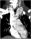
Left cervical aortic arch (CAA) in 27-year-old-woman presenting with pulsatile mass in left neck. Coronal maximum intensity projection image shows apex of left CAA is above level of clavicle. AAo = ascending aorta

References

    1. Grathwohl KW, Afifi AY, Dillard TA, Olson JP, Heric BR. Vascular rings of the thoracic aorta in adults. Am Surg. 1999;65:1077–1083. - PubMed
    1. Lee EY, Siegel MJ, Hildebolt CF, Gutierrez FR, Bhalla S, Fallah JH. MDCT evaluation of thoracic aortic anomalies in pediatric patients and young adults: comparison of axial, multiplanar, and 3D images. AJR Am J Roentgenol. 2004;182:777–784. - PubMed
    1. Gilkeson RC, Ciancibello L, Zahka K. Multidetector CT evaluation of congenital heart disease in pediatric and adult patients. AJR Am J Roentgenol. 2003;180:973–980. - PubMed
    1. Edwards JE. Anomalies of the derivatives of the aortic arch system. Med Clin North Am. 1948;32:925–949. - PubMed
    1. Stewart JR, Kincaid OW, Edwards JE. An atlas of vascular rings and related malformations of the aortic arch system. Illinois: Springfield; 1964. pp. 12–131.

MeSH terms