

Structure of the human telomere in K+ solution: a stable basket-type G-quadruplex with only two G-tetrad layers
- PMID: 19271707
- PMCID: PMC2662591
- DOI: 10.1021/ja807503g
Structure of the human telomere in K+ solution: a stable basket-type G-quadruplex with only two G-tetrad layers
Abstract
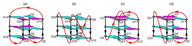
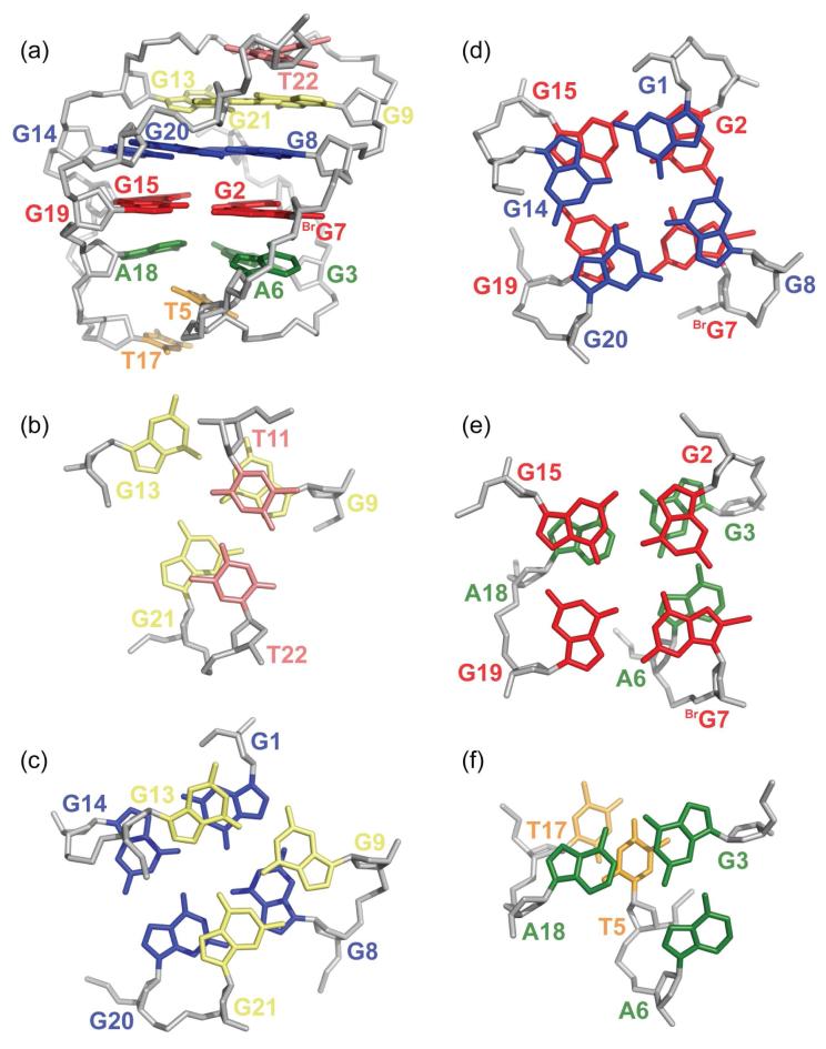
Previously, it has been reported that human telomeric DNA sequences could adopt in different experimental conditions four different intramolecular G-quadruplexes each involving three G-tetrad layers, namely, Na(+) solution antiparallel-stranded basket form, K(+) crystal parallel-stranded propeller form, K(+) solution (3 + 1) Form 1, and K(+) solution (3 + 1) Form 2. Here we present a new intramolecular G-quadruplex adopted by a four-repeat human telomeric sequence in K(+) solution (Form 3). This structure is a basket-type G-quadruplex with only two G-tetrad layers: loops are successively edgewise, diagonal, and edgewise; glycosidic conformations of guanines are syn x syn x anti x anti around each tetrad. Each strand of the core has both a parallel and an antiparallel adjacent strands; there are one narrow, one wide, and two medium grooves. Despite the presence of only two G-tetrads in the core, this structure is more stable than the three-G-tetrad intramolecular G-quadruplexes previously observed for human telomeric sequences in K(+) solution. Detailed structural elucidation of Form 3 revealed extensive base pairing and stacking in the loops capping both ends of the G-tetrad core, which might explain the high stability of the structure. This novel structure highlights the conformational heterogeneity of human telomeric DNA. It establishes a new folding principle for G-quadruplexes and suggests new loop sequences and structures for targeting in human telomeric DNA.
Figures


Similar articles
-
Structure of a two-G-tetrad intramolecular G-quadruplex formed by a variant human telomeric sequence in K+ solution: insights into the interconversion of human telomeric G-quadruplex structures.Nucleic Acids Res. 2010 Jan;38(3):1009-21. doi: 10.1093/nar/gkp1029. Epub 2009 Nov 27. Nucleic Acids Res. 2010. PMID: 19946019 Free PMC article.
-
Human telomeric sequence forms a hybrid-type intramolecular G-quadruplex structure with mixed parallel/antiparallel strands in potassium solution.Nucleic Acids Res. 2006 May 19;34(9):2723-35. doi: 10.1093/nar/gkl348. Print 2006. Nucleic Acids Res. 2006. PMID: 16714449 Free PMC article.
-
Structure of the intramolecular human telomeric G-quadruplex in potassium solution: a novel adenine triple formation.Nucleic Acids Res. 2007;35(7):2440-50. doi: 10.1093/nar/gkm009. Epub 2007 Mar 29. Nucleic Acids Res. 2007. PMID: 17395643 Free PMC article.
-
Human telomere, oncogenic promoter and 5'-UTR G-quadruplexes: diverse higher order DNA and RNA targets for cancer therapeutics.Nucleic Acids Res. 2007;35(22):7429-55. doi: 10.1093/nar/gkm711. Epub 2007 Oct 2. Nucleic Acids Res. 2007. PMID: 17913750 Free PMC article. Review.
-
Polymorphism of human telomeric quadruplex structures.Biochimie. 2008 Aug;90(8):1172-83. doi: 10.1016/j.biochi.2008.02.026. Epub 2008 Mar 8. Biochimie. 2008. PMID: 18373984 Free PMC article. Review.
Cited by
-
A direct view of the complex multi-pathway folding of telomeric G-quadruplexes.Nucleic Acids Res. 2016 Dec 15;44(22):11024-11032. doi: 10.1093/nar/gkw1010. Epub 2016 Oct 30. Nucleic Acids Res. 2016. PMID: 27799468 Free PMC article.
-
Structure of a two-G-tetrad intramolecular G-quadruplex formed by a variant human telomeric sequence in K+ solution: insights into the interconversion of human telomeric G-quadruplex structures.Nucleic Acids Res. 2010 Jan;38(3):1009-21. doi: 10.1093/nar/gkp1029. Epub 2009 Nov 27. Nucleic Acids Res. 2010. PMID: 19946019 Free PMC article.
-
Structure of human telomere G-quadruplex in the presence of a model drug along the thermal unfolding pathway.Nucleic Acids Res. 2018 Dec 14;46(22):11927-11938. doi: 10.1093/nar/gky1092. Nucleic Acids Res. 2018. PMID: 30407585 Free PMC article.
-
Complexity of Guanine Quadruplex Unfolding Pathways Revealed by Atomistic Pulling Simulations.J Chem Inf Model. 2023 Aug 14;63(15):4716-4731. doi: 10.1021/acs.jcim.3c00171. Epub 2023 Jul 17. J Chem Inf Model. 2023. PMID: 37458574 Free PMC article.
-
Mapping the sequences of potential guanine quadruplex motifs.Nucleic Acids Res. 2011 Jul;39(12):4917-27. doi: 10.1093/nar/gkr104. Epub 2011 Feb 26. Nucleic Acids Res. 2011. PMID: 21357607 Free PMC article.
References
Publication types
MeSH terms
Substances
Associated data
- Actions
- Actions
Grants and funding
LinkOut - more resources
Full Text Sources
Other Literature Sources
Medical
Miscellaneous
