Review
. 2009 Jul;11(7):1603-14.
doi: 10.1089/ars.2009.2536.

Aggregation of copper-zinc superoxide dismutase in familial and sporadic ALS

Affiliations
Review

Aggregation of copper-zinc superoxide dismutase in familial and sporadic ALS

Madhuri Chattopadhyay et al. Antioxid Redox Signal. 2009 Jul.

Abstract

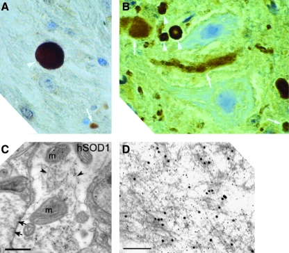
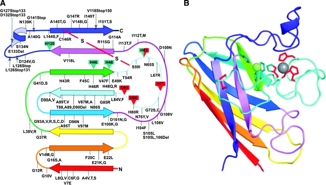
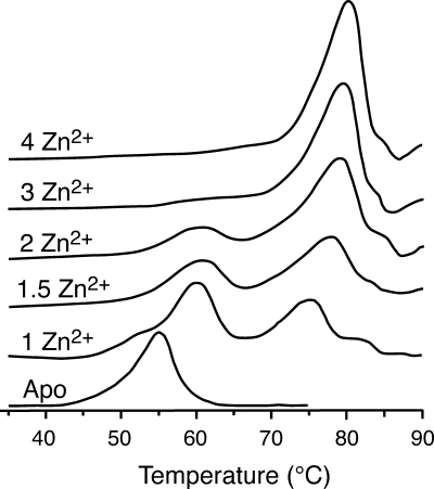
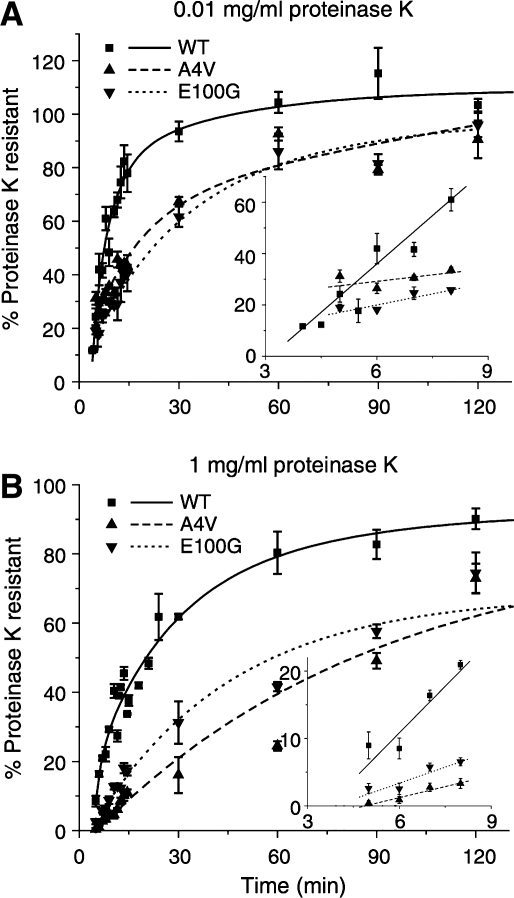
Amyotrophic lateral sclerosis (ALS) is a progressive, fatal neurodegenerative disease characterized by the selective death of motor neurons. While the most common form of ALS is sporadic and has no known cause, a small subset of cases is familial because of underlying genetic mutations. The best-studies example of familial ALS is that caused by mutations in the protein copper-zinc superoxide dismutase. The formation of SOD1-rich inclusions in the spinal cord is an early and prominent feature of SOD1-linked familial ALS in human patients and animal models of this disease. These inclusions have been shown to consist of SOD1-rich fibrils, suggesting that the conversion of soluble SOD1 into amyloid fibrils may play an important role in the etiology of familial ALS. SOD1 is also present in inclusions found in spinal cords of sporadic ALS patients, allowing speculations to arise regarding a possible involvement of SOD1 in the sporadic form of this disease. We here review the recent research on the significance, causes, and mechanisms of SOD1 fibril formation from a biophysical perspective.
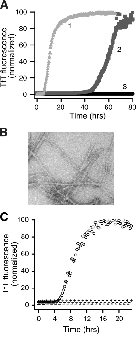

Figures

FIG. 1.
FIG. 1.
SOD1-containing inclusions and fibrils from human patients and transgenic mice. (A) An inclusion from the spinal cord of a fALS patient expressing a frameshift mutation at position 126 in SOD1, who died from disease, shows strong SOD1 staining (white arrowheads) when reacted with an antibody raised against an SOD1 peptide (Reprinted by permission of Bruijn et al. (15)). (B) SOD1-positive inclusions (arrowheads) from the spinal cords of end-stage transgenic mice expressing SOD1 mutants G85R (left) visualized by staining with an antibody recognizing both mouse and human SOD1 (Reprinted by permission of Bruijn et al. (adapted, 15). (C) Postembedding immunogold electron microscopy shows the fibrillar nature of inclusions (arrowheads) that are immunoreactive for both ubiquitin and human SOD1 in dendritic processes in the spinal cords of G93A mice. The letter ‘m’ denotes mitochondria. Scale bar corresponds to 500 nm. (Reprinted by permission of Jaarsma et al. (37)). (D) Fibrillar aggregates in G93A mice are recognized by antibodies that recognize human SOD1 only. (Immunogold electron microscopy, scale bar corresponds to 500 nm. [Reprinted by permission of Basso et al. (10)]. (For interpretation of the references to color in this figure legend, the reader is referred to the web version of this article at www.liebertonline.com/ars).
FIG. 2.
FIG. 2.
Secondary structural representation of SOD1. A diagram showing the locations of fALS-associated mutations (A) and the structure of a monomer of SOD1 (B) colored to match the drawing on the left. Copper ligands are shown in green and zinc ligands shown in red. Copper and zinc ions are shown as green and gray spheres, respectively, and the intrasubunit disulfide bond is shown in red. Point mutation, deletions, and insertions are indicated with a line, whereas mutations that cause C-terminal truncations are shown as scissor cuts at the point of the stop codon [Reprinted by permission of Valentine et al. (67)]. (For interpretation of the references to color in this figure legend, the reader is referred to the web version of this article at www.liebertonline.com/ars).
FIG. 3.
FIG. 3.
DSC scans of apo and zinc derivatives of hSOD1 in phosphate buffer, pH 7.0, showing the transition from two peaks when two or fewer equivalents of zinc are bound to one peak when three or four equivalents of zinc are bound per dimer [adapted from Potter et al. (55)].
FIG. 4.
FIG. 4.
The slower folding and dimerization kinetics for fALS mutants compared to wild type SOD1, as shown by proteinase K sensitivity. (A) Rates of protease resistance for wild-type SOD1 and mutants A4V and E100G at low protease concentration, showing the folding rate of the initial hydrophobic nucleus. (B) Rates of protease resistance for the same proteins at high protease concentration, showing the rate of formation of the dimeric form. Insets show the initial time points [Reprinted by permission of Bruns et al. (17)].
FIG. 5.
FIG. 5.
Fibrillation of SOD1 is induced by mildly reducing conditions or small amounts of disulfide-reduced, apo SOD1 protein. (A) Increase in TfT florescence indicates fibrillation of 50 μM apo SOD1 in 10 mM potassium phosphate buffer, pH 7.4, incubated with constant agitation at 37°C in 96-well plates with 50 mM DTT (line 1, light gray), 1 M GdmHCl (line 2, medium gray), or no addition (line 3, black). (B) Electron micrograph of SOD1 fibrils generated in the presence of 5 mM DTT. (C) Disulfide-reduced SOD1 with free thiol groups initiates fibrillation in the absence of reducing agent. SOD1SH-SH and SOD1S-S generated during the lag phase of a fibrillation reaction in 5 mM DTT were purified by HPLC, either not treated or treated with N-ethylmaleimide (NEM) to block free thiols, and added to fibrillation reactions without reducing agent. Apo-SOD1 (47.5 μM) in phosphate buffer was incubated with 2.5 μM of SOD1 in the following forms: SOD12SH, ○; SOD1S-S, ▵; SOD12SH-4NEM, −; or SOD1S-S-2NEM, +. [Adapted and reprinted by permission of Chattopadhyay et al. (20)].

References

    1. Antonyuk S. Elam JS. Hough MA. Strange RW. Doucette PA. Rodriguez JA. Hayward LJ. Valentine JS. Hart PJ. Hasnain SS. Structural consequences of the familial amyotrophic lateral sclerosis SOD1 mutant His46Arg. Prot Sc. 2005;14:1201–1213. - PMC - PubMed
    1. Arnesano F. Banci L. Bertini I. Martinelli M. Furukawa Y. O'Halloran TV. The unusually stable quaternary structure of human Cu, Zn-superoxide dismutase 1 is controlled by both metal occupancy and disulfide status. J Biol Chem. 2004;279:47998–48003. - PubMed
    1. Assfalg M. Banci L. Bertini I. Turano P. Vasos PR. Superoxide dismutase folding/unfolding pathway: role of the metal Ions in modulating structural and dynamical features. J Mol Biol. 2003;330:145–158. - PubMed
    1. Banci L. Bertini I. Boca M. Girotto S. Martinelli M. Valentine JS. Vieru M. SOD1 and amyotrophic lateral sclerosis: Mutations and oligomerization. PLoS One. 2008;3:e1677. - PMC - PubMed
    1. Banci L. Bertini I. Cantini F. D'Amelio N. Gaggelli E. Human SOD1 before harboring the catalytic metal: Solution structure of copper depleted, disulfide-reduced form. J Biol Chem. 2006;281:2333–2337. - PubMed

Publication types

Substances