. 2009 Apr;20(4):303-13.
doi: 10.1089/hum.2008.124.

Treatment with LL-37 peptide enhances antitumor effects induced by CpG oligodeoxynucleotides against ovarian cancer

Affiliations

Treatment with LL-37 peptide enhances antitumor effects induced by CpG oligodeoxynucleotides against ovarian cancer

Chi-Mu Chuang et al. Hum Gene Ther. 2009 Apr.

Abstract

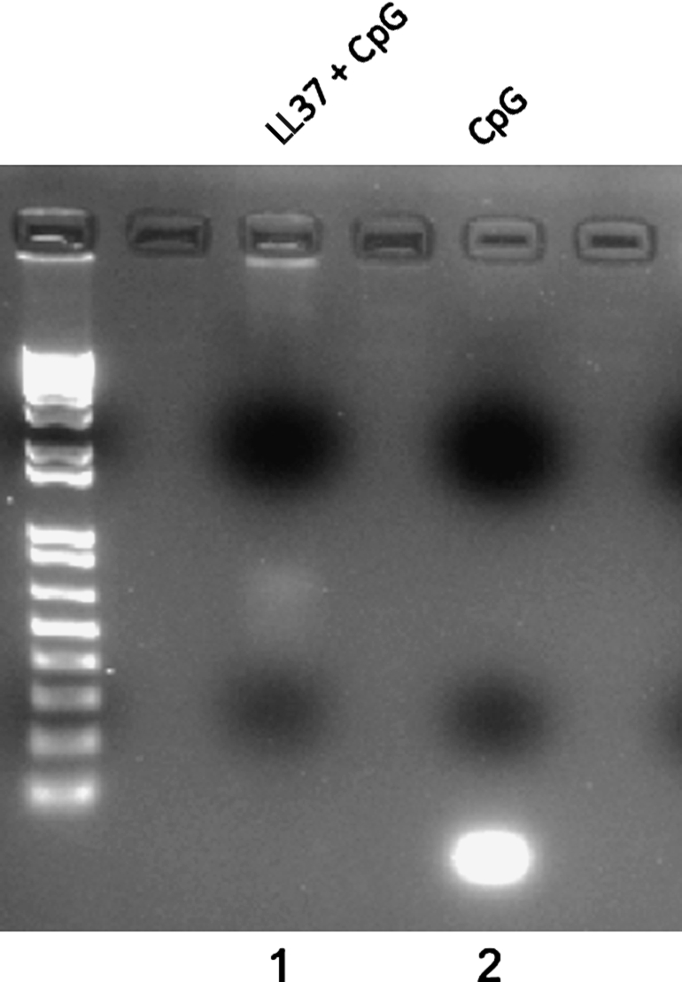
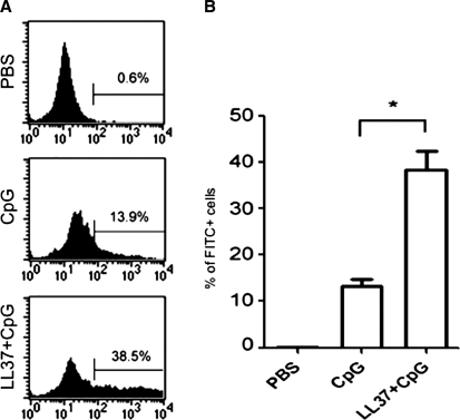
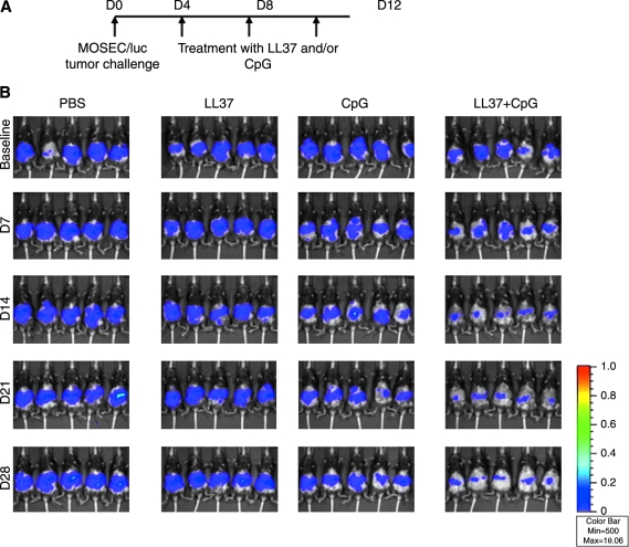
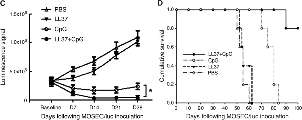
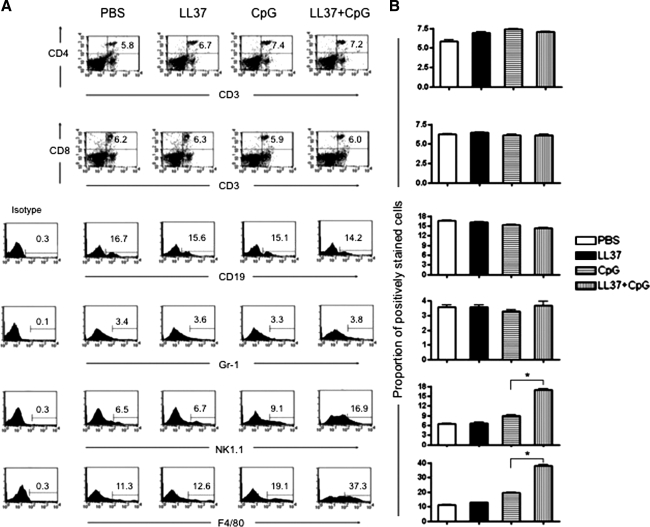
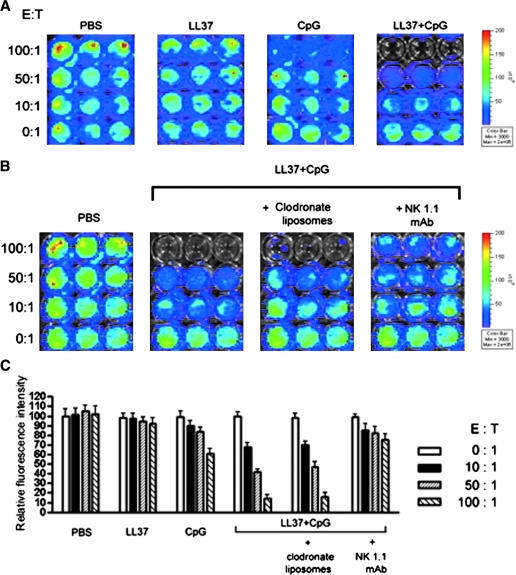
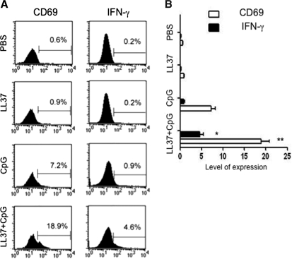
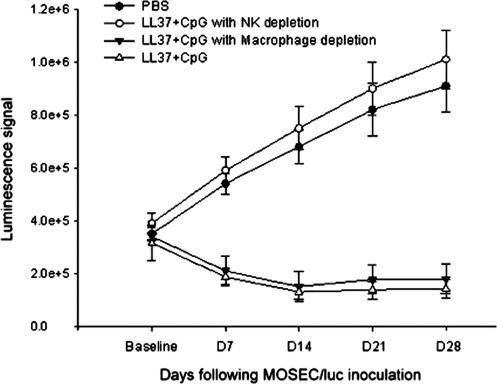
There is an urgent need for innovative therapies against ovarian cancer, one of the leading causes of death from gynecological cancers in the United States. Immunotherapy employing Toll-like receptor (TLR) ligands, such as CpG oligodeoxynucleotides (CpG-ODN), may serve as a potentially promising approach in the control of ovarian tumors. The CpG-ODN requires intracellular delivery into the endosomal compartment, where it can bind to TLR9 in order to activate the immune system. In the current study, we aim to investigate whether the antimicrobial polypeptide from the cathelicidin family, LL-37, could enhance the immunostimulatory effects of CpG-ODN by increasing the uptake of CpG-ODN into the immune cells, thus enhancing the antitumor effects against ovarian cancer. We found that treatment with the combination of CpG-ODN and LL-37 generated significantly better therapeutic antitumor effects and enhanced survival in murine ovarian tumor-bearing mice compared with treatment with CpG-ODN or LL-37 alone. We also observed that treatment with the combination of CpG-ODN and LL-37 enhanced proliferation and activation of natural killer (NK) cells, but not CD4(+) or CD8(+) T cells, in the peritoneal cavity. Furthermore, in vivo antibody depletion experiments indicated that peritoneal NK cells played a critical role in the observed antitumor effects. Thus, our data suggest that the combination of CpG-ODN with LL-37 peptide may lead to the control of ovarian tumors through the activation of innate immunity.


Figures

FIG. 1.
FIG. 1.
Gel retardation assay demonstrating that LL-37 forms a complex with CpG. LL-37 was mixed with CpG at a 1:5 (peptide–DNA) mass ratio (lane 1) and incubated for 30 min. Complex formation was assayed by running samples on gels. CpG alone was loaded as a control (lane 2). Note: Addition of LL-37 to CpG-ODN leads to retardation of the migration of CpG-ODN.
FIG. 2.
FIG. 2.
Flow cytometric analysis to characterize the uptake of FITC-labeled CpG into peritoneal effector cells with or without LL-37. Single-cell suspensions (1 × 106/ml) were made from peritoneal cells harvested from naive mice and seeded into a 24-well plate. PBS, FITC-conjugated CpG-ODN (10 μg/ml), or FITC-conjugated CpG-ODN combined with LL-37 (50 μg/ml) were added to each of triplicate wells and incubated at 37°C for 1 hr. Cells were then recovered, washed with phosphate-buffered saline, and analyzed by flow cytometry. Analysis was performed on gated lymphocytes. (A) Representative flow cytometry for each indicated reagent. (B) Bar graph depicting the percentage of FITC-positive cells (*p < 0.05). Data shown are representative of two separate experiments performed (mean ± SD).
FIG. 3.
FIG. 3.
In vivo tumor treatment experiments. Groups of C57BL/6 mice (five per group) were inoculated with murine ovarian cancer cells (2 × 105 per mouse) that express luciferase (MOSEC/luc). Four, 8, and 12 days after tumor inoculation, each mouse was intraperitoneally administered PBS, LL-37 alone (100 μg/dose per mouse), CpG-ODN alone (30 μg/dose per mouse), or CpG-ODN plus LL-37. Mice were imaged with the IVIS imaging system series 200 to monitor tumor growth. Bioluminescence signals were acquired for 1 min. (A) Schematic diagram of the treatment regimen. (B) Luminescence images of representative mice challenged with MOSEC/luc cells and treated according to the various treatment regimens. (C) Line graph depicting the luminescence intensity in MOSEC/luc tumor-bearing mice treated according to the various treatment regimens (means ± SD) (*p < 0.05). (D) Kaplan–Meier survival analysis of MOSEC/luc tumor-bearing mice treated according to the various treatment regimens. For gross morphological analysis of the tumor-bearing mice treated with the various regimens, see Supplementary Fig.1 at www.liebertonline.com/hum.
FIG. 3.
FIG. 3.
In vivo tumor treatment experiments. Groups of C57BL/6 mice (five per group) were inoculated with murine ovarian cancer cells (2 × 105 per mouse) that express luciferase (MOSEC/luc). Four, 8, and 12 days after tumor inoculation, each mouse was intraperitoneally administered PBS, LL-37 alone (100 μg/dose per mouse), CpG-ODN alone (30 μg/dose per mouse), or CpG-ODN plus LL-37. Mice were imaged with the IVIS imaging system series 200 to monitor tumor growth. Bioluminescence signals were acquired for 1 min. (A) Schematic diagram of the treatment regimen. (B) Luminescence images of representative mice challenged with MOSEC/luc cells and treated according to the various treatment regimens. (C) Line graph depicting the luminescence intensity in MOSEC/luc tumor-bearing mice treated according to the various treatment regimens (means ± SD) (*p < 0.05). (D) Kaplan–Meier survival analysis of MOSEC/luc tumor-bearing mice treated according to the various treatment regimens. For gross morphological analysis of the tumor-bearing mice treated with the various regimens, see Supplementary Fig.1 at www.liebertonline.com/hum.
FIG. 4.
FIG. 4.
Flow cytometric analysis to determine numbers of the various cell types in the peritoneal cavity after LL-37 plus CpG-ODN treatment. Groups of naive C57BL/6 mice (five per group) were treated either with PBS, LL-37 alone, CpG-ODN alone, or CpG-ODN plus LL-37 as described in Fig. 3. Two days after the last treatment, cells from the peritoneal cavity were harvested, counted, washed once in FACScan buffer, and stained with surface markers for innate and adaptive effectors including PE-conjugated anti-CD4 (L3T4), PE-conjugated anti-CD8 (53-6.7), FITC-conjugated anti-GR-1 (RB6-8C5), PE-conjugated anti-CD19 (1D3), PE-conjugated anti-NK1.1 (PK136), and PE–Cy5-conjugated anti-F4/80 (BM8). Numbers of the various peritoneal cell types were determined by flow cytometric analysis. (A) Representative flow cytometric analysis demonstrating numbers of the various peritoneal cell types after treatment with LL-37 plus CpG-ODN. (B) Bar graph depicting numbers of the various peritoneal cell types after treatment with LL-37 plus CpG-ODN. *p < 0.05 (means ± SD). Data shown are representative of two experiments performed.
FIG. 5.
FIG. 5.
In vitro cytotoxicity assay using luminescence imaging. (A) Representative luminescence images depicting the cytotoxicity of MOSEC/luc tumor cells. Groups of mice (three per group) were treated with PBS, LL-37 alone, CpG-ODN alone, or CpG-ODN plus LL-37 on days 0, 4, and 8. Two days after the last treatment (day 10), cells from the peritoneal cavity were harvested, washed, and counted. MOSEC/luc cells (at 60–80% confluence) were seeded into a round-bottom 96-well microplate at 5 × 103 per well in complete medium. After 2 hr, counted cells from the peritoneal cavity were then added to each well in triplicate at titrated effector-to-target ratios (0:1, 10:1, 50:1, and 100:1). Sixteen hours after incubation at 37°C with 5% CO2, the plates were imaged for bioluminescence activity. (B) Representative luminescence images depicting the cytotoxicity of MOSEC/luc tumor cells after depletion of macrophages or NK cells. Groups of mice (three per group) were administered CpG-ODN plus LL-37 on days 0, 4, and 8. Mice were depleted of peritoneal macrophages, using clodronate liposomes, or of NK cells, using NK1.1 antibody, on days 0 and 4. Undepleted mice were used as negative controls. Two days after the last treatment (day 10), cells from the peritoneal cavity were added to MOSEC/luc cells followed by luminescence imaging as described previously. (C) Bar graph depicting the relative fluorescence intensities of the MOSEC/luc tumor cells in the various treatment groups. Data shown are representative of two experiments performed.
FIG. 6.
FIG. 6.
Flow cytometric analysis demonstrating the expression of CD69 and IFN-γ on peritoneal NK cells stimulated with LL-37 plus CpG-ODN. Cells from the peritoneal cavity were harvested from naive mice, and seeded into round-bottom 24-well plates at a density of 1 × 106/well and stimulated with either PBS, LL-37 alone (50 μg/ml), CpG-ODN alone (10 μg/ml), or CpG-ODN plus LL-37. Stimulated cells were then retrieved after 16 hr of incubation and stained for CD69 and IFN-γ. NK cells were gated and analyzed by flow cytometric analysis. (A) Representative flow cytometric data demonstrating the expression of CD69 and IFN-γ on NK cells stimulated with the various reagents. (B) Bar graph depicting the level of expression of CD69 and IFN-γ on NK cells stimulated with the various reagents. *p < 0.05; **p <0.01. Data shown are representative of two experiments performed.
FIG. 7.
FIG. 7.
In vivo antibody depletion experiment. Groups of C57BL/6 mice (five per group) were inoculated with MOSEC/luc (2 × 105 per mouse) and treated with PBS or CpG-ODN plus LL-37 on days 4, 8, and 12 as depicted in Fig. 3. Mice were either depleted of peritoneal macrophages, using clodronate liposomes, or of NK cells, using anti-mouse NK1.1 monoclonal antibody (PK136), 1 day before and 3 days after the first treatment and thereafter once per week until the end of follow-up as described in Materials and Methods. Undepleted mice were used as a negative control. Mice were imaged with the IVIS imaging system series 200 every 7 days. Bioluminescence signals were acquired for 1 min. Shown is a line graph depicting the quantification of luminescence activity in the tumors of tumor-challenged mice treated with PBS or with CpG-ODN plus LL-37 and depleted of peritoneal macrophages or NK cells (means ± SD).

Similar articles

Cited by

References

    1. Aderem A. Ulevitch R.J. Toll-like receptors in the induction of the innate immune response. Nature. 2000;406:782–787. - PubMed
    1. Akira S. Takeda K. Toll-like receptor signalling. Nat. Rev. 2004;4:499–511. - PubMed
    1. Akira S. Takeda K. Kaisho T. Toll-like receptors: Critical proteins linking innate and acquired immunity. Nat. Immunol. 2001;2:675–680. - PubMed
    1. Ballas Z.K. Modulation of NK cell activity by CpG oligodeoxynucleotides. Immunologic Res. 2007;39:15–21. - PubMed
    1. Ballas Z.K. Rasmussen W.L. Krieg A.M. Induction of NK activity in murine and human cells by CpG motifs in oligodeoxynucleotides and bacterial DNA. J. Immunol. 1996;157:1840–1845. - PubMed

Publication types

MeSH terms