. 2009 Mar 9:6:26.
doi: 10.1186/1742-4690-6-26.

Suppression of HIV-1 replication by microRNA effectors

Affiliations

Suppression of HIV-1 replication by microRNA effectors

Christine Chable-Bessia et al. Retrovirology. .

Abstract

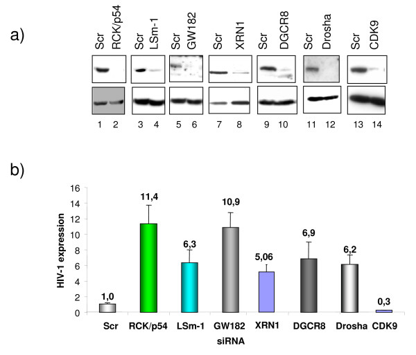
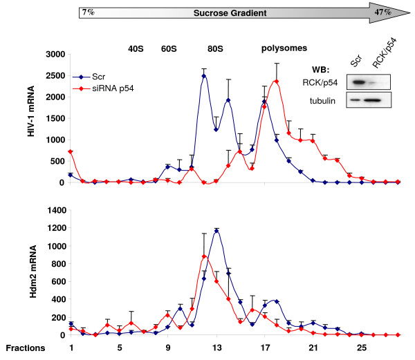
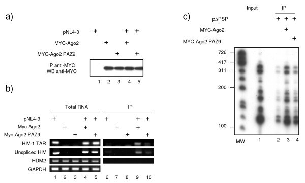
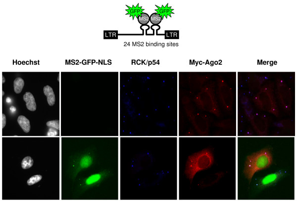
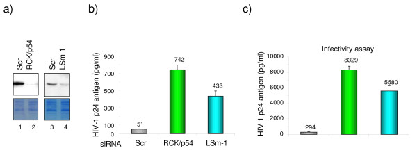
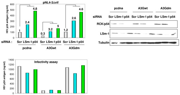
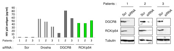
The rate of HIV-1 gene expression is a key step that determines the kinetics of virus spread and AIDS progression. Viral entry and gene expression were described to be the key determinants for cell permissiveness to HIV. Recent reports highlighted the involvement of miRNA in regulating HIV-1 replication post-transcriptionally. In this study we explored the role of cellular factors required for miRNA-mediated mRNA translational inhibition in regulating HIV-1 gene expression. Here we show that HIV-1 mRNAs associate and co-localize with components of the RNA Induced Silencing Complex (RISC), and we characterize some of the proteins required for miRNA-mediated silencing (miRNA effectors). RCK/p54, GW182, LSm-1 and XRN1 negatively regulate HIV-1 gene expression by preventing viral mRNA association with polysomes. Interestingly, knockdown of RCK/p54 or DGCR8 resulted in virus reactivation in PBMCs isolated from HIV infected patients treated with suppressive HAART.


Figures

Figure 1
Figure 1
miRNA effectors are repressors of HIV-1 replication. HeLa cells were transfected with siRNA as indicated. 48 hours post transfection, cells were analyzed for RCK/p54, LSm-1, GW182, XRN1, DGCR8, DROSHA and CDK9 expression by Western blotting (a), or infected with a single round infectious virus (HIV-1-VSV-luc 200 ng/ml) and cell extracts were measured for luciferase activity 48 hours after infection (b). Results are presented as fold HIV production relative to Scr transfected cells, and data are representative of three independent experiments.
Figure 2
Figure 2
RCK/p54 restricts HIV-1 mRNA association with polysomes. Cytoplasmic extracts from HeLa cells that were transfected with the indicated siRNA and infected with HIV-1-VSVG-luc were run on glycerol gradient (7% to 47%). Fractions were collected and their RNA contents were monitored by measuring absorbance at 254 nm. HIV-1 mRNA (top panel) and Hdm2 mRNA (lower panel) were quantified in all the fractions by Q-RT-PCR using specific oligonucleotides.
Figure 3
Figure 3
HIV-1 mRNAs associate with Argonaute 2. 293 cells were transfected with HIV-1 molecular clone pNL4-3, Myc-Ago2 or Myc-AgoPAZ9 as indicated. 48 hours later cells were harvested and cytoplasmic extracts were prepared. Total RNA was purified from a fraction of harvested cells while the rest was subjected to immunoprecipitation using anti-Myc antibody. After washing, a fraction was used to analyze the amount of Myc-Ago2 and Myc-Ago2PAZ9 immunoprecipitated by Western blotting (a), and the rest of the Myc-IPs was used for RNA extraction. HIV-1 mRNAs (TAR and unspliced), Hdm2 and GAPDH mRNA were quantified from total RNA (b, left panel) or from Myc immunoprecipitated mRNPs (b, right panel) by RT-PCR using specific oligonucleotides. c) Experiment was performed as in fig 3 except that 293 cells were transfected with HIV-1 ΔPSP which contains a partial gag/pol deletion but retains all the mRNA splicing sites [66], and 32P-labelled nucleotides were used in the PCR reaction. PCR products were visualized by autoradiography.
Figure 4
Figure 4
HIV-1 mRNA co-localizes with RCK/p54 and Ago2. HeLa cells were transfected with Myc-Ago2 expression vector either alone (top panels) or co-transfected with HIV-1 vector containing 24 repeats of MS2 binding sites and MS2-GFP expression vectors [64,65] (lower panels). Endogenous RCK/p54 and transfected Myc-Ago2 were visualized using specific primary antibodies and appropriate secondary antibodies coupled with Cy5 (shown in blue) and Cy3 (red) respectively. HIV-1 RNA bound to MS2-GFP is shown in green. Green, blue and red merged images are shown.
Figure 5
Figure 5
Disruption of P-bodies through knockdown of RCK/p54 and LSm-1 leads to enhanced production of infectious HIV-1 virions. HeLa CD4+ cells were transfected with siRNA as indicated. 48 hours post transfection cells were analyzed for RCK/p54 and LSm-1 expression by Western blotting (a) and infected with equal amounts of HIV-1 (200 ng/ml). b) Virus production was monitored 48 hours post infection by measuring p24 antigen in culture supernatant. c) To analyze the infectivity of new progeny virions, equal volumes of supernatant from siRNA transfected Hela CD4+cells were used to re-infect HeLa CD4+ cells. P24 antigen was measured in culture supernatant 48 hours post infection.
Figure 6
Figure 6
RNAi effectors and APOBEC 3G-mediated HIV-1 repression involve different pathways. HeLa CD4+ cells were transfected with the indicated siRNA. 48 hours later cells were analyzed for RCK/p54 and LSm-1 expression (right panel) or co-transfected with 1 μg of pNL4-3Δvif (lacking vif gene) and pcDNA or expression vectors for wild-type APOBEC3G or APOBEC3G double mutant lacking both deaminase and antiviral activity, A3G H65R/H257R [63]. HIV-1 production was measured 24 hours post-transfection in culture supernatant by quantifying p24 antigen (top left panel). Numbers on the top of the columns are fold increase relative to the respective Scr. Numbers on the top of Scr samples in A3Gwt and A3Gdm represent fold increase relative to Scr in pCDNA transfected cells. Infectivity assay was performed using equal amounts of p24 antigen to infect HeLa CD4+ cells. HIV-1 p24 antigen was measured 48 hours post infection (lower left panel). A representative experiment out of five is shown.
Figure 7
Figure 7
Implication of RNAi in HIV-1 latency. PBMCs were isolated from three patients undergoing active HAART. Isolated PBMCs were transfected with the indicated siRNA and either analyzed for RCK/p54, DGCR8 and DROSHA expression by Western blotting 48 hours after transfection (right panel) or co-cultured with activated PBMCs obtained from healthy donors. Virus replication was monitored every 3 to 4 days post co-culture by measuring p24 antigen in culture supernatant. Shown is the amount of p24 antigen at day 15 post co-culture. No virus was isolated from Scr transfected-PBMCs for up to 27 days.

References

    1. Brodersen P, Sakvarelidze-Achard L, Bruun-Rasmussen M, Dunoyer P, Yamamoto YY, Sieburth L, Voinnet O. Widespread translational inhibition by plant miRNAs and siRNAs. Science. 2008;320:1185–1190. doi: 10.1126/science.1159151. - DOI - PubMed
    1. Filipowicz W, Bhattacharyya SN, Sonenberg N. Mechanisms of post-transcriptional regulation by microRNAs: are the answers in sight? Nat Rev Genet. 2008;9:102–114. doi: 10.1038/nrg2290. - DOI - PubMed
    1. Tam OH, Aravin AA, Stein P, Girard A, Murchison EP, Cheloufi S, Hodges E, Anger M, Sachidanandam R, Schultz RM, Hannon GJ. Pseudogene-derived small interfering RNAs regulate gene expression in mouse oocytes. Nature. 2008;453:534–538. doi: 10.1038/nature06904. - DOI - PMC - PubMed
    1. Stefani G, Slack FJ. Small non-coding RNAs in animal development. Nat Rev Mol Cell Biol. 2008;9:219–230. doi: 10.1038/nrm2347. - DOI - PubMed
    1. Okamura K, Lai EC. Endogenous small interfering RNAs in animals. Nat Rev Mol Cell Biol. 2008;9:673–678. doi: 10.1038/nrm2479. - DOI - PMC - PubMed

Publication types

MeSH terms

LinkOut - more resources