. 2009;11(2):R15.
doi: 10.1186/bcr2234. Epub 2009 Mar 9.

T-cell metagene predicts a favorable prognosis in estrogen receptor-negative and HER2-positive breast cancers

Affiliations

T-cell metagene predicts a favorable prognosis in estrogen receptor-negative and HER2-positive breast cancers

Achim Rody et al. Breast Cancer Res. 2009.

Abstract

Introduction: Lymphocyte infiltration (LI) is often seen in breast cancer but its importance remains controversial. A positive correlation of human epidermal growth factor receptor 2 (HER2) amplification and LI has been described, which was associated with a more favorable outcome. However, specific lymphocytes might also promote tumor progression by shifting the cytokine milieu in the tumor.

Methods: Affymetrix HG-U133A microarray data of 1,781 primary breast cancer samples from 12 datasets were included. The correlation of immune system-related metagenes with different immune cells, clinical parameters, and survival was analyzed.

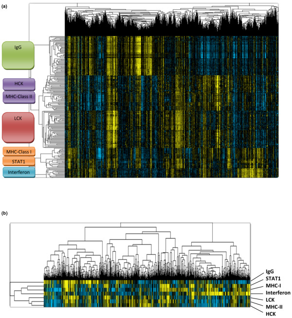
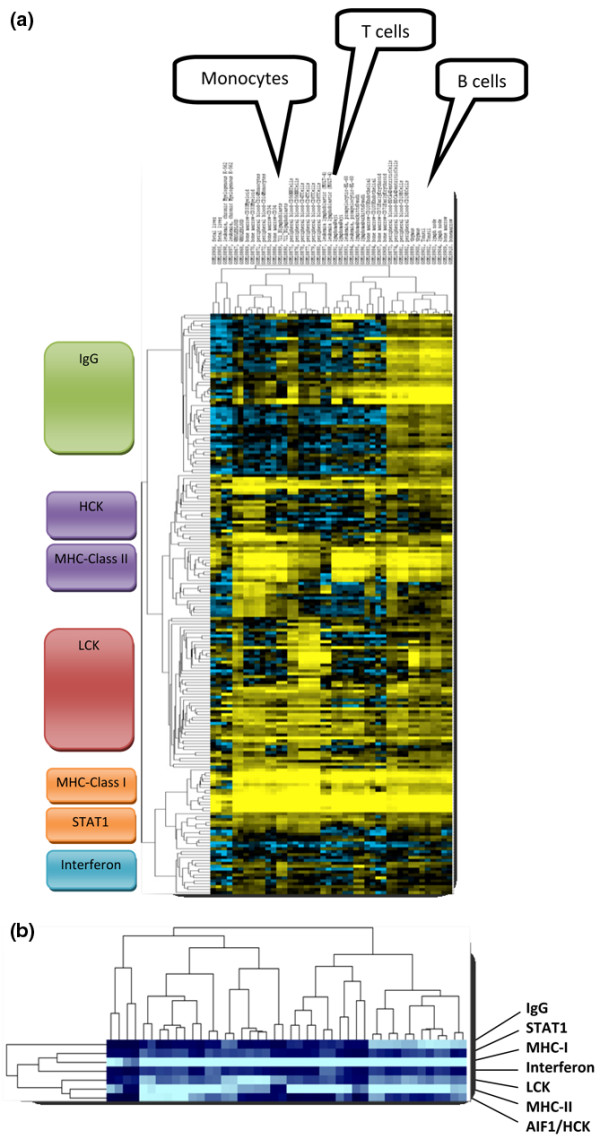
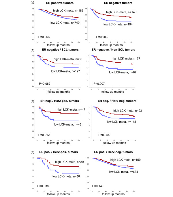
Results: A large cluster of nearly 600 genes with functions in immune cells was consistently obtained in all datasets. Seven robust metagenes from this cluster can act as surrogate markers for the amount of different immune cell types in the breast cancer sample. An IgG metagene as a marker for B cells had no significant prognostic value. In contrast, a strong positive prognostic value for the T-cell surrogate marker (lymphocyte-specific kinase (LCK) metagene) was observed among all estrogen receptor (ER)-negative tumors and those ER-positive tumors with a HER2 overexpression. Moreover ER-negative tumors with high expression of both IgG and LCK metagenes seem to respond better to neoadjuvant chemotherapy.

Conclusions: Precise definitions of the specific subtypes of immune cells in the tumor can be accomplished from microarray data. These surrogate markers define subgroups of tumors with different prognosis. Importantly, all known prognostic gene signatures uniformly assign poor prognosis to all ER-negative tumors. In contrast, the LCK metagene actually separates the ER-negative group into better or worse prognosis.


Figures

Figure 1
Figure 1
Identification of immune-system-related metagenes. (a) To identify metagenes for the principal expression vectors we selected those gene clusters that encompassed at least 10 elements and displayed a minimal average correlation of 0.7 from the larger data matrix of 569 ProbeSets (see Additional data file 3). Expression of these selected 199 ProbeSets among the 1,230 breast cancer samples is shown. HCK, hemopoietic cell kinase; LCK, lymphocyte-specific kinase; MHC, major histocompatibility complex; STAT1, signal transducer and activator of transcription 1. (b) Seven metagenes were derived as mean values of all 199 ProbeSets from the seven clusters.
Figure 2
Figure 2
Expression of the metagene clusters in immunological cell types. (a) The 199 ProbeSets from Figure 1a were used to cluster 44 samples of isolated cells and tissues with immune-system-related functions that were profiled on Affymetrix U133A arrays by Su and colleagues [GEO:GSE1133] [31]. In each case, two samples for the following cell/tissue types are presented from left to right: fetal liver (1,2), K-562 (3,4), whole blood (5,6), CD33 myeloid (7,8), CD14 monocytes (9,10), CD34 (11,12), B lymphoblasts (13,14), CD56 natural killer cells (15,16), CD4 T cells (17,18), CD8 T cells (19,20), MOLT-4 (21,22), Raji (23,24), HL-60 (25,26), Daudi (27,28), CD105 (29,30), CD71 (31,32), BDCA4 dendritic cells (33,34), CD19 B cells (35,36), thymus (37,38), tonsil (39,40), lymph node (41,42), bone marrow (43,44). Details about the respective samples are given in Additional data file 10. HCK, hemopoietic cell kinase; LCK, lymphocyte-specific kinase; MHC, major histocompatibility complex; STAT1, signal transducer and activator of transcription 1. (b) Representation of the seven metagenes that were derived from the 199 ProbeSets as in Figure 1b.
Figure 3
Figure 3
Verification of microarray results by histological examination. (a) Example of the verification of lymphocytic infiltration by immunohistochemistry (Frankfurt dataset). Consecutive sections of a tumor sample with high expression of both IgG and lymphocyte-specific kinase (LCK) metagenes stained with antibodies against either CD20 or CD3 to detect B lymphocytes and T lymphocytes, respectively. (b) Validation of the correlation of immune-system-related metagenes and lymphocytic infiltration in independent data. Expression of different metagenes compared with pathological information on lymphocytic infiltration (LI score) from the London dataset (Desmedt and colleagues [26], n = 35). P values determined using the Kruskal–Wallis H test. STAT1, signal transducer and activator of transcription 1.
Figure 4
Figure 4
Prognostic value of the lymphocyte-specific kinase metagene in subgroups of breast cancer patients. Samples of the combined dataset were stratified according to the highest quartile of expression of the lymphocyte-specific kinase (LCK) metagene. Kaplan–Meier analyses of disease-free survival were performed in different tumor subgroups according to estrogen receptor (ER), human epidermal growth factor receptor 2 (HER2), and stem-cell like (SCL) status. (a) The LCK metagene had a highly significant prognostic value among ER-negative samples. This high prognostic value was observed in all ER-negative samples independently of (b) their expression of SCL markers or (c) their HER2 status. (d) In addition, a high prognostic value of LCK metagene expression was also found in ER-positive HER2-positive samples.

References

    1. Coussens LM, Werb Z. Inflammation and cancer. Nature. 2002;420:860–867. doi: 10.1038/nature01322. - DOI - PMC - PubMed
    1. Meylan E, Tschopp J, Karin M. Intracellular pattern recognition receptors in the host response. Nature. 2006;442:39–44. doi: 10.1038/nature04946. - DOI - PubMed
    1. de Visser KE, Eichten A, Coussens LM. Paradoxical roles of the immune system during cancer development. Nat Rev Cancer. 2006;6:24–37. doi: 10.1038/nrc1782. - DOI - PubMed
    1. Coronella-Wood JA, Hersh EM. Naturally occurring B-cell responses to breast cancer. Cancer Immunol Immunother. 2003;52:715–738. doi: 10.1007/s00262-003-0409-4. - DOI - PMC - PubMed
    1. Wong PY, Staren ED, Tereshkova N, Braun DP. Functional analysis of tumor-infiltrating leukocytes in breast cancer patients. J Surg Res. 1998;76:95–103. doi: 10.1006/jsre.1998.5301. - DOI - PubMed

Publication types

MeSH terms

Substances